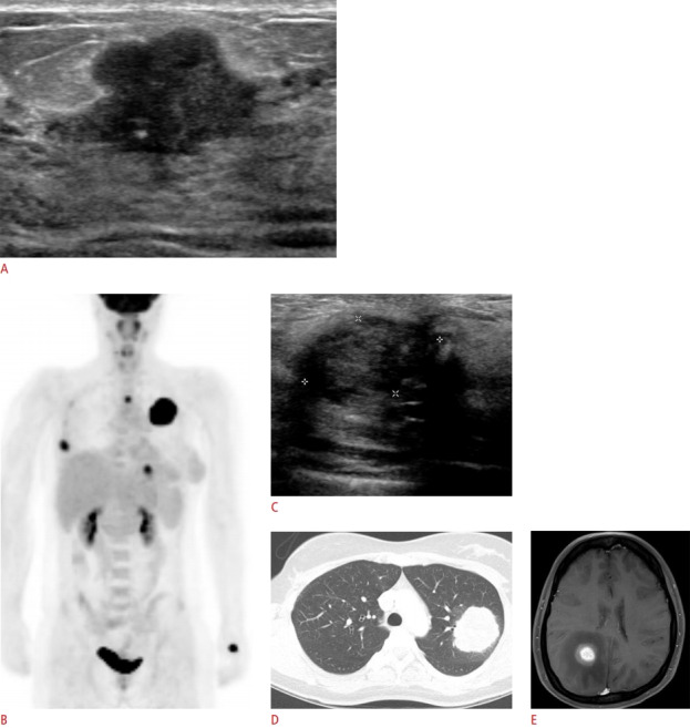
Fig. 11. Pregnancy-associated breast cancer with local recurrence and distant metastases in a 30-year-old pregnant woman (intrauterine pregnancy, 37 weeks).

A. Ultrasonography shows an irregular-shaped, hypoechoic mass with an angular margin. The patient underwent right breastconserving surgery after delivery, and the histologic findings revealed invasive ductal carcinoma. B-E. Local recurrence and distant metastases occured 30 months after breast-conserving surgery for initial pregnancy-associated breast cancer. C. Ultrasonography shows a newly developed, irregular hypoechoic mass at the surgical site, demonstrated by core needle biopsy to be recurrent invasive ductal carcinoma. Positron emission tomography-computed tomography and chest computed tomography indicate the presence of lung and bone metastases (B, D), and brain magnetic resonance imaging indicates the presence of brain metastasis (E).
