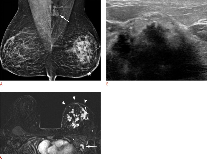
Fig. 13. Pregnancy-associated breast cancer in a 30-year-old woman (10 months postpartum) with a palpable mass in her left breast for the prior 2 months.
A. Bilateral mediolateral oblique mammography show global asymmetry with multiple irregular-shaped masses and diffuse skin thickening (asterisk) in the left breast. Enlarged lymph nodes are also seen in the left axilla (white arrow). B. Ultrasonography shows multiple irregular-shaped masses with posterior acoustic shadowing. C. Post-contrast T1-weighted positron emission tomography-magnetic resonance image shows multiple irregular enhancing masses with diffuse skin thickening (arrowheads) in the left breast and enlarged lymph nodes (arrow) in the left axilla. These findings are consistent with inflammatory breast cancer. Core needle biopsy revealed invasive ductal carcinoma.

