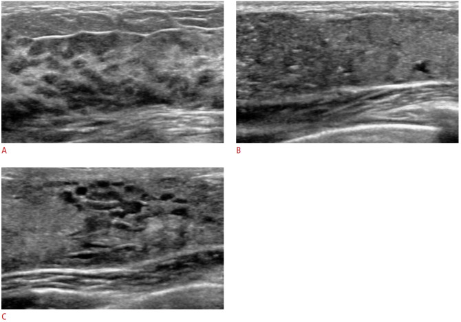
Fig. 2. Ultrasonography of physiological changes during pregnancy and lactation.
A. Ultrasonography of a 34-year-old pregnant woman (intrauterine pregnancy, 23 weeks) shows diffuse enlargement of the nonfatty glandular component with diffuse hypoechogenicity. B, C. Ultrasonography of a 30-year-old lactating woman show diffuse enlargement of the glandular component with multiple hyperechogenic dots and a prominent ductal system with ductal dilatation.

