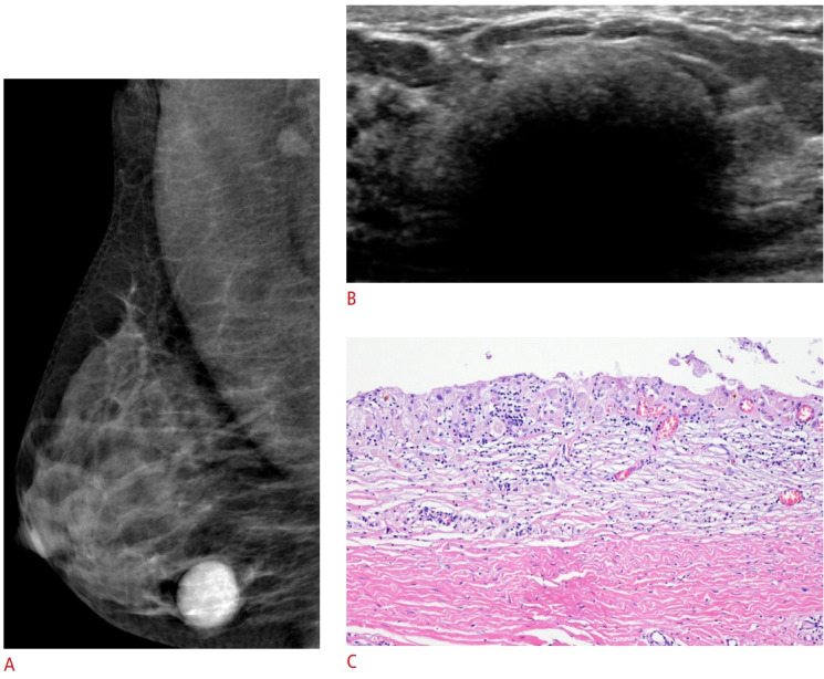
Fig. 6. Galactocele in a 40-year-old woman with a palpable mass.

The palpable mass was detected during lactation and examined 6 months after cessation of breastfeeding. A. A mediolateral oblique mammogram shows an oval-shaped, circumscribed hyperdense mass. B. Ultrasonography shows an oval-shaped mass with echogenic contents and posterior acoustic shadowing. Ultrasound-guided core needle biopsy and subsequent excision were performed. C. The lining epithelium of cystically dilated duct is desquamated. Chronic inflammatory changes with lymphohistiocytic infiltration and fibrosis in the wall can be observed (H&E, ×100). Considering the clinical history and input from a pathologist, the lesion was hypothesized to have originated as a galactocele relatively long ago. It was accompanied by chronic inflammation secondary to leakage.
