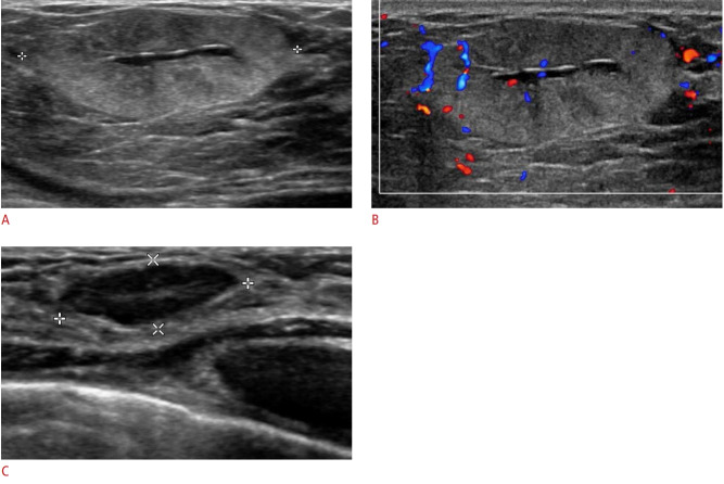
Fig. 8. Lactating adenoma in a 29-year-old lactating woman with a palpable mass.
A. Ultrasonography shows an oval, circumscribed hyperechoic mass with a central hypoechoic area and posterior acoustic enhancement. B. Color Doppler ultrasonography shows internal vascularity. Ultrasound-guided core needle biopsy revealed a lactating adenoma. C. On a follow-up ultrasonography 8 months after cessation of lactation, the lactating adenoma had decreased in both size and internal echogenicity.

