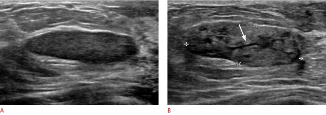
Fig. 9. Fibroadenoma with lactational changes in a 36-year-old lactating woman.
A. Ultrasonography obtained before pregnancy shows an oval, circumscribed, hypoechoic mass, which a biopsy showed to be fibroadenoma. B. In the lactational period, the pre-existing fibroadenoma changed in response to hormonal stimulation. The fibroadenoma shows mixed heterogeneous echogenicity with a prominent ductal pattern (arrow).

