PLOS ONE logoLink to PLOS ONE
. 2020 Jun 25;15(6):e0234401. doi: 10.1371/journal.pone.0234401

Regional differences in hamstring muscle damage after a marathon

Ayako Higashihara 1,*, Kento Nakagawa 2, Takayuki Inami 1, Mako Fukano 3, Satoshi Iizuka 2, Toshihiro Maemichi 2, Satoru Hashizume 4, Takaya Narita 5, Norikazu Hirose 2
Editor: Jeremy P Loenneke6
PMCID: PMC7316338  PMID: 32584826

Abstract

Previous studies suggest that marathon running induces lower extremity muscle damage. This study aimed to examine inter- and intramuscular differences in hamstring muscle damage after a marathon using transverse relaxation time (T2)–weighted magnetic resonance images (MRI). 20 healthy collegiate marathon runners (15 males) were recruited for this study. T2-MRI was performed before (PRE) and at 1 (D1), 3 (D3), and 8 days (D8) after marathon, and the T2 values of each hamstring muscle at the distal, middle, and proximal sites were calculated. Results indicated that no significant intermuscular differences in T2 changes were observed and that, regardless of muscle, the T2 values of the distal and middle sites increased significantly at D1 and D3 and recovered at D8, although those values of the proximal site remained constant. T2 significantly increased at distal and middle sites of the biceps femoris long head on D1 (p = 0.030 and p = 0.004, respectively) and D3 (p = 0.007 and p = 0.041, respectively), distal biceps femoris short head on D1 (p = 0.036), distal semitendinosus on D1 (p = 0.047) and D3 (p = 0.010), middle semitendinosus on D1 (p = 0.005), and distal and middle sites of the semimembranosus on D1 (p = 0.008 and p = 0.040, respectively) and D3 (p = 0.002 and p = 0.018, respectively). These results suggest that the distal and middle sites of the hamstring muscles are more susceptible to damage induced by running a full marathon. Conditioning that focuses on the distal and middle sites of the hamstring muscles may be more useful in improving recovery strategies after prolonged running.

Introduction

Prolonged running, such as in a marathon, has become a popular sports activity for health promotion. However, marathon races are often reported to induce muscle damage in the lower extremity muscles, manifesting as decreased muscle strength [14], occurrence of delayed onset muscle soreness [4, 5], and increased plasma creatine kinase (CK) levels [1, 2, 4], which usually peak at 1–3 days after the race and take several days to recover. To better understand the location of muscle damage in the leg muscles induced by a marathon, previous studies using ultrasound elastography have demonstrated that the thigh and lower leg muscles became harder after a marathon [6, 7], that the amount of change in muscle mechanical properties (e.g., muscle hardness) does not occur uniformly within the lower extremity muscles, and that the recovery time for each muscle varies. Muscle hardness in the vastus lateralis, biceps femoris, and soleus muscles returned to baseline at 8 days after the race, whereas the rectus femoris and gastrocnemius medial muscle hardness did not recover even after 8 days [6]. A study which determines each muscle damage after prolonged running can provide evidence for recreational and competitive runners about the time required for the recovery of each lower extremity muscle between training sessions and/or races.

Muscle functional magnetic resonance imaging (mfMRI) allows for the examination of differences in the intensity and/or pattern of exercise-induced muscle damage. This method relies on an exercise-induced increase in the proton transverse relaxation time (T2) of the muscle on MRI, which could provide information about the water content of muscle tissues. A delayed T2 increase, which occurs at 1–5 days after exercise, is a consequence of muscle damage (inflammatory oedema) induced by eccentric contractions [8]; a high correlation has been reported between changes in T2 and plasma CK levels measured within several days after eccentric exercise [9]. In addition, since T2 values are mapped out across cross-sectional images of muscles, mfMRI can determine muscle damage even in deep muscles; this high spatial resolution overcomes the limitations associated with ultrasound imaging. By using these advantages, a previous study examined the location of muscle damage within the quadriceps femoris muscles induced by downhill running and suggested that muscle damage is specifically located at the proximal and middle sites of the vastus intermedius muscle compared to other knee extensors [10]. Considering these findings, it may be possible that different patterns of running-induced muscle damage may also exist in each section of the hamstring muscles.

The hamstring muscles are composed of three muscles: the semimembranosus (SM), semitendinosus (ST), and biceps femoris (BF); the BF has a short (BFsh) and a long head (BFlh). The BFsh arises from the femur and shares a common distal tendon with BFlh, making it a monoarticular muscle that spans the knee joint only. By contrast, the SM, ST, and BFlh all arise from the ischial tuberosity on the pelvis and thus are biarticular muscles that span the hip and knee joints. During running, the hamstring muscles play an important role in decelerating the flexing hip and rapidly extending the knee [11]; biarticular hamstring muscles contract eccentrically in the terminal swing phase of running [12, 13]. In addition, hamstring muscles are known to be susceptible to muscle strain. Repetitive eccentric contractions associated with running may lead to the accumulation of eccentrically induced muscle damage [14, 15]. Such accumulation could, in turn, leave the hamstring muscles more at risk of strain injury [14]. Considering the fact that hamstring muscles are susceptible to muscle strain, identification of the sites in the hamstring muscles that are significantly damaged by prolonged running would contribute to an effective strategy for recovery focusing on injury prevention. In addition, this will provide evidence about the time required for the recovery of each site in the hamstring muscle between training sessions and/or competitions. Nevertheless, to our knowledge, there have been no studies on the location of muscle damage in the hamstring muscles induced by repetitive eccentric contractions after marathon running.

Thus, this study aimed to identify the sites in the hamstring muscles that are significantly damaged by prolonged running (full marathon) using mfMRI. For this purpose, we measured running-induced changes in T2 values at the proximal, middle, and distal sites of each hamstring muscle. We hypothesized that the location of muscle damage following running would be greatest in the BFlh muscle, based on previous kinematic analyses of running, considering that the peak muscle–tendon stretch is greatest in the BFlh muscle among the biarticular hamstring muscles during the terminal swing phase of running [12, 13], which occurs simultaneously with high muscle activity [16]. In addition, given that the knee joint performs greater negative (eccentric) work than the hip joint during running [17], we hypothesized that muscle damage would be more pronounced in the distal site of the biarticular hamstring muscles.

Materials and methods

Subjects

Twenty collegiate runners (males, 15; females, 5) without lower extremity injuries were recruited for this study. Mean age, height, and body mass of participants were 20.5±1.4 years, 168.6±7.1 cm, and 59.4±7.1 kg, respectively. Participants were members of a track and field club who had registered for a marathon race (Fujisan Marathon in Japan, 2016–2017; 42.195 km) and had no history of lower limb injuries.

This study was approved by the local human research ethics committee of Waseda University (2014–246), complied with their requirements for human experimentation, and conformed to the principles of the Declaration of Helsinki. The purpose, procedures, and risks of the study were communicated to the participants, and written informed consent was obtained from each participant. Participants were asked not to perform exercise, such as running, and not to receive special care for recovery, such as a massage, during the observation period after marathon.

Procedures

Data collection

All participants completed the marathon. Maximal isometric knee flexion torque and T2-weighted magnetic resonance (MR) images of the thigh were obtained before (2 days before the race [PRE]) and at 1 (D1), 3 (D3), and 8 (D8) days after the marathon race. The course altitude was approximately 850 m above sea level from the start to approximately 22 km, increased to around 910 m, was almost flat from 23 km to 34 km, declined to 850 m from 34 km to 36 km, and was flat from 36 km to finish.

T2-MRI

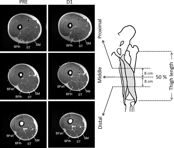
Thigh T2-MRI (Fig 1) was obtained with an MR scanner (Signa EXCITE 1.5T; GE Medical Systems, Waukesha, WI). Before scanning, participants lay quietly for 10 min. Ink lines were drawn transversely across the middle (50%) of the thigh (i.e., the distance from the great trochanter of the femur to the articular cleft between the femoral and tibial condyles) and oil capsules were put as markers on the skin surface at the lateral side. Participants lay supine with their legs fully extended and muscles relaxed in a magnet bore. The images were acquired with the following parameters: echo times, 25, 50, 75, and 100 ms; repetition time, 2000 ms; matrix, 256×160; field of view, 240 mm; slice thickness, 10 mm; gap, 10 mm. Images were analysed with ImageJ software (National Institute of Health, Bethesda, MD). Regions of interest were drawn in each slice by manually tracing the border of the anatomical cross-sectional area of each of the hamstring muscles at the middle (50%), proximal (8 cm proximal from the middle site), and distal (8 cm distal from the middle site) thigh sites (Fig 1). Care was taken to exclude noncontractile tissues, such as intramuscular fat and blood vessels. The T2 values for each pixel within the BFlh, BFsh, ST, and SM muscles were calculated, and the mean value was computed for each slice. As the BFsh is a monoarticular muscle with an origin at a more distal site than those of the other three muscles, only the middle and distal sites were analysed. T2 relaxation time was calculated by least-squares analysis, fitting the signal intensity at each of the four echo times (n×25 ms: 25, 50, 75, and 100 ms) to a monoexponential decay using the following equation:

Sn=S0exp(TEn/T2)(n=25,50,75,and100ms),

where TE is the echo time, S0 is the signal intensity at 0 ms, and Sn is the signal intensity at TEn.

Fig 1. MRI measurement sites.

Fig 1

Examples of T2-MRI at the proximal, middle, and distal sites scanned before (PRE) and at 1 day (D1) after a full marathon race. BFlh, biceps femoris long head; BFsh, biceps femoris short head; MRI, magnetic resonance imaging; SM, semimembranosus; ST, semitendinosus.

The aforementioned analyses were performed three times for each slice and the average value was used for further analysis. The coefficient of variation of the three measurements for T2 values was 0.8±0.8%. The intraclass correlation coefficient of the measurements was 0.964. The absolute T2 values (ms) were used in between-time comparisons for each site. In addition, as the timing of T2 manifestations could vary among sites and individuals, peak T2 values (peak ΔT2, in ms) were calculated by subtracting the baseline (PRE) value from the peak value after marathon for each site, irrespective of the time point at which peak value occurred, and was used for between-site comparisons.

Maximal isometric knee flexion torque

After MR scanning, participants performed isometric right knee flexion with maximal effort while lying down in a prone position. The experimenter fixed the participant’s knee joint at 90° with a hand-held dynamometer (microFET2; Hoggan Scientific, Salt Lake, UT); the participants performed two isometric contractions against the experimenter’s force. They were asked to develop torque gradually over 5 s and reach the maximum, and they received verbal encouragement to sustain the maximum effort for 2 s from the examiner. Two peak torque values were averaged for the statistical analysis.

Statistical analyses

All statistical analyses were conducted using SPSS version 14.0 (IBM Corp., Armonk, NY). Changes in T2 values were compared by a three-way analysis of variance (ANOVA) with repeated measures (muscle × site × time point). When significant interaction effects were found, Bonferroni’s post hoc test was performed to compare changes from the PRE for each site. Absolute peak ΔT2 values were compared among the sites using one-way ANOVA (11 sites) followed by Bonferroni post hoc testing. Changes in maximal knee flxexion torque were compared by a one-way (4 time points) repeated measures ANOVA followed by Bonferroni post hoc testing. Partial η2 (for ANOVA) was calculated as indices of effect size (ES) for the ANOVA. To report the ES for post hoc comparisons, the mean change value from the PRE divided by the standard deviation of the change value was calculated [18]. Statistical significance was set at p<0.05.

Results

Marathon time

The average marathon completion time was 4 h, 7 min, 55 s ± 47 min, 39 s.

T2-MRI

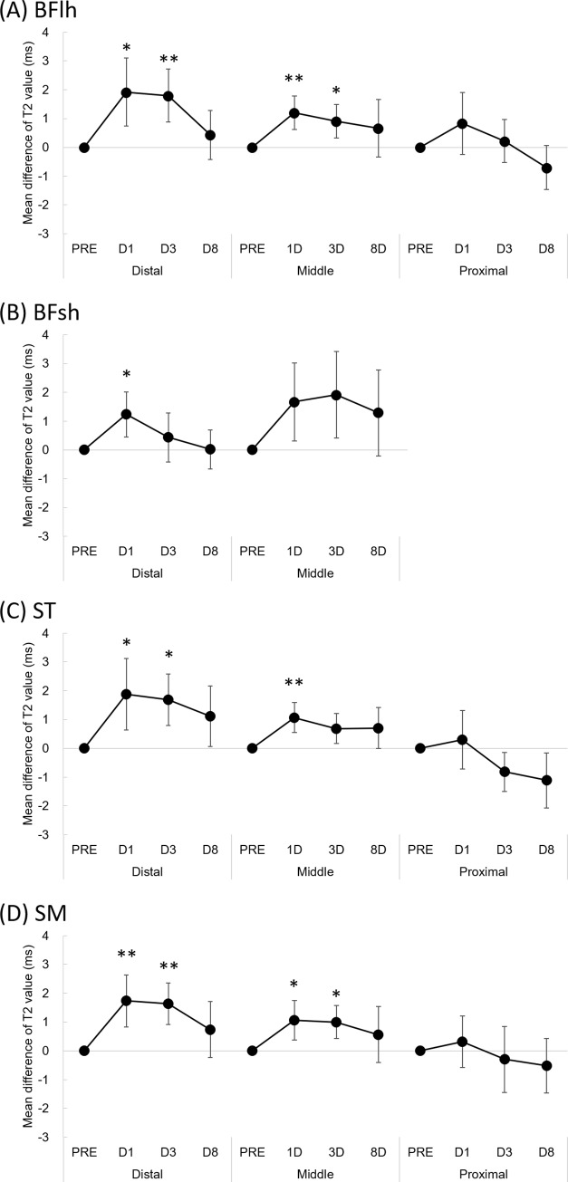
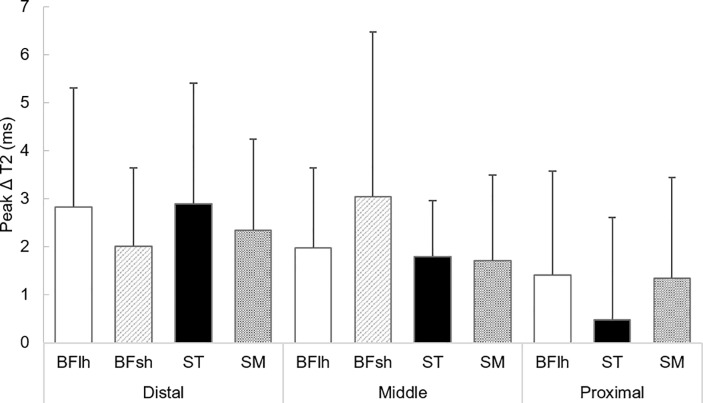
Three-way ANOVA analysis for T2 values indicated some interaction effects: muscle × site (F = 3.393, p = 0.006) and site × time point (F = 5.729, p<0.001) (Table 1). The absolute T2 values for each site are shown in the Table 2. Fig 2 shows the mean difference and 95% confidence intervals in T2 value from the baseline (PRE) for each muscle. Bonferroni’s post hoc analysis indicated that, regardless of muscle, the T2 values of the distal and middle sites increased significantly at D1 and D3 and recovered at D8 (no significant difference on D8 when compared with PRE), although those values of the proximal site remained constant. T2 significantly increased at distal and middle sites of the biceps femoris long head on D1 (p = 0.030 and p = 0.004, respectively) and D3 (p = 0.007 and p = 0.041, respectively), distal biceps femoris short head on D1 (p = 0.036), distal semitendinosus on D1 (p = 0.047) and D3 (p = 0.010), middle semitendinosus on D1 (p = 0.005), and distal and middle sites of the semimembranosus on D1 (p = 0.008 and p = 0.040, respectively) and D3 (p = 0.002 and p = 0.018, respectively). There was no significant difference in the T2 value of the middle BFsh muscle and the proximal site of the BFlh, ST, and SM at any time point compared with that of PRE. Fig 3 shows the peak ΔT2 value for each site. No significant differences in the peak ΔT2 values after the full marathon were observed among sites.

Table 1. Results of statistical analyses for the T2 values.
Source Three-way ANOVA
df F-value p-value η2
Site 2 14.950 0.000 0.125 **
Muscle 3 21.158 0.000 0.233 **
Time point 3 27.508 0.000 0.116 **
Site × muscle 5 3.393 0.006 0.075 *
Site × time point 6 5.729 0.000 0.052 **
Muscle × time point 9 0.368 0.950 0.005 n.s.
Site × muscle × time point 15 0.861 0.609 0.020 n.s.

* p < 0.01

** p < 0.001; n.s. not significantly different

Table 2. Time-dependent change in the absolute value of T2 values (Mean ± SD) measured before (PRE) and 1 day (D1), 3 days (D3) and 8 days (D8) after the marathon race.

Effect sizes are shown in parentheses.

PRE D1 D3 D8
BFlh
Distal 31.7 ± 1.2 33.6 ± 2.7* [0.71] 33.5 ± 2.0** [0.86] 32.1 ± 1.9 [0.22]
Middle 32.2 ± 1.1 33.4 ± 1.5** [0.90] 33.1 ±1.4* [0.68] 32.8 ± 2.3 [0.29]
Proximal 35.5 ±1.8 36.3 ±2.1 [0.34] 35.7 ± 1.7 [0.13] 34.8 ± 1.3 [0.41]
BFsh
Distal 32.2 ± 1.6 33.9 ± 2.9* [0.69] 34.1 ± 3.2 [0.22] 33.5 ± 3.4 [0.01]
Middle 32.8 ± 1.9 34.0 ± 2.5 [0.54] 33.2 ± 2.3 [0.56] 32.8 ± 2.2 [0.38]
ST
Distal 30.4 ± 1.6 32.3 ± 2.7* [0.66] 32.1 ± 2.0* [0.82] 31.5 ± 2.4 [0.47]
Middle 30.9 ± 1.1 32.0 ± 1.5** [0.89] 31.6 ± 1.7 [0.58] 31.6 ± 2.0 [0.43]
Proximal 32.2 ± 2.0 32.5 ± 1.4 [0.3] 31.3 ± 1.5 [0.53] 31.0 ±1.2 [0.51]
SM
Distal 31.5 ± 1.5 33.2 ± 2.5** [0.84] 33.1 ± 1.6** [1.00] 32.2 ± 2.2 [0.33]
Middle 31.7 ± 1.1 32.7 ± 1.4* [0.68] 32.7 ± 1.3* [0.76] 32.3 ± 1.9 [0.26]
Proximal 33.8 ± 2.9 34.1 ± 3.1 [0.15] 33.5 ± 3.2 [0.11] 33.3 ± 2.2 [0.24]

*Significantly different from PRE at p < 0.05.

**Significantly different from PRE at p < 0.01. BFlh, biceps femoris long head; BFsh, biceps femoris short head; ST, semitendinosus; SM, semimembranosus.

Fig 2.

Fig 2

Mean difference in T2 value from the baseline value (PRE) of the biceps femoris long head (A), biceps femoris short head (B), semitendinosus (C), and semimembranosus (D) muscles. Error bars indicate 95% confidence intervals. *Significantly different from PRE at p < 0.05. **Significantly different from PRE at p < 0.01. D1, 1 day after marathon; D3, 3 days after marathon; D8, 8 days after marathon. BFlh, biceps femoris long head; BFsh, biceps femoris short head; ST, semitendinosus; SM, semimembranosus.

Fig 3. Peak T2 changes from PRE (ΔT2) within 8 days after a full marathon.

Fig 3

Values are mean±standard deviation. BFlh, biceps femoris long head; BFsh, biceps femoris short head; ST, semitendinosus; SM, semimembranosus.

Maximal isometric knee flexion torque

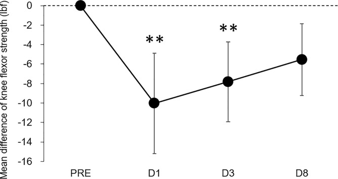
One-way ANOVA revealed a significant main effect of time on maximal knee flexion torque (F = 8.491, p<0.001, partial η2 = 0.309). Bonferroni post-hoc testing showed that the knee flexion torque significantly decreased compared with the baseline (PRE) value on D1 (−22.7%, p = 0.007, ES = 0.85) and D3 (−17.7%, p = 0.009, ES = 0.83) after marathon; however, there was no significant difference on D8 (−5.5%, p = 0.050, ES = 0.65) compared with PRE (Fig 4).

Fig 4. Mean difference in maximal isometric knee flexion torque from the baseline (PRE).

Fig 4

Error bars indicate 95% confidence intervals. D1, 1 day after marathon; D3, 3 days after marathon; D8, 8 days after marathon.

Discussion

We determined whether there are inter- and intramuscular differences in damage to the hamstring muscles after a full marathon using T2-MRI. The main findings of the present study were that no significant intermuscular differences in the magnitude of T2 change after a marathon were found and that T2 significantly increased at the distal and middle sites of the biarticular hamstring muscles on D1 and D3 after the marathon race; however, no significant difference in the T2 value of the proximal site was observed at any time point when compared with PRE. These results suggest that running a full marathon induces muscle damage (inflammatory oedema) appearing as a T2 increase in the hamstring muscles and that the magnitude of muscle damage induced by prolonged running would be greater in the distal and middle sites than in the proximal site of the hamstring muscles.

Marathon running resulted in significant decreases in maximal knee flexion torque on D1 and D3 (Fig 4). This result is similar to that of a previous study, which showed a significant reduction in maximal voluntary knee flexion torque following a full marathon [4]. However, no significant differences in the magnitude of T2 change (peak ΔT2) after running were found among muscles (Fig 3). These results did not support our hypothesis that muscle damage would be greatest in the BFlh muscle. We drafted this hypothesis based on previous observations, which suggested that the magnitude of muscle–tendon strain during contraction is the more relevant parameter for muscle damage [19] and that peak muscle–tendon stretch is greatest in the BFlh during running compared to the ST and SM [12, 13]. Schache et al. [12] investigated the peak muscle–tendon stretch of the biarticular hamstring muscles at different running speeds and demonstrated that the peak stretch was greater for BFlh compared to ST and SM across a range of running speeds. They also found that there was no statistical difference in the magnitude of the maximum stretch between these muscles during slower speed below 18 km/h. The participants in the present study completed a full marathon at an average time of 4 h, 7 min, 55 s ± 47 min, 39 s (i.e., at an average speed of 11 km/h). Thus, the findings of Schache et al. [12] would support the result of the current study; however, the degree of muscle damage induced by prolonged running, such as after a full marathon, may not simply be explained based on the intermuscular difference in stretching magnitude: one possibility is the alternate muscle activity among the synergists due to the fatigue during prolonged running, while the previous study suggested the alterations in the neuromuscular activation patterns among the quadriceps muscles during the repetitive fatiguing exercise [20]. This phenomenon may occur in the hamstring muscles during marathon running. However, given the results of the mfMRI analysis indicating the intensity and/or pattern of exercise-induced muscle damage after exercise, whether the alternate muscle activity among the hamstring muscles occurs during the marathon race is uncertain. Even though it is a speculation, is worthy of further examination.

T2 values significantly increased at the distal and middle sites of the BFlh, ST, and SM muscles on D1 and D3 after running (Table 2, Fig 2), with a pronounced effect size found, whereas the T2 value of the proximal site at any time point did not significantly increase compared with PRE. This suggests that the distal and middle sites of the biarticular hamstring muscles are more susceptible to damage after a marathon. The amount of negative work performed by the muscle would be the best predictor of the magnitude of damage, as indicated by force deficits following active or passive muscle stretching [21]. Therefore, a possible explanation for the present results is that the location of muscle damage after prolonged running is primarily influenced by the intensity of repetitive negative work performed by the lower extremity. During running, the knee joint performs greater negative work than the hip joint (e.g., −0.41 J/kg for the knee and −0.10 J/kg for the hip at a running speed of 3.5 m/s) [17]. In addition, a previous study reported that during eccentric knee flexion, the greatest tissue motion in the BFlh muscle is observed along the distal musculotendinous junction [22]. Considering these findings and the fact that a greater degree of muscle tissue strain during eccentric contraction causes muscle damage [19], the magnitude of muscle damage induced by prolonged running would be greater in the distal than in the proximal portions of the hamstring muscles.

A previous study suggested that the accumulation of eccentrically-induced muscle damage associated with running may leave the hamstring muscles more vulnerable to strain injury [14]. The aetiology and risk of hamstring strain injury is closely related to high-speed running [23], and transiently elevated high-speed running exposure (<24 km/h) at 7–14 days prior to injury increases the likelihood of injury [24, 25]. However, sports in which a high rate of hamstring strain injury has been reported [26], such as Australian football, require prolonged and high-intensity intermittent performance, and players cover long distances during a game (12,939±1145 m for total distance) at various speeds; movement distance with a higher-speed running (>14.5 km/h) accounts only for 30% of the total distance [27]. The present results indicated that repetitive eccentric contractions associated with lower speed (at an average speed of 11 km/h) but prolonged running, such as in a marathon, produce inflammatory oedema at the distal and middle sites of biarticular hamstring muscles. In addition, considering that this microscopic damage in hamstring muscles takes several days to recover, accumulation of prior running exposure even at lower speeds may contribute to the susceptibility of strain injury for some time after. This hypothesis is yet to be explored. Moreover, the magnitude of muscle damage is largely reduced when the same or similar eccentric exercise is repeated within several weeks [28]. Therefore, differences in training and competition level may affect the magnitude of muscle damage induced by prolonged running. This should be regarded as a limitation; future research should ascertain whether similar findings are also seen in runners from different competitive levels or other competitive athletes.

In this study, we demonstrate that the distal and middle sites of the biarticular hamstring muscles are more susceptible to damage induced after running a marathon. Based on our results, conditioning that focuses on the distal and middle sites of the hamstring muscles may be more useful in improving recovery strategies as well as for preventing injury after prolonged running. In addition, our results showed that inflammatory oedema appearing as T2 increase takes several days to recover. Therefore, accumulation of low-intensity running sessions may contribute to the susceptibility of strain injury afterwards. However, our results are inconsistent with the clinical observations that the proximal muscle–tendon junction of the BFlh muscles is more commonly affected in acute hamstring muscle injury [23, 29]. This discrepancy could be explained by the possible difference in the neuromechanical behaviour and joint kinetics between high-speed running, in which an injury occurs, and prolonged running. Therefore, further research is required to identify the regional damage patterns after repetitive high-speed running relative to the injury site. It is likely that such studies can provide further insights into the mechanisms of muscle strain injury during high-speed running.

In conclusion, the results indicated that marathon running induces a significant damage to hamstring muscles; however, the amount of change in inflammatory oedema appearing as T2 increase did not differ among hamstring muscles. Furthermore, site-dependent changes in T2 values after a full marathon were observed within muscles; that is, pronounced inflammatory oedema was observed in the distal and middle sites of the biarticular hamstring muscles. Our findings provide a better understanding of the site-specific muscle damage patterns of the hamstring muscle induced by prolonged running.

Data Availability

All relevant data are uploaded to Zenodo (DOI: 10.5281/zenodo.3734098).

Funding Statement

This work was supported by the Mizuno Sports Promotion Foundation.

References

  • 1.Clifford T, Allerton DM, Brown MA, Harper L, Horsburgh S, Keane KM, et al. Minimal muscle damage after a marathon and no influence of beetroot juice on inflammation and recovery. Appl Physiol Nutr Metab. 2017;42(3):263–70. 10.1139/apnm-2016-0525 [DOI] [PubMed] [Google Scholar]
  • 2.Hill JA, Howatson G, van Someren KA, Walshe I, Pedlar CR. Influence of compression garments on recovery after marathon running. J Strength Cond Res. 2014;28(8):2228–35. 10.1519/JSC.0000000000000469 [DOI] [PubMed] [Google Scholar]
  • 3.Petersen K, Hansen CB, Aagaard P, Madsen K. Muscle mechanical characteristics in fatigue and recovery from a marathon race in highly trained runners. Eur J Appl Physiol. 2007;101(3):385–96. 10.1007/s00421-007-0504-x [DOI] [PubMed] [Google Scholar]
  • 4.Takayama F, Aoyagi A, Shimazu W, Nabekura Y. Effects of marathon running on aerobic fitness and performance in recreational runners one week after a race. J Sports Med (Hindawi Publ Corp). 2017;2017:9402386 10.1155/2017/9402386 [DOI] [PMC free article] [PubMed] [Google Scholar]
  • 5.Howatson G, McHugh MP, Hill JA, Brouner J, Jewell AP, van Someren KA, et al. Influence of tart cherry juice on indices of recovery following marathon running. Scand J Med Sci Sports. 2010;20(6):843–52. 10.1111/j.1600-0838.2009.01005.x [DOI] [PubMed] [Google Scholar]
  • 6.Inami T, Nakagawa K, Yonezu T, Fukano M, Higashihara A, Iizuka S, et al. Tracking of time-dependent changes in muscle hardness after a full marathon. J Strength Cond Res. 2019;33(12):3431–7. 10.1519/JSC.0000000000002495 [DOI] [PubMed] [Google Scholar]
  • 7.Nakagawa K, Inami T, Yonezu T, Kenmotsu Y, Narita T, Kawakami Y, et al. Unstable rocker shoes promote recovery from marathon-induced muscle damage in novice runners. Scand J Med Sci Sports. 2018;28(2):621–9. 10.1111/sms.12911 [DOI] [PubMed] [Google Scholar]
  • 8.Ochi E, Tsuchiya Y, Nosaka K. Differences in post-exercise T2 relaxation time changes between eccentric and concentric contractions of the elbow flexors. Eur J Appl Physiol. 2016;116(11–12):2145–54. 10.1007/s00421-016-3462-3 [DOI] [PubMed] [Google Scholar]
  • 9.Larsen RG, Ringgaard S, Overgaard K. Localization and quantification of muscle damage by magnetic resonance imaging following step exercise in young women. Scand J Med Sci Sports. 2007;17(1):76–83. 10.1111/j.1600-0838.2006.00525.x [DOI] [PubMed] [Google Scholar]
  • 10.Maeo S, Ando Y, Kanehisa H, Kawakami Y. Localization of damage in the human leg muscles induced by downhill running. Sci Rep. 2017;7(1):5769 10.1038/s41598-017-06129-8 [DOI] [PMC free article] [PubMed] [Google Scholar]
  • 11.Schache AG, Dorn TW, Blanch PD, Brown NA, Pandy MG. Mechanics of the human hamstring muscles during sprinting. Med Sci Sports Exerc. 2012;44(4):647–58. Epub 2011/09/14. 10.1249/MSS.0b013e318236a3d2 [DOI] [PubMed] [Google Scholar]
  • 12.Schache AG, Dorn TW, Wrigley TV, Brown NA, Pandy MG. Stretch and activation of the human biarticular hamstrings across a range of running speeds. Eur J Appl Physiol. 2013;113(11):2813–28. Epub 2013/09/10. 10.1007/s00421-013-2713-9 [DOI] [PubMed] [Google Scholar]
  • 13.Thelen DG, Chumanov ES, Hoerth DM, Best TM, Swanson SC, Li L, et al. Hamstring muscle kinematics during treadmill sprinting. Med Sci Sports Exerc. 2005;37(1):108–14. 10.1249/01.mss.0000150078.79120.c8 [DOI] [PubMed] [Google Scholar]
  • 14.Fyfe JJ, Opar DA, Williams MD, Shield AJ. The role of neuromuscular inhibition in hamstring strain injury recurrence. J Electromyogr Kinesiol. 2013;23(3):523–30. Epub 2013/02/14. 10.1016/j.jelekin.2012.12.006 . [DOI] [PubMed] [Google Scholar]
  • 15.Morgan DL. New insights into the behavior of muscle during active lengthening. Biophys J. 1990;57(2):209–21. 10.1016/S0006-3495(90)82524-8 [DOI] [PMC free article] [PubMed] [Google Scholar]
  • 16.Higashihara A, Nagano Y, Ono T, Fukubayashi T. Relationship between the peak time of hamstring stretch and activation during sprinting. Eur J Sport Sci. 2016;16(1):36–41. Epub 2014/11/02. 10.1080/17461391.2014.973913 [DOI] [PubMed] [Google Scholar]
  • 17.Schache AG, Blanch PD, Dorn TW, Brown NA, Rosemond D, Pandy MG. Effect of running speed on lower limb joint kinetics. Med Sci Sports Exerc. 2011;43(7):1260–71. Epub 2010/12/07. 10.1249/MSS.0b013e3182084929 [DOI] [PubMed] [Google Scholar]
  • 18.Dankel SJ and Loenneke JP. Effect sizes for paired data should use the change score variability rather than the pre-test variability. J Strength Cond Res. 2018. 10.1519/jsc.0000000000002946 [DOI] [PubMed] [Google Scholar]
  • 19.Lieber RL, Friden J. Muscle damage is not a function of muscle force but active muscle strain. J Appl Physiol. 1993;74(2):520–6. Epub 1993/02/01. 10.1152/jappl.1993.74.2.520 [DOI] [PubMed] [Google Scholar]
  • 20.Akima H, Foley JM, Prior BM, Dudley GA, Meyer RA. Vastus lateralis fatigue alters recruitment of musculus quadriceps femoris in humans. J Appl Physiol. 2002;92(2):679–84. Epub 2002/01/18. 10.1152/japplphysiol.00267.2001 [DOI] [PubMed] [Google Scholar]
  • 21.Brooks SV, Zerba E, Faulkner JA. Injury to muscle fibres after single stretches of passive and maximally stimulated muscles in mice. The Journal of physiology. 1995;488 (Pt 2):459–69. [DOI] [PMC free article] [PubMed] [Google Scholar]
  • 22.Silder A, Reeder SB, Thelen DG. The influence of prior hamstring injury on lengthening muscle tissue mechanics. J Biomech. 2010;43(12):2254–60. Epub 2010/05/18. 10.1016/j.jbiomech.2010.02.038 [DOI] [PMC free article] [PubMed] [Google Scholar]
  • 23.Askling CM, Tengvar M, Saartok T, Thorstensson A. Acute first-time hamstring strains during slow-speed stretching: clinical, magnetic resonance imaging, and recovery characteristics. Am J Sports Med. 2007;35(10):1716–24. Epub 2007/06/15. 10.1177/0363546507303563 [DOI] [PubMed] [Google Scholar]
  • 24.Duhig S, Shield AJ, Opar D, Gabbett TJ, Ferguson C, Williams M. Effect of high-speed running on hamstring strain injury risk. Br J Sports Med. 2016. Epub 2016/06/12. 10.1136/bjsports-2015-095679 [DOI] [PubMed] [Google Scholar]
  • 25.Ruddy JD, Pollard CW, Timmins RG, Williams MD, Shield AJ, Opar DA. Running exposure is associated with the risk of hamstring strain injury in elite Australian footballers. Br J Sports Med. 2018;52(14):919–28. 10.1136/bjsports-2016-096777 . [DOI] [PubMed] [Google Scholar]
  • 26.Orchard JW, Seward H, Orchard JJ. Results of 2 decades of injury surveillance and public release of data in the Australian Football League. Am J Sports Med. 2013;41(4):734–41. Epub 2013/03/06. 10.1177/0363546513476270 [DOI] [PubMed] [Google Scholar]
  • 27.Coutts AJ, Quinn J, Hocking J, Castagna C, Rampinini E. Match running performance in elite Australian Rules Football. Journal of science and medicine in sport / Sports Medicine Australia. 2010;13(5):543–8. 10.1016/j.jsams.2009.09.004 [DOI] [PubMed] [Google Scholar]
  • 28.McHugh MP. Recent advances in the understanding of the repeated bout effect: the protective effect against muscle damage from a single bout of eccentric exercise. Scand J Med Sci Sports. 2003;13(2):88–97. 10.1034/j.1600-0838.2003.02477.x [DOI] [PubMed] [Google Scholar]
  • 29.Crema MD, Guermazi A, Tol JL, Niu J, Hamilton B, Roemer FW. Acute hamstring injury in football players: Association between anatomical location and extent of injury-A large single-center MRI report. J Sci Med Sport. 2016;19(4):317–22. Epub 2015/04/30. 10.1016/j.jsams.2015.04.005 [DOI] [PubMed] [Google Scholar]

Decision Letter 0

Jeremy P Loenneke

27 Mar 2020

PONE-D-20-03671

Regional differences in hamstring muscle damage after a marathon

PLOS ONE

Dear Ms. Higashihara,

Thank you for submitting your manuscript to PLOS ONE.  I sincerely apologize for the delay in reviewing your manuscript. After careful consideration, we feel that it has merit but does not fully meet PLOS ONE’s publication criteria as it currently stands. Therefore, we invite you to submit a revised version of the manuscript that addresses the points raised during the review process.

Please carefully consider the following points along with that of the other reviewers prior to resubmitting: 

1) Per PLOS ONE policy, the authors  must make the data available to the extend that the analysis can be reproduced. Currently, this has not been done. 

2) The analysis does not appear to match your study purpose. Please alter this prior to resubmitting. The document states that the study aimed to identify sites (proximal, distal, middle) in the hamstring muscles (biceps femoris, semitendinosus, etc) in the days following a marathon (Pre, d1, d3, d8). It appears that the purpose was to determine differences between all of these but the current model does not appear to account for that. It seems that you have a Muscle x Site x Time design. This, I think, would better address your research question. 

3) please explicitly state how Cohen's d was calculated.   

4) It might be helpful for you to represent Figure 2 as change scores (variability of the change in 95% CI). This would seemingly better highlight the variability of interest. For example, the variability of the sample at Pre is not relevant to the research question. What is relevant, is the variability of the change at that site following the marathon. 

We would appreciate receiving your revised manuscript by May 11 2020 11:59PM. When you are ready to submit your revision, log on to https://www.editorialmanager.com/pone/ and select the 'Submissions Needing Revision' folder to locate your manuscript file.

If you would like to make changes to your financial disclosure, please include your updated statement in your cover letter.

To enhance the reproducibility of your results, we recommend that if applicable you deposit your laboratory protocols in protocols.io, where a protocol can be assigned its own identifier (DOI) such that it can be cited independently in the future. For instructions see: http://journals.plos.org/plosone/s/submission-guidelines#loc-laboratory-protocols

Please include the following items when submitting your revised manuscript:

  • A rebuttal letter that responds to each point raised by the academic editor and reviewer(s). This letter should be uploaded as separate file and labeled 'Response to Reviewers'.

  • A marked-up copy of your manuscript that highlights changes made to the original version. This file should be uploaded as separate file and labeled 'Revised Manuscript with Track Changes'.

  • An unmarked version of your revised paper without tracked changes. This file should be uploaded as separate file and labeled 'Manuscript'.

Please note while forming your response, if your article is accepted, you may have the opportunity to make the peer review history publicly available. The record will include editor decision letters (with reviews) and your responses to reviewer comments. If eligible, we will contact you to opt in or out.

We look forward to receiving your revised manuscript.

Kind regards,

Jeremy P Loenneke

Academic Editor

PLOS ONE

Journal Requirements:

When submitting your revision, we need you to address these additional requirements.

1. Please ensure that your manuscript meets PLOS ONE's style requirements, including those for file naming. The PLOS ONE style templates can be found at

http://www.journals.plos.org/plosone/s/file?id=wjVg/PLOSOne_formatting_sample_main_body.pdf and http://www.journals.plos.org/plosone/s/file?id=ba62/PLOSOne_formatting_sample_title_authors_affiliations.pdf

2. Thank you for submitting the above manuscript to PLOS ONE. During our internal evaluation of the manuscript, we found significant text overlap between your submission and the following previously published works:

https://link.springer.com/article/10.1007%2Fs00421-013-2713-9

https://onlinelibrary.wiley.com/doi/full/10.1111/sms.12880

We would like to make you aware that copying extracts from previous publications, especially outside the methods section, word-for-word is unacceptable. In addition, the reproduction of text from published reports has implications for the copyright that may apply to the publications.

Please revise the manuscript to rephrase the duplicated text, cite your sources, and provide details as to how the current manuscript advances on previous work. Please note that further consideration is dependent on the submission of a manuscript that addresses these concerns about the overlap in text with published work.

We will carefully review your manuscript upon resubmission, so please ensure that your revision is thorough.

[Note: HTML markup is below. Please do not edit.]

Reviewers' comments:

Reviewer's Responses to Questions

Comments to the Author

1. Is the manuscript technically sound, and do the data support the conclusions?

The manuscript must describe a technically sound piece of scientific research with data that supports the conclusions. Experiments must have been conducted rigorously, with appropriate controls, replication, and sample sizes. The conclusions must be drawn appropriately based on the data presented.

Reviewer #1: Yes

Reviewer #2: Yes

**********

2. Has the statistical analysis been performed appropriately and rigorously?

Reviewer #1: Yes

Reviewer #2: Yes

**********

3. Have the authors made all data underlying the findings in their manuscript fully available?

The PLOS Data policy requires authors to make all data underlying the findings described in their manuscript fully available without restriction, with rare exception (please refer to the Data Availability Statement in the manuscript PDF file). The data should be provided as part of the manuscript or its supporting information, or deposited to a public repository. For example, in addition to summary statistics, the data points behind means, medians and variance measures should be available. If there are restrictions on publicly sharing data—e.g. participant privacy or use of data from a third party—those must be specified.

Reviewer #1: No

Reviewer #2: Yes

**********

4. Is the manuscript presented in an intelligible fashion and written in standard English?

PLOS ONE does not copyedit accepted manuscripts, so the language in submitted articles must be clear, correct, and unambiguous. Any typographical or grammatical errors should be corrected at revision, so please note any specific errors here.

Reviewer #1: Yes

Reviewer #2: Yes

**********

5. Review Comments to the Author

Please use the space provided to explain your answers to the questions above. You may also include additional comments for the author, including concerns about dual publication, research ethics, or publication ethics. (Please upload your review as an attachment if it exceeds 20,000 characters)

Reviewer #1: This study used mfMRI to examine muscle damage in sites along the hamstring muscles following marathon running. The “experiments, statistics, and other analyses” appear to have been “performed to a high technical standard and are described in sufficient detail” as required by PLOS ONE. The conclusions are logical and the article is well-written. This study provides novel and useful original research. This reviewer sees no reason for this article to not be published as is.

Reviewer #2: Overall, the manuscript is well written. The study was nicely designed and the results were also convincing. This reviewer has only a couple of minor questions that need to be clarified.

1. Why muscle soreness was not measured given the fact that muscle soreness is also a commonly used indirect marker for exercise-induced muscle damage?

2. Line 50:

“recovery of the rectus femoris and gastrocnemius medial muscle hardness was not observed “

Does that mean the rectus femoris and gastrocnemius medial muscle got permanent muscle damage? If not, what does the sentence mean?

3. Line 106-107: Participants were asked not to perform exercise, such as running…”

How long (e.g. # of days prior and after the marathon running) were the participants not allowed to perform exercise? Were these participants trained or untrained people? If they were trained people, did the exercise prohibition of from this study induce any detraining or other effect(s)?

4. Line 113: The baseline measurements were obtained 2 days before the race. Why 2 days prior specifically? Why not 1 day or any other time before the race?

5. Line 160 and 162: Why two different post-hoc testings (Bonferroni and Dunnett)? Why not use only one of them and keep it consistent?

6. Lines 218-226. The discussion of the non-significant intermuscular damage after marathon running needs to be expanded and more thorough. If this cannot be explained only by stretching magnitude, what else could be the mechanism/explanation?

**********

6. PLOS authors have the option to publish the peer review history of their article (what does this mean?). If published, this will include your full peer review and any attached files.

If you choose “no”, your identity will remain anonymous but your review may still be made public.

Do you want your identity to be public for this peer review? For information about this choice, including consent withdrawal, please see our Privacy Policy.

Reviewer #1: No

Reviewer #2: No

[NOTE: If reviewer comments were submitted as an attachment file, they will be attached to this email and accessible via the submission site. Please log into your account, locate the manuscript record, and check for the action link "View Attachments". If this link does not appear, there are no attachment files to be viewed.]

While revising your submission, please upload your figure files to the Preflight Analysis and Conversion Engine (PACE) digital diagnostic tool, https://pacev2.apexcovantage.com/. PACE helps ensure that figures meet PLOS requirements. To use PACE, you must first register as a user. Registration is free. Then, login and navigate to the UPLOAD tab, where you will find detailed instructions on how to use the tool. If you encounter any issues or have any questions when using PACE, please email us at figures@plos.org. Please note that Supporting Information files do not need this step.

PLoS One. 2020 Jun 25;15(6):e0234401. doi: 10.1371/journal.pone.0234401.r002

Author response to Decision Letter 0


17 Apr 2020

Responses to Editor:

We appreciate your insightful comments that helped improve the quality of our manuscript. We have revised the manuscript in accordance with your suggestions and hope that the revised manuscript is eligible for publication.

Our answers to the comments raised in the editorial report are as follows.

1) Per PLOS ONE policy, the authors must make the data available to the extend that the analysis can be reproduced. Currently, this has not been done.

We have uploaded all relevant data to Zenodo at https://zenodo.org/record/3659388#.XoL9Roj7RPY.

2) The analysis does not appear to match your study purpose. Please alter this prior to resubmitting. The document states that the study aimed to identify sites (proximal, distal, middle) in the hamstring muscles (biceps femoris, semitendinosus, etc) in the days following a marathon (Pre, d1, d3, d8). It appears that the purpose was to determine differences between all of these but the current model does not appear to account for that. It seems that you have a Muscle x Site x Time design. This, I think, would better address your research question.

Thank you for your insightful comments regarding data analysis. We have conducted three-way ANOVA analysis (muscle × site × time point) for the T2 values and added Table 1 to display the results of statistical analysis in the revised manuscript. Three-way ANOVA found some interaction effects: muscle × site (F = 3.393, p = 0.006) and site × time point (F = 5.729, p < 0.001). Post hoc analysis indicated that, regardless of muscle, the T2 values of the distal sites increased significantly at D1 and D3, although those values of the medial and proximal sites remained constant. Regarding the intermuscular differences, the results of the peak T2 changes from PRE (ΔT2) shows no significant intermuscular differences in T2 changes. This would support the result that intermuscular differences in magnitude of the hamstring muscle damage was not observed after marathon race.

The results were slightly altered; however, the directions of the discussion and conclusion are almost the same to the previous version.

3) please explicitly state how Cohen's d was calculated.

We have added the formula that was used for the calculation of the Cohen’s d values as follow:

[Lines 163-169] To report the ES for post hoc comparisons, Cohen’s d was calculated (Eq. (1)) and interpreted as large (≥ 0.80), medium (0.50–0.79), small (0.20–0.49), and trivial (<0.20) [18]:

d= (y ®_B (q)-y ®_A (q))/√((s_A^2 (q)+s_B^2 (q))/2) (1)

where, y ®_A (q) and y ®_b (q) are the pointwise means for the values of the baseline and each time point, and s_A (q) and s_B (q) are the pointwise standard deviations for the baseline and each time point, respectively.

4) It might be helpful for you to represent Figure 2 as change scores (variability of the change in 95% CI). This would seemingly better highlight the variability of interest. For example, the variability of the sample at Pre is not relevant to the research question. What is relevant, is the variability of the change at that site following the marathon.

Thank you for your insightful suggestion regarding the figure. We have revised the Figure 2 in accordance with your suggestions. We also revised the Figure 4 showing maximal isometric knee flexion torque as well. Error bars indicate 95% confidence intervals in Figures 2 and 4.

Revised Figure2 Revised Figure 4

Responses to Reviewer #1:

Reviewer #1: This study used mfMRI to examine muscle damage in sites along the hamstring muscles following marathon running. The “experiments, statistics, and other analyses” appear to have been “performed to a high technical standard and are described in sufficient detail” as required by PLOS ONE. The conclusions are logical and the article is well-written. This study provides novel and useful original research. This reviewer sees no reason for this article to not be published as is.

We appreciate your comments. We hope our findings provide more useful information in improving recovery strategies after prolonged running.

Responses to Reviewer #2:

Reviewer #2: Overall, the manuscript is well written. The study was nicely designed and the results were also convincing. This reviewer has only a couple of minor questions that need to be clarified.

We appreciate your insightful comments that have helped improve the quality of our manuscript. We have revised the manuscript in accordance with your suggestions and hope that the revised manuscript is eligible for publication.

Our answers to the comments raised in the editorial report are as follows:

1. Why muscle soreness was not measured given the fact that muscle soreness is also a commonly used indirect marker for exercise-induced muscle damage?

This study aimed to examine inter- and intramuscular differences in hamstring muscle damage after a marathon using mfMRI. Since mfMRI can determine muscle damage even in deep muscles (e.g., biceps femoris short head in this study), this high spatial resolution overcomes the limitations associated with the muscle soreness. Therefore, the present study examined the location of muscle damage within the hamstring muscles by using mfMRI.


2. Line 50:

“recovery of the rectus femoris and gastrocnemius medial muscle hardness was not observed “

Does that mean the rectus femoris and gastrocnemius medial muscle got permanent muscle damage? If not, what does the sentence mean?

Your understanding is correct. The rectus femoris and gastrocnemius medial muscle hardness significantly increased in 1 day and 3 days after the marathon race and did not recover to the baseline value even after 8 days. We have revised the sentence in the manuscript (Lines 47-48).

3. Line 106-107: Participants were asked not to perform exercise, such as running…”

How long (e.g. # of days prior and after the marathon running) were the participants not allowed to perform exercise? Were these participants trained or untrained people? If they were trained people, did the exercise prohibition of from this study induce any detraining or other effect(s)?

The participants were members of a college track and field club. They were prohibited from taking part in sports activity, training, or using any recovery practice such as massage for 8 days after the race to not induce any effect to the measurement. However, due to the fatigue after the full-marathon race, most participants in this study were not able to participate in sports activities during the observation period. Therefore, the exercise prohibition would be a recovery period from fatigue and it would not induce detraining effect.

4. Line 113: The baseline measurements were obtained 2 days before the race. Why 2 days prior specifically? Why not 1 day or any other time before the race?

The participants had registered for a full marathon race which took place far away from where they live; thus, they (examiner as well) had to move to the venue the day before the race. Therefore, the MR scanning was conducted 2 days before the race.

5. Line 160 and 162: Why two different post-hoc testings (Bonferroni and Dunnett)? Why not use only one of them and keep it consistent?

We used two different post-hoc tests depending on the different purpose of each comparison. Changes in T2 values and maximal knee flexion torque were compared by a three-way ANOVA (muscle × site × time point) and one-way ANOVA (4 time points), respectively. We performed Dunnett’s post hoc testing to compare the changes in these values from the baseline (PRE). On the other hand, absolute peak ΔT2 value for each site were compared by a one-way repeated measures ANOVA followed by Bonferroni post hoc testing because we intended to investigate the differences in each 11 sites.

6. Lines 218-226. The discussion of the non-significant intermuscular damage after marathon running needs to be expanded and more thorough. If this cannot be explained only by stretching magnitude, what else could be the mechanism/explanation?

We appreciate your suggestion that helps for improving our manuscript. We have expanded the discussion regarding this in accordance your suggestions as follow:

[Lines 215-230] Schache et al. [12] investigated the peak muscle–tendon stretch of the biarticular hamstring muscles at different running speeds and demonstrated that the peak stretch was greater for BFlh compared to ST and SM across a range of running speeds. They also found that there was no statistical difference in the magnitude of the maximum stretch between these muscles during slower speed below 18 km/h. The participants in the present study completed a full marathon at an average time of 4 h, 7 min, 55 s ± 47 min, 39 s (i.e., at an average speed of 11 km/h). Thus, the findings of Schache et al. [12] would support the result of the current study; however, the degree of muscle damage induced by prolonged running, such as after a full marathon, may not simply be explained based on the intermuscular difference in stretching magnitude: one possibility is the alternate muscle activity among the synergists due to the fatigue during prolonged running, while the previous study suggested the alterations in the neuromuscular activation patterns among the quadriceps muscles during the repetitive fatiguing exercise [20]. This phenomenon may occur in the hamstring muscles during marathon running. However, given the results of the mfMRI analysis indicating the intensity and/or pattern of exercise-induced muscle damage after exercise, whether the alternate muscle activity among the hamstring muscles occurs during the marathon race is uncertain. Even though it is a speculation, is worthy of further examination.

Decision Letter 1

Jeremy P Loenneke

11 May 2020

PONE-D-20-03671R1

Regional differences in hamstring muscle damage after a marathon

PLOS ONE

Dear Ms. Higashihara,

Thank you for submitting your manuscript to PLOS ONE. After careful consideration, we feel that it has merit but does not fully meet PLOS ONE’s publication criteria as it currently stands. Therefore, we invite you to submit a revised version of the manuscript that addresses the points raised during the review process.

I thank the authors for addressing the reviewers concerns on what is certainly a very interesting paper. It is clear the authors have worked hard to improve this manuscript. However, I have a few additional comments that need to be addressed as it relates to your statistical analyses/data interpretation.

  1. I appreciate that you put the 95% CI on your figure but it still capturing the wrong variability. For example, in your peak T2 figure, you have the mean change and the 95% CI of the change (from Pre). That’s useful because that is the variability of the change as opposed to the variability of the sample at a given time point. So your other figures should begin at “0” and then show the change at each time point and the 95% CI of that change at each time point. So for strength…the variability would be reported as

Pre: 0

D1:-10 (-15.5, -4.5)

D3: -7.8 (-12.2, -3.4)

D8: -5.5 (-9.5, -1.5)

  1. Related to the previous comment, in the manuscript you state that you performed a Dunnet’s post-hoc test…I am not sure that is typical of a repeated measures (within, within) design…are you sure you didn’t run a Bonferroni correction?

  1. Related to the first comment, your effect size is calculated using what some would consider the wrong variability. For example, you are interested in stating the mean difference relative to the variability of that difference. Your calculation does not include that.  Based on how you are using the effect size, the calculation is fairly easy (change score divided by SD of the change score)…see below for strength as an example

Change to D1: 0.85

Change to D3: 0.83

Change to D8: 0.65

The values are pretty close to what you have but you are using the variability of the sample as opposed to the variability of the change…this can often really impact the effect size estimate (discussed here https://www.ncbi.nlm.nih.gov/pubmed/30358698).

  1. In the abstract (line 29) you are saying that T2 significantly increased at the distal biceps femoris head within each individual muscle and have p values for each one. It would be more in line with your analyses if you just stated that the distal site swelled more compared to the middle or proximal sites because this did not differ across muscles...so in essence all of the muscles are pooled together at each site and compared.  This is also true in the results section. It would be clearer if you emphasized that this was a site x time effect (not a site x time x muscle) you were describing.

  1. I thank the authors for addressing the 3-way interaction as this is directly related to their research question. The authors should be a bit clearer in the statistical analyses section for how they dealt with the proximal site for the BFsh…since there are no values for this site, how was this handed in the analysis?

We would appreciate receiving your revised manuscript by Jun 25 2020 11:59PM. When you are ready to submit your revision, log on to https://www.editorialmanager.com/pone/ and select the 'Submissions Needing Revision' folder to locate your manuscript file.

If you would like to make changes to your financial disclosure, please include your updated statement in your cover letter.

To enhance the reproducibility of your results, we recommend that if applicable you deposit your laboratory protocols in protocols.io, where a protocol can be assigned its own identifier (DOI) such that it can be cited independently in the future. For instructions see: http://journals.plos.org/plosone/s/submission-guidelines#loc-laboratory-protocols

Please include the following items when submitting your revised manuscript:

  • A rebuttal letter that responds to each point raised by the academic editor and reviewer(s). This letter should be uploaded as separate file and labeled 'Response to Reviewers'.

  • A marked-up copy of your manuscript that highlights changes made to the original version. This file should be uploaded as separate file and labeled 'Revised Manuscript with Track Changes'.

  • An unmarked version of your revised paper without tracked changes. This file should be uploaded as separate file and labeled 'Manuscript'.

Please note while forming your response, if your article is accepted, you may have the opportunity to make the peer review history publicly available. The record will include editor decision letters (with reviews) and your responses to reviewer comments. If eligible, we will contact you to opt in or out.

We look forward to receiving your revised manuscript.

Kind regards,

Jeremy P Loenneke

Academic Editor

PLOS ONE

[Note: HTML markup is below. Please do not edit.]

Reviewers' comments:

Reviewer's Responses to Questions

Comments to the Author

1. If the authors have adequately addressed your comments raised in a previous round of review and you feel that this manuscript is now acceptable for publication, you may indicate that here to bypass the “Comments to the Author” section, enter your conflict of interest statement in the “Confidential to Editor” section, and submit your "Accept" recommendation.

Reviewer #2: All comments have been addressed

**********

2. Is the manuscript technically sound, and do the data support the conclusions?

The manuscript must describe a technically sound piece of scientific research with data that supports the conclusions. Experiments must have been conducted rigorously, with appropriate controls, replication, and sample sizes. The conclusions must be drawn appropriately based on the data presented.

Reviewer #2: (No Response)

**********

3. Has the statistical analysis been performed appropriately and rigorously?

Reviewer #2: (No Response)

**********

4. Have the authors made all data underlying the findings in their manuscript fully available?

The PLOS Data policy requires authors to make all data underlying the findings described in their manuscript fully available without restriction, with rare exception (please refer to the Data Availability Statement in the manuscript PDF file). The data should be provided as part of the manuscript or its supporting information, or deposited to a public repository. For example, in addition to summary statistics, the data points behind means, medians and variance measures should be available. If there are restrictions on publicly sharing data—e.g. participant privacy or use of data from a third party—those must be specified.

Reviewer #2: (No Response)

**********

5. Is the manuscript presented in an intelligible fashion and written in standard English?

PLOS ONE does not copyedit accepted manuscripts, so the language in submitted articles must be clear, correct, and unambiguous. Any typographical or grammatical errors should be corrected at revision, so please note any specific errors here.

Reviewer #2: (No Response)

**********

6. Review Comments to the Author

Please use the space provided to explain your answers to the questions above. You may also include additional comments for the author, including concerns about dual publication, research ethics, or publication ethics. (Please upload your review as an attachment if it exceeds 20,000 characters)

Reviewer #2: (No Response)

**********

7. PLOS authors have the option to publish the peer review history of their article (what does this mean?). If published, this will include your full peer review and any attached files.

If you choose “no”, your identity will remain anonymous but your review may still be made public.

Do you want your identity to be public for this peer review? For information about this choice, including consent withdrawal, please see our Privacy Policy.

Reviewer #2: No

[NOTE: If reviewer comments were submitted as an attachment file, they will be attached to this email and accessible via the submission site. Please log into your account, locate the manuscript record, and check for the action link "View Attachments". If this link does not appear, there are no attachment files to be viewed.]

While revising your submission, please upload your figure files to the Preflight Analysis and Conversion Engine (PACE) digital diagnostic tool, https://pacev2.apexcovantage.com/. PACE helps ensure that figures meet PLOS requirements. To use PACE, you must first register as a user. Registration is free. Then, login and navigate to the UPLOAD tab, where you will find detailed instructions on how to use the tool. If you encounter any issues or have any questions when using PACE, please email us at figures@plos.org. Please note that Supporting Information files do not need this step.

PLoS One. 2020 Jun 25;15(6):e0234401. doi: 10.1371/journal.pone.0234401.r004

Author response to Decision Letter 1


22 May 2020

Responses to Editor:

We appreciate your insightful comments that helped improve the quality of our manuscript. We have conducted statistical reanalysis and revised the manuscript in accordance with your suggestions and hope that the revised manuscript is eligible for publication.

Our answers to the comments raised in the editorial report are as follows.

1) I appreciate that you put the 95% CI on your figure but it still capturing the wrong variability. For example, in your peak T2 figure, you have the mean change and the 95% CI of the change (from Pre). That’s useful because that is the variability of the change as opposed to the variability of the sample at a given time point. So your other figures should begin at “0” and then show the change at each time point and the 95% CI of that change at each time point. So for strength…the variability would be reported as

Pre: 0

D1:-10 (-15.5, -4.5)

D3: -7.8 (-12.2, -3.4)

D8: -5.5 (-9.5, -1.5)

2) Related to the previous comment, in the manuscript you state that you performed a Dunnet’s post-hoc test…I am not sure that is typical of a repeated measures (within, within) design…are you sure you didn’t run a Bonferroni correction?

We appreciate your insightful suggestion. We apologize for our misunderstanding regarding the variability. We have revised the Figure 2 and Figure 4. In addition, after consideration, we have conducted the Bonferroni’s post-hoc tests in accordance with your suggestions and added the Table 2 that shows the T2 value for each site. The results were slightly altered (reverted to same result with previous version): the T2 values of the distal and middle sites increased significantly at D1 and D3 and recovered at D8, although those values of the proximal site remained constant. Although the results have been altered, we thought there is no need to change the directions of the discussion and conclusion from the previous version.

3) Related to the first comment, your effect size is calculated using what some would consider the wrong variability. For example, you are interested in stating the mean difference relative to the variability of that difference. Your calculation does not include that. Based on how you are using the effect size, the calculation is fairly easy (change score divided by SD of the change score) see below for strength as an example

Change to D1: 0.85

Change to D3: 0.83

Change to D8: 0.65

The values are pretty close to what you have but you are using the variability of the sample as opposed to the variability of the change…this can often really impact the effect size estimate (discussed here https://www.ncbi.nlm.nih.gov/pubmed/30358698).

Thank you for your insightful suggestion regarding the effect size. We have calculated the effect size as the mean change value from the PRE divided by the standard deviation of the change value and added these values in the Table 2 (for absolute T2 values). We also calculated the effect size using the same formula for the knee flexor torque in accordance with your suggestion. In addition, we referred the paper that you suggested (Dankel and Loenneke, 2018) in the revised manuscript.

4) In the abstract (line 29) you are saying that T2 significantly increased at the distal biceps femoris head within each individual muscle and have p values for each one. It would be more in line with your analyses if you just stated that the distal site swelled more compared to the middle or proximal sites because this did not differ across muscles...so in essence all of the muscles are pooled together at each site and compared. This is also true in the results section. It would be clearer if you emphasized that this was a site x time effect (not a site x time x muscle) you were describing.

Thank you for your comments. Because the results have been changed due to statistic reanalysis, we have revised the abstract and result section as follows:

[Line 29-32]

Results indicated that no significant intermuscular differences in T2 changes were observed and that, regardless of muscle, the T2 values of the distal and middle sites increased significantly at D1 and D3 and recovered at D8, although those values of the proximal site remained constant. T2 significantly increased at distal and middle sites of the biceps femoris long head on D1 (p=0.030 and p=0.004, respectively) and D3 (p=0.007 and p=0.041, respectively), distal biceps femoris short head on D1 (p=0.036), distal semitendinosus on D1 (p=0.047) and D3 (p=0.010), middle semitendinosus on D1 (p=0.005), and distal and middle sites of the semimembranosus on D1 (p=0.008 and p=0.040, respectively) and D3 (p=0.002 and p=0.018, respectively).

[Lines 174-180]

Bonferroni’s post hoc analysis indicated that, regardless of muscle, the T2 values of the distal and middle sites increased significantly at D1 and D3 and recovered at D8 (no significant difference on D8 when compared with PRE), although those values of the proximal site remained constant. T2 significantly increased at distal and middle sites of the biceps femoris long head on D1 (p=0.030 and p=0.004, respectively) and D3 (p=0.007 and p=0.041, respectively), distal biceps femoris short head on D1 (p=0.036), distal semitendinosus on D1 (p=0.047) and D3 (p=0.010), middle semitendinosus on D1 (p=0.005), and distal and middle sites of the semimembranosus on D1 (p=0.008 and p=0.040, respectively) and D3 (p=0.002 and p=0.018, respectively). There was no significant difference in the T2 value of the middle BFsh muscle and the proximal site of the BFlh, ST, and SM at any time point compared with that of PRE.

5) I thank the authors for addressing the 3-way interaction as this is directly related to their research question. The authors should be a bit clearer in the statistical analyses section for how they dealt with the proximal site for the BFsh…since there are no values for this site, how was this handed in the analysis?

Thank you for your suggestion regarding the statistical analysis. We conducted the 3-way ANOVA excluding the proximal site of the BFsh by adjusting the degrees of freedom (df) (Site × muscle = 5). Table 1 in the manuscript shows the results of statistical analysis including df.

Decision Letter 2

Jeremy P Loenneke

27 May 2020

Regional differences in hamstring muscle damage after a marathon

PONE-D-20-03671R2

Dear Dr. Higashihara,

We are pleased to inform you that your manuscript has been judged scientifically suitable for publication and will be formally accepted for publication once it complies with all outstanding technical requirements.

Within one week, you will receive an e-mail containing information on the amendments required prior to publication. When all required modifications have been addressed, you will receive a formal acceptance letter and your manuscript will proceed to our production department and be scheduled for publication.

Shortly after the formal acceptance letter is sent, an invoice for payment will follow. To ensure an efficient production and billing process, please log into Editorial Manager at https://www.editorialmanager.com/pone/, click the "Update My Information" link at the top of the page, and update your user information. If you have any billing related questions, please contact our Author Billing department directly at authorbilling@plos.org.

If your institution or institutions have a press office, please notify them about your upcoming paper to enable them to help maximize its impact. If they will be preparing press materials for this manuscript, you must inform our press team as soon as possible and no later than 48 hours after receiving the formal acceptance. Your manuscript will remain under strict press embargo until 2 pm Eastern Time on the date of publication. For more information, please contact onepress@plos.org.

With kind regards,

Jeremy P Loenneke

Academic Editor

PLOS ONE

Additional Editor Comments (optional):

Reviewers' comments:

Acceptance letter

Jeremy P Loenneke

15 Jun 2020

PONE-D-20-03671R2

Regional differences in hamstring muscle damage after a marathon

Dear Dr. Higashihara:

I'm pleased to inform you that your manuscript has been deemed suitable for publication in PLOS ONE. Congratulations! Your manuscript is now with our production department.

If your institution or institutions have a press office, please let them know about your upcoming paper now to help maximize its impact. If they'll be preparing press materials, please inform our press team within the next 48 hours. Your manuscript will remain under strict press embargo until 2 pm Eastern Time on the date of publication. For more information please contact onepress@plos.org.

If we can help with anything else, please email us at plosone@plos.org.

Thank you for submitting your work to PLOS ONE and supporting open access.

Kind regards,

PLOS ONE Editorial Office Staff

on behalf of

Dr. Jeremy P Loenneke

Academic Editor

PLOS ONE

Associated Data

    This section collects any data citations, data availability statements, or supplementary materials included in this article.

    Data Availability Statement

    All relevant data are uploaded to Zenodo (DOI: 10.5281/zenodo.3734098).


    Articles from PLoS ONE are provided here courtesy of PLOS

    RESOURCES