

ABSTRACT
Working equids rely on sound, balanced hooves, but data describing the typical morphology of the legs and feet of working donkeys are currently lacking. To address this gap in knowledge, the front and hind feet of twenty healthy working donkeys were measured and compared. Hoof width, weight-bearing lengths, heel width, dorsal hoof wall length and lateral and medial heel length of the hoof wall were determined, as well as toe angle, heel angle, hoof pastern axis, coronary band angle and a measure of ‘ground surface size’. Viewed from the ground surface of the foot, front feet were more rounded and significantly larger than hind feet. Measures of medial-lateral balance and toe-heel angle ratio were within the recommended healthy guidelines for horses. Hoof pastern axis was broken forward for the studied animals, which supports previous research suggesting that a broken forward hoof pastern axis is normal for donkeys, although further study would be required to confirm whether this conformation is natural. Significant correlations were found between estimated body mass and hoof width in both the front and hind feet. These measurements provide valuable insight into the relationship between hoof and body characteristics, which may aid the development of guidelines for the trimming and management of working donkey hooves. Further study is, however, advised to confirm natural hoof conformation.
Keywords: donkey in Egypt, farrier guideline, hoof morphology
In horses (Equus caballus), clinical studies have identified the importance of correct foot conformation in preventing an array of injuries and diseases [16]. Misalignment of the dorsal hoof wall and the dorsal aspect of the pastern, resulting in a broken hoof pastern axis, is well reported [15, 17]. Imbalances between the medial-lateral and dorsal-palmar aspects of the hoof are also common and can result in sheared heels, hoof cracks or lameness [16].
Maintenance of the equine foot, particularly trimming, is essential to ensure soundness and prevent the onset of diseases relating to hoof deformity [16]. Guidelines for hoof trimming and its effects are well documented for horses [12, 14, 16]. Despite the strong influence of trimming on hoof shape and size, guidelines for the correct trimming of the hooves of donkeys (Equus asinus) are limited [5, 9, 23]. Thus, in the absence of detailed data on donkey-specific hoof morphology, these guidelines tend to be based on guidance for horses.
However, hoof morphology in the donkey is markedly different to that of horses [4, 21]. Although previous guidelines suggest that a donkey’s hoof pastern axis should be straight [9], more recent hoof conformation guidelines suggest that a ‘limited’ broken forward hoof pastern axis, as compared to that of a horse, may be normal [24, 25]. Overall, there are few studies available to quantify donkey-specific hoof morphometrics [4, 7, 20, 21]. Morphology also differs between donkey breeds [11], but the currently available research on hoof morphometrics focuses on endangered breeds such as the Amiata donkey [20] and may not be applicable to the Egyptian donkey breeds. Historically, donkeys were thought to have been domesticated in Egypt before spreading to other areas of the world along trade routes [3, 18], and they remain a vital part of domestic life in Egypt today, with a population of 1.7 million [8]. An investigation of foot conformation for Egyptian donkeys would, therefore, be useful to establish suitable foot trimming guidelines.
Previous research demonstrates a link between hoof parameters and the animal’s body mass [21] which has been used to develop hoof maintenance guidelines for horses [26] and, along with height, may provide a useful metric for refining the guidelines for donkeys. This link is, however, currently based on very limited data [21]. In this paper, we aim to
1) contribute baseline data for ‘normal’ values of the front and hind hooves of donkeys,
2) compare the dimensions of the front and hind limbs to inform foot-specific maintenance,
3) assess how donkey hoof dimensions differ from guideline values for horses for balance and alignment,
4) assess the links between hoof morphology and both body mass and height to inform hoof trimming guidelines.
Materials and Methods
We assessed twenty healthy donkeys with morphologically non-abnormal foot conformation in the brick kilns in Giza Province, Egypt. All donkeys were actively involved in the transportation of bricks by cart or pack. Donkey age ranged from 3 to 17 years (mean=10.4). Donkeys were selected opportunistically, and all owners gave verbal consent for assessments. At the time of our assessment, none of the donkeys showed signs of lameness; this was assessed by trotting them in a straight line on a hard and even surface. There was no evidence of pathological disease within the hoof capsule. Approximate estimated body mass was calculated using body mass (kg)=(heart girth [cm]2.575 × height [cm]0.240)/3,968 [23].
Hoof trimming
The same person gave the feet of all donkeys a light rasp prior to measurements being taken to ensure that the start and end points for each measurement were clear, but hooves were not fully trimmed, ensuring that measurements taken represented the natural hoof dimensions in a population of healthy, sound working donkeys that did not currently display any signs of lameness.
Hoof measurements
All hoof measurements were recorded using standardized methodology, as described in Kane et al. [10], Turner[26] and Souza et al. [21]. Donkeys stood square on a concrete surface with equal weight-bearing on all four limbs. A calibration ruler and flexible tape meter were used to measure the hooves on each limb of each donkey. From the lateral view, measurements were i) heel length, i.e. the hair line of the palmar/plantar coronet to the ground bearing surface along the outline of trailing edge of its heel wall, and ii) dorsal hoof wall length (HWL), measured from the hair line of the coronet to the toe on the midline of the foot (Fig. 1). Viewed from the ground surface, measurements were i) weight-bearing length (WBL) medial, i.e. the distance parallel to the long axis of the hoof from the buttress of the medial heel to a line perpendicular to the long axis of the hoof across the toe; ii) WBL lateral, i.e. the distance parallel to the long axis of the hoof from the buttress of the lateral heel to a line perpendicular to the long axis of the hoof across the toe; iii) heel width, i.e. measured as the distance between the lateral and medial buttresses; and iv) hoof width, i.e. the width across the ground surface of the hoof at its widest point, perpendicular to the long axis of the hoof (Fig. 2). All angles were measured using digital photographic images and specialist software (AutoCAD 2013, v19, Autodesk Inc., San Rafael, CA, U.S.A.). The camera (Lumix DMC-FT5, Panasonic, Osaka, Japan) was centred between the dorsal and palmar aspects of the coronary band at a focal distance of 0.75 m. Angles were i) toe angle, i.e. the palmar/plantar angle between the dorsal slope of the hoof and the ground surface; ii) heel angle, i.e. the palmar/planter angle between the palmar/planter aspect of the bulb and the ground surface; iii) coronary band angle, i.e. the angle subtended between the coronary band and the ground surface; and iv) hoof pastern axis, i.e. the angle from the dorsal surface of the hoof wall to the dorsal surface of the pastern, according to Dyson et al. [6] (Fig. 3). Ground surface size was calculated as the sum of the hoof width and the mean value of the lateral and medial weight bearing lengths, in accordance with the ‘hoof size’ measure defined by Stachurska et al. [22]. This measure was included in the analysis as it has been found to correlate strongly with body trait measures in horses [22]. Measurements were taken from 24 front limbs and 18 hind limbs of 20 donkeys. It was not possible to measure all hooves from all donkeys in the analysis due to the limited time available with each animal; this was due to the necessity of donkey handlers to return their equines to work, as well as a need to minimise risk to the assessor and stress to the equines when the animals were not calm enough for an assessment to be completed for all four hooves. All measurements were taken by the same person.
Fig. 1.
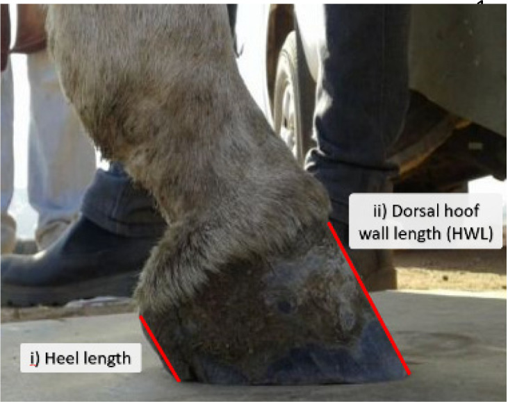
Photograph of the ground surface of the right front limb. The red lines indicate how the i) weight bearing length (WBL) lateral, ii) weight bearing length (WBL) medial, iii) heel width and iv) hoof width were measured.
Fig. 2.
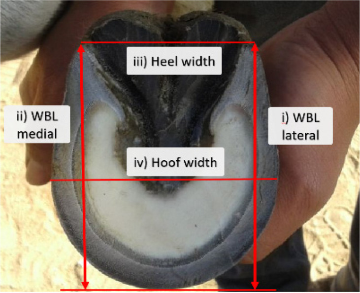
Photograph of the ground surface of the right front limb. The red lines indicate how the i) weight bearing length (WBL) lateral, ii) weight bearing length (WBL) medial, iii) heel width and iv) hoof width were measured.
Fig. 3.
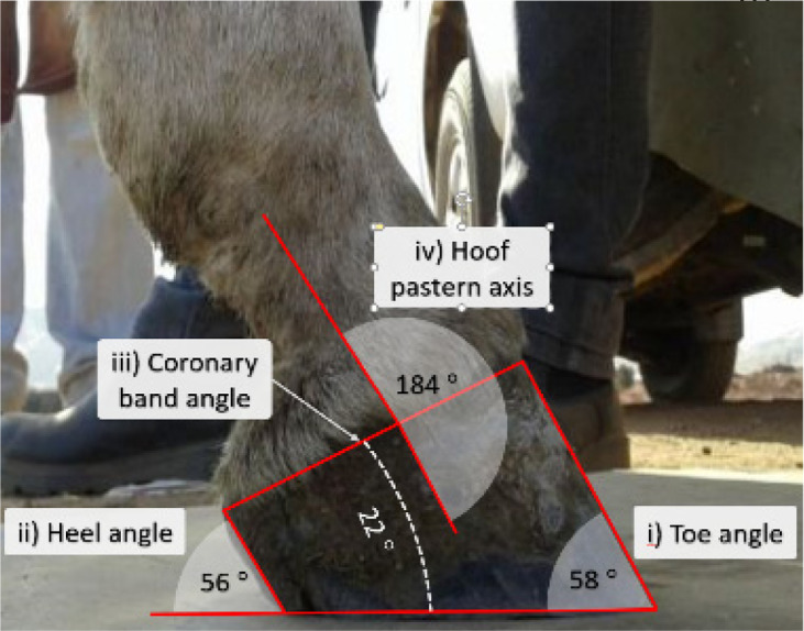
Lateral view of the right front limb of a donkey. The red lines indicate the lines from which angles were measured. The angles were i) toe angle, ii) heel angle, iii) coronary band angle and iv) hoof pastern axis. Values represent example angles for each measurement.
Statistical analyses
Differences in hoof parameters between front and hind limbs were tested using independent two-sample t-tests. All t-tests followed an F-test for equal variance.
To test whether donkeys’ hooves were broken forward as suggested by previous research (i.e. hoof pastern axis >180°), one-sample t-tests with equal variance and µ=180 were conducted for hoof pastern axis in front limbs and hind limbs.
To assess whether the hooves conformed to the same thresholds as horses in the range of medial-lateral balance (i.e. horse’s hooves are considered balanced when the difference in length between medial and lateral hoof wall quarters is ≤0.5 cm [12]), the difference between lateral and medial heel lengths was calculated as lateral length− medial length. The mean difference and 95% confidence intervals were calculated using the t-distribution [19], and hooves were considered to be in medial-lateral balance when the 95% confidence intervals were within the range of −0.5 to 0.5.
To assess whether hoof dimensions conformed to the same thresholds as the hooves of horses in terms of palmar support, the difference between toe and heel angles was tested using a paired t-test, with a pair consisting of the toe and heel angles measured from the same hoof.
Pearson’s correlation coefficient (r) was used to assess whether each of the measured hoof parameters correlated with the height at the withers or estimated body mass (Table 2).
For all statistical tests, results were considered statistically significant if P<0.05. All statistical analyses were performed using the software package R version 3.4.3.
Results
Estimated body mass ranged from 150 to 241 kg (mean ± SD: 186 ± 24 kg); body height ranged from 110 to 122 cm (mean ± SD: 115 ± 5 cm); and heart girth ranged from 113 to 134 cm (mean ± SD: 122 ± 6 cm). Mean measures of hoof dimensions are presented in Table 1 .
Table 1. Mean colostral IgG concentrations in various breeds at the time of parturition.
| Horse breed | Measurement (mean) | P value | ||||
|---|---|---|---|---|---|---|
| Front foot (± SD) | Hind foot (± SD) | MD | ||||
| Medial heel length (cm) | 3.51 | 0.40 | 3.41 | 0.36 | 0.10 | 0.399 |
| Lateral heel length (cm) | 3.52 | 0.41 | 3.39 | 0.33 | 0.13 | 0.291 |
| Lateral heel length (cm) | 6.73 | 0.46 | 6.84 | 0.57 | –0.11 | 0.494 |
| WBL medial (cm) | 8.14 | 0.60 | 7.48 | 0.49 | 0.66 | <0.001 |
| WBL lateral (cm) | 8.09 | 0.74 | 7.66 | 0.51 | 0.44 | 0.038 |
| Heel width (cm) | 6.34 | 0.58 | 6.27 | 0.55 | 0.07 | 0.692 |
| Hoof width (cm) | 7.20 | 0.51 | 6.33 | 0.37 | 0.87 | <0.001 |
| Toe angle (˚) | 58.25 | 5.56 | 59.61 | 3.48 | –1.36 | 0.367 |
| Heel angle (˚) | 54.71 | 5.61 | 56.89 | 3.64 | –2.18 | 0.159 |
| Coronary band angle (˚) | 25.33 | 3.19 | 29.83 | 3.90 | –4.50 | <0.001 |
| Hoof pastern axis (˚) | 198.80 | 7.74 | 193.10 | 9.86 | 5.69 | 0.042 |
| Ground surface size (cm) | 15.33 | 0.94 | 13.89 | 0.72 | 1.43 | <0.001 |
Units for each measurement are provided in the first column. MD, mean difference; HWL, hoof wall length; WBL, weight bearing length. SD, standard deviation. Results that were significant at the 0.05 level are indicated in bold.
Regarding hoof shape, front hoof capsules appeared to be more rounded and wider than hind hoof capsules. These findings were supported by statistical analyses, with hoof width, weight-bearing lengths (both lateral and medial) and ground surface size being consistently larger in the front hooves than the hind hooves (Table 1).
Equal variance was found between front and hind limbs for all measurements using the full dataset. Significant differences were found between front and hind limbs in the hoof pastern axis and coronary band angle; the hoof pastern axis and coronary slope band were larger and smaller, respectively, in front feet than in hind feet (Table 1).
No significant differences were found in heel width, heel length (lateral or medial), dorsal HWL, heel angle or toe angle between the front and hind hooves (Table 1).
The hoof pastern axis was significantly wider than 180° in both front limbs (t=11.9, df=23, P<0.001) and hind limbs (t=5.6, df=17, P<0.001).
The difference between lateral and medial heel lengths was significantly less than 0.5 cm in both front limbs (n=24, mean difference=0.006 cm, 95% CI= −0.05 to 0.07 cm) and hind limbs (n=18, mean difference= −0.02 cm, 95% CI= −0.09 to 0.06 cm).
The heel angle was significantly lower than the toe angle in the front limbs (−3.5°; 95% CI= −2.1 to −4.9°; t=−5.2, df=23, P<0.001) and hind limbs (−2.7°; 95% CI= −1.1 to −4.3°; t=−3.6, df=17, P<0.001).
The height at the withers showed a significant positive correlation with hoof width in the front limb and a significant negative correlation with lateral heel length in the hind limb (Table 2 ). Estimated body mass correlated positively with hoof width in both front and hind limbs, ground surface size in the hind limbs and lateral WBL in the hind limbs (Table 2).
Table 2. Pearson’s r correlation coefficients to assess the relationship between height at the withers, estimated body mass, hoof size and toe angle with specified hoof parameters measured from the front (n=24) and hind (n=18) hooves of 20 donkeys.
| Horse breed | Front foot | Hind hoof | |||||||
|---|---|---|---|---|---|---|---|---|---|
| r | P value | −95% CI | +95% CI | r | P value | −95% CI | +95% CI | ||
| Height at the withers | Medial heel length | –0.02 | 0.935 | –0.42 | 0.39 | –0.46 | 0.052 | –0.77 | 0.00 |
| Lateral heel length | –0.12 | 0.584 | –0.50 | 0.30 | –0.51 | 0.031 | –0.79 | –0.06 | |
| Dorsal HWL | 0.24 | 0.261 | –0.18 | 0.59 | 0.31 | 0.208 | –0.18 | 0.68 | |
| WBL medial | 0.34 | 0.100 | –0.07 | 0.66 | 0.46 | 0.055 | –0.01 | 0.76 | |
| WBL lateral | 0.17 | 0.436 | –0.25 | 0.53 | 0.43 | 0.074 | –0.05 | 0.75 | |
| Heel width | –0.09 | 0.688 | –0.47 | 0.33 | –0.10 | 0.705 | –0.54 | 0.39 | |
| Hoof width | 0.44 | 0.031 | 0.05 | 0.72 | 0.28 | 0.257 | –0.21 | 0.66 | |
| Ground surface size | 0.40 | 0.055 | –0.01 | 0.69 | 0.45 | 0.060 | –0.02 | –0.02 | |
| Height at the withers | Medial heel length | 0.30 | 0.151 | –0.11 | 0.63 | –0.03 | 0.897 | –0.49 | 0.44 |
| Lateral heel length | 0.28 | 0.182 | –0.14 | 0.62 | –0.10 | 0.706 | –0.54 | 0.39 | |
| Dorsal HWL | 0.33 | 0.110 | –0.08 | 0.65 | 0.39 | 0.111 | –0.10 | 0.72 | |
| WBL medial | 0.18 | 0.390 | –0.24 | 0.55 | 0.36 | 0.139 | –0.12 | 0.71 | |
| WBL lateral | 0.25 | 0.246 | –0.17 | 0.59 | 0.60 | 0.009 | 0.18 | 0.83 | |
| Heel width | 0.25 | 0.246 | –0.17 | 0.59 | 0.33 | 0.177 | –0.16 | –0.16 | |
| Hoof width | 0.47 | 0.021 | 0.08 | 0.73 | 0.65 | 0.004 | 0.26 | 0.86 | |
| Ground surface size | 0.40 | 0.055 | –0.01 | –0.01 | 0.67 | 0.002 | 0.30 | 0.87 | |
CI, confidence interval; HWL, hoof wall length; WBL, weight bearing length. Results that were significant at the 0.05 level are indicated in bold.
Discussion
Our results support previous findings from Souza et al. [21] indicating that the ground surface area of the front hooves is significantly larger than that of the hind hooves. Whilst we found hoof width and both lateral and medial weight bearing lengths to be significantly larger in front hooves, the difference in hoof width between front and hind hooves was more substantial than the difference in length, giving the front hooves a more rounded conformation. Similar findings have been reported in horses [1, 13, 22].
Incorrect alignment of the hoof pastern axis in horse digits can lead to a number of detrimental effects relating to incorrect load bearing, such as stresses to the deep digital flexor tendon [14]. In horses, alignment is considered to be correct when the hoof pastern axis is straight (180°) [15, 17]. This guidance is reflected in hoof trimming guidelines for donkeys, which recommend a straight hoof pastern axis [9, 23]. However, we found the hoof pastern axis to be significantly broken forward. Whilst recent guidance and hoof morphometric studies suggest the hoof pastern axis may be broken forward in comparison to a horse [4, 25], the degree to which the hoof pastern axis may be broken forward and still considered ‘normal’ is described as ‘limited’ [24]. Further, whilst the donkeys in the current study did not show any signs of lameness at the time of examination, a longitudinal study with a larger sample size using donkeys that have not been subjected to work is recommended. This addition would confirm whether or not a broken forward pastern axis is natural, rather than a result of working conditions, and whether it leads to lameness or other issues over time. At the very least, our findings suggest that hoof trimming guidelines for horses may not be suitable for donkeys, but further study is strongly recommended before donkey-specific guidelines can be developed with regard to the alignment of the hoof pastern.
The toe and heel angles have an important role in preventing tissue strain during load bearing of the foot [6]. Previous guidance for horses’ hooves suggests that the heel angle can be up to 5° lower than the toe angle [2, 6]. Although the heel angle is usually lower than the toe angle in donkeys [21], the magnitude of this difference has not been formally tested. Our findings suggest that the heel angle is an average of 3° lower than the toe angle in healthy donkeys (3.5° for front limbs, 2.7° for hind limbs), although further testing would be required to identify whether this represents the threshold value.
In horses, the heels are considered to be in medial-lateral balance when the difference in length between lateral and medial hoof wall quarters is ≤0.5 cm [12]. We found the mean difference between medial and lateral heel lengths to be significantly smaller than this threshold, suggesting that donkeys are in medial-lateral balance within the current suggested threshold of ≤0.5 cm. Further data, including measurements from lame donkeys, would be needed to confirm whether this threshold could be raised.
Hoof trimming guidelines provide appropriate dorsal HWLs based on estimated body mass for all breeds of horses except racing Standardbreds [2, 26]. In our study, dorsal HWL did not correlate with either estimated body mass or height. Estimated body mass did, however, correlate with hoof width, lateral WBL and ground surface size in the hind limb and hoof width in the front limb. We suggest, therefore, that the link between estimated body mass and measures of ground surface size may present the most promising starting point with which to develop species-specific guidelines for sound hoof dimensions in donkeys.
Acknowledgments
We are extremely grateful to all owners who permitted examination of their donkeys.
References
- 1.Back W. 2013. The role of the hoof and shoeing. In: Equine Locomotion (Back, W., and Clayton, H.M. eds.), Elsevier Health Sciences, London. [Google Scholar]
- 2.Balch O., White K., Butler D. 1991. Factors involved in the balancing of equine hooves. J. Am. Vet. Med. Assoc. 198: 1980–1989. [PubMed] [Google Scholar]
- 3.Bough J. 2011. Donkeys in human history, mythology and religion. pp. 45–75. In: Donkey, Reaktion Book Ltd., London. [Google Scholar]
- 4.Collins S.N., Dyson S.J., Murray R.C., Burden F., Trawford A. 2011. Radiological anatomy of the donkey’s foot: objective characterisation of the normal and laminitic donkey foot. Equine Vet. J. 43: 478–486. [DOI] [PubMed] [Google Scholar]
- 5.Crane M. 2007. Hoof disorders of the donkey. In: 10th Geneva Congress of Equine Medicine and Surgery, Organization (Chuit, P.A., and Montavon, S., eds.) Geneva.
- 6.Dyson S.J., Tranquille C.A., Collins S.N., Parkin T.D.H., Murray R.C. 2011. External characteristics of the lateral aspect of the hoof differ between non-lame and lame horses. Vet. J. 190: 364–371. [DOI] [PubMed] [Google Scholar]
- 7.El-Shafaey E.A., Salem M.G., Mosbah E., Zaghloul A.E. 2016. Morphometric evaluation of relevant radiographic parameters of the forefeet of clinically normal donkeys (Equus Asinus). J. Hell. Vet. Med. Soc. 68: 467–478. [Google Scholar]
- 8.FAO (Food and Agriculture Organization of the United Nations). 2017. Statistical databases. http://www.fao.org/faostat/en/#data/QA [accessed on May 8, 2019].
- 9.Fowler J. 1995. Trimming donkey feet. Equine Vet. Educ. 7: 18–21. [Google Scholar]
- 10.Kane A.J., Stover S.M., Gardner I.A., Bock K.B., Case J.T., Johnson B.J., Anderson M.L., Barr B.C., Daft B.M., Kinde H., Larochelle D., Moore J., Mysore J., Stoltz J., Woods L., Read D.H., Ardans A.A. 1998. Hoof size, shape, and balance as possible risk factors for catastrophic musculoskeletal injury of Thoroughbred racehorses. Am. J. Vet. Res. 59: 1545–1552. [PubMed] [Google Scholar]
- 11.Kosťuková M., Černohorská H., Bihuncová I., Oravcová I., Sobotková E., Jiskrová I. 2015. Characteristics of morphological parameters of donkeys in the Czech Republic. Acta Univ. Agric. Silvic. Mendel. Brun. 63: 419–424. [Google Scholar]
- 12.Kummer M., Geyer H., Imboden I., Auer J., Lischer C. 2006. The effect of hoof trimming on radiographic measurements of the front feet of normal Warmblood horses. Vet. J. 172: 58–66. [DOI] [PubMed] [Google Scholar]
- 13.Łuszczynski J., Pieszka M., Paszkot W. 2007. The trial of founding the dependence between the size of hoof sole and biometric measurements of Hucul horses. Lucr. Stiint. Zooteh. Biotehnol. 40: 181–187. [Google Scholar]
- 14.O’Grady S.E. 2009. Guidelines for trimming the equine foot: a review. pp. 218–225. In: 55th Annual Convention of the American Association of Equine Practitioners. AAEP Proceedings, Lexington.
- 15.O’Grady S.E. 2013. How to evaluate the equine hoof capsule. AAEP Proceedings59: 54–61.
- 16.O’Grady S.E., Poupard D.A. 2001. Physiological horseshoeing–an overview. Equine Vet. Educ. 28: 426–430. [Google Scholar]
- 17.Parks A.H. 2003. Foot balance, conformation, and lameness. pp. 250–261. In: Diagnosis and Management of Lameness in the Horse, 1st ed. (Ross, M.W., and Dyson, S.J. eds.), W. B. Saunders, Philadelphia. [Google Scholar]
- 18.Rossel S., Marshall F., Peters J., Pilgram T., Adams M.D., O’Connor D. 2008. Domestication of the donkey: timing, processes, and indicators. Proc. Natl. Acad. Sci. U.S.A. 105: 3715–3720. [DOI] [PMC free article] [PubMed] [Google Scholar]
- 19.Hazra A. 2017. Using the confidence interval confidently. J. Thorac. Dis. 9: 4125–4130. [DOI] [PMC free article] [PubMed] [Google Scholar]
- 20.Sargentini C., Tocci R., Andrenelli L., Giorgetti A. 2012. Preliminary studies on hoof characteristics in Amiata donkey. Ital. J. Anim. Sci. 11: 123–127. [Google Scholar]
- 21.Souza A.F., Kunz J.R., Laus R., Moreira M.A., Muller T.R., Fonteque J.H. 2016. Biometrics of hoof balance in equids. Anim. Sci. Pap. Rep. 68: 825–831. [Google Scholar]
- 22.Stachurska A., Kolstrung R., Pięta M., Silmanowicz P. 2011. Hoof size as related to body size in the horse (Equus caballus). Arch. Anim. Breed. 29: 213–222. [Google Scholar]
- 23.Svendson E.D. 2008. The Professional Handbook of the Donkey, 4th ed. (Duncan, J., and Hadrill, D. eds.), Whittet Books Ltd., Essex. [Google Scholar]
- 24.The Donkey Sanctuary 2018. The Clinical Companion of the Donkey, 1st ed. Troubador Publishing, Leicester. [Google Scholar]
- 25.Thiemann A., Rickards K. 2013. Donkey hoof disorders and their treatment. In Pract. 35: 134–140. [Google Scholar]
- 26.Turner T.A. 1992. The use of hoof measurements for the objective assessment of hoof balance. pp. 389–395. In: 38th AAEP Proceedings.