Abstract
Background
Treating the lower face with neuromodulators and targeting the masseter muscle can reduce masseteric hypertrophy but can also change the facial shape. A novel observation after the treatment of the masseter muscle with incobotulinumtoxin Type A was the increase in temporal volume.
Aim
Objectively assess temporal volume increase following treatment of masseteric hypertrophy using incobotulinumtoxin Type A.
Methods
Nine female patients with a mean age of 35.11 years ± 9.1 [Asian (11.1%) and Caucasian (88.9%)] were treated with incobotulinumtoxin Type A for masseteric hypertrophy. Masseteric prominence and temporal volume were assessed by two independent raters, and temporal fossa volume was measured via 3‐dimensional volumetric imaging.
Results
Independent of the neuromodulator injection technique (ie, single‐injection versus multi‐injection), a reduction in masseteric hypertrophy occurred represented by a decrease in the masseter prominence scale. In addition, the treatment resulted in a significant improvement of the temporal volume scale and an increase in the measured volume of the temporal fossa. None of the presented measurements were statistically significantly different between the two utilized injection techniques.
Conclusions
This study supports using a full‐face approach when performing aesthetic treatments. Anatomical concepts can help to guide treatments: the compensatory increase in temporalis function after masseter muscle treatment resulted in an increased in temporal fossa volume. The findings presented herein should not be considered as a new concept for treating the temporal fossa but rather as an additional possibility for increasing the temporal volume.
Keywords: facial anatomy, incobotulinumtoxin type A, masseteric hypertrophy, neuromodulator, temporal hollowing, temporal volume
1. INTRODUCTION
The increasing desire for a smooth and rhytid‐free facial appearance in today's society is reflected by the increasing demand for neuromodulator injections, as reported by the American Society of Plastic Surgeons. According to their national plastic surgery statistics, the number of Botulinum Toxin Type A treatments increased by 845% between the years 2000 and 2018. 1 Besides the treatment of facial rhytids, neuromodulator injections are successfully utilized to treat bruxism, temporomandibular joint disorders, and various types of masseteric hypertrophy. 2
One beneficial side effect of treating the masseter muscle with neuromodulators is the change in facial shape. When assessed from the frontal view, the shape of the each individual's face can be described according to various terms, such as heart‐shaped, square, pear, rectangle, round, oval, diamond, and oblong, and can significantly drive the desire for aesthetic intervention. 3 , 4 Reducing the masseteric volume can alter the width proportions between the upper, middle and lower face and thus influence the overall facial shape.
Another beneficial side effect of treating the masseter muscle with neuromodulators could be a change in muscular balance between the muscles of mastication, which are composed of the masseter, temporalis, and medial and lateral pterygoids. Reducing the contractility of the masseter muscle could result in a compensatory increase of function of the other muscles of mastication, which could increase in volume and thus alter the shape of the face. This could potentially be used for treating temporal hollowing. 5
A recent study compared the noninferiority of two different injection techniques for treating masseteric hypertrophy, 6 using incobotulinumtoxin Type A in a sample of n = 30 patients. In some subjects, the authors also observed a change in temporal volume. As the outcomes of that particular study were the comparison of two injection techniques with regard to efficacy and safety, an objective analysis of the change in temporal volume following neuromodulator injection into the masseter muscles was not performed.
The scope of the present study is to objectively analyze the change in temporal volume following neuromodulator treatment of the masseter muscle.
2. MATERIAL AND METHODS
2.1. Study sample
Thirty consecutive patients were screened for this study. Inclusion criteria were as follows: female gender, aged older than 18 years, having a palpable and visible hypertrophy of the masseter muscle, accepting the obligation not to receive any other facial procedures during the 6‐month follow‐up period of the study, no previous facial fillers for 12 months prior to the study, no previous facial fillers of the jawline 18 months prior to the study, no previous neuromodulator injection into the masseter muscle for the last 12 months prior to the study and willingness to participate in this study.
Exclusion criteria were as follows: current pregnancy or lactation, hypersensitivity or allergy to incobotulinumtoxin type A, known disorders of normal muscle contractility (eg, myasthenia gravis or Lambert‐Eaton Syndrome), presence of infection at the site of injection (ie, mandibular angle), inability to comply with follow‐up examinations, abstain from facial procedures during the 6‐month follow‐up period of the study and having a score of ≥2 on the Merz Aesthetics Scale for jowling. 7 A detailed description of the inclusion/exclusion criteria of the screened sample has been published previously. 7
This study received approval from the independent review board “Veritas IRB” and can be tracked at: www.clinicaltrials.gov (Identification‐number: NCT03376464). The procedures were performed in adherence to the Declaration of Helsinki (1996), in accordance with regional laws and good clinical practice for studies in human subjects. 6 Written and informed consent was obtained by all participants prior to inclusion into the study.
Out of thirty screened patients, nine were included in the final analysis. The mean age of the final sample (n = 9) was 35.11 years ± 9.1 and consisted of n = 1 Asian (11.1%) and n = 8 Caucasian (88.9%) females.
2.2. Injection procedure
The commercially available incobotulinumtoxin Type A [Xeomin® (Bocouture), Merz Pharmaceuticals] was used for all injections procedures. Two different injection techniques were randomly used for both sides of the same patients: single‐injection technique (SIT) and multi‐injection technique (MIT). Both injection techniques were previously described and utilized 40 U of neuromodulator product for each masseter muscle and 80 U of product per patient. 7
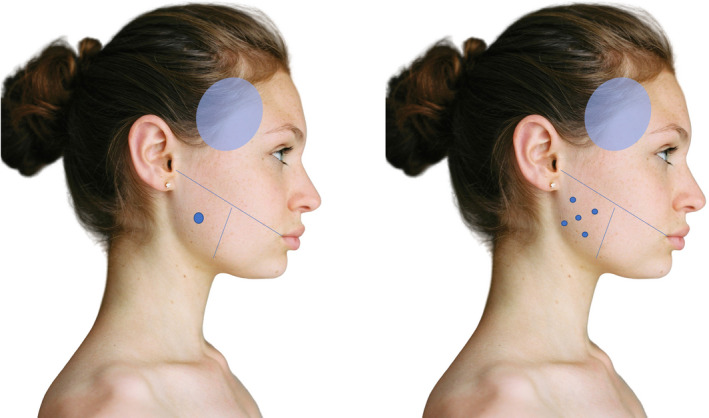
Utilizing the SIT, 40 U of incobotulinumtoxin Type A was injected into the region where the three masseter heads overlap, which was identified via palpation as the thickest area of the masseter muscle and corresponded to an approximate location of 1 cm anterior and 1 cm superior from the mandibular angle (Figure 1). Four patients (44.4%) were injected with the SIT.
Figure 1.

Schematic illustration of the injection location (blue dots) for the single‐injection (SIT) and the multi‐injection technique (MIT). The blue area represents the area for objective volume measurements
Utilizing the MIT, 8 U of incobotulinumtoxin Type A was injected into five distinct areas across the palpable extent of the masseter muscle, at 1 cm intervals, totaling 40 U of neuromodulator product (Figure 1). Five patients (55.6%) were injected with the MIT.
2.3. Clinical assessment
Bilateral temporal volume was assessed by two independent, blinded reviewers before and after the neuromodulator injections into the masseter muscle via the temporal volume scale according to the following classification: 0 = No volume loss; 1 = Mild volume loss; 2 = Moderate volume loss; 3 = Severe Volume loss; 4 = Very severe volume loss. 8 In addition, masseter prominence was rated using a 10‐point photonumeric scale that rates the severity of masseter prominence from I (none) to V (much) and the position of the maximum masseter prominence (low or high). 7
Intra‐rater agreement coefficients were calculated for both reviewers based on baseline images, to ensure their ability to consistently rate the same image. To accomplish this, reviewers graded all baseline images during a single session and returned two weeks later to grade the same images again (ie, test and re‐test). Baseline images were randomly assigned during each session, to eliminate memory bias. A linear weighted Kappa (κ) was used as a measure of association for analyzing the ordinal data collected from both raters. 9 Reviewers remained blinded to each other's scores and were assessed separately.
2.4. Imaging
Three‐dimensional (3D) surface scanning procedures were performed using the Vectra H1 camera system (Canfield Scientific Inc). Prior to the respective injection procedures, a full‐face baseline scan of each patient was performed (= baseline scan). An additional full‐face scan was performed 16 weeks after the neuromodulator injections (= follow–up scan). Obtained 3D images were imported into the Mirror Software Suite® (Canfield Scientific Inc) for further analysis. Each follow‐up scan was aligned to its respective baseline scan to compute differences in temporal volume. Applying the automated algorithm of the Mirror Software Suite®, values for changes in volume (ie, difference between follow‐up and baseline scans) for each temple were calculated in cc (Figure 2).
Figure 2.

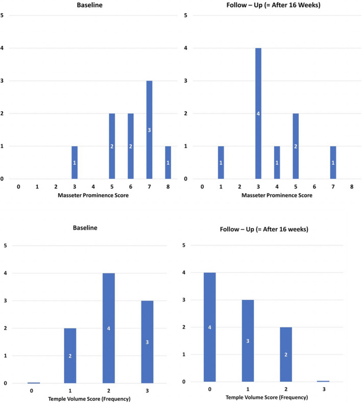
Bar graphs of the masseteric prominence scale 7 and temporal volume scale 8 at baseline and 16 wk after the treatment
2.5. Statistical analysis
Mann‐Whitney U tests were used to test for differences in the temporal volume and masseteric prominence scale between treatment groups and between baseline and follow‐up visits. Student's T tests were used to evaluate differences of the volume changes between injection techniques and between baseline and follow–up. Spearman's correlations were run to assess correlations between masseteric prominence scale improvement and temporal volume score improvement. Statistical analyses were performed using SPSS Statistics 23 (IBM), and results were considered statistically significant at a probability level of ≤.05 to guide conclusions.
3. RESULTS
3.1. Intra‐ and inter–rater agreement
Intra‐rater agreement coefficients (rs = .764, .835) and inter‐rater agreement coefficients (κ = .664; SE: 0.152; 95% CI: 0.37‐0.96) were high for both raters when grading temporal volume.
3.2. Masseteric prominence scale
The median baseline value for the masseteric prominence scale was [median value (interquartile range)] 6.0 (5.0, 7.0).
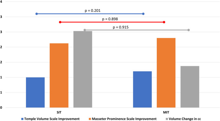
The median follow‐up value for the masseteric prominence scale was 3.5 (1.5, 4.7), representing a statistically significant improvement after the treatment with P = .005. Stratifying for the two injection techniques revealed that SIT resulted in a follow‐up value of 3.5 (1.5, 4.7) with P = .046 and that MIT resulted in a follow‐up value of 3.0 (3.0, 6.0) with P = .046. No statistically significant difference in masseteric prominence scale follow‐up values between the two different injection techniques was detected with P = .730.
3.3. Temporal volume scale
The median baseline value for the temporal volume scale was 2.0 (1.0, 3.0; Figure 2). The median follow‐up value for the temporal volume scale was 1.0 (0.0, 1.5), representing a statistically significant improvement after the treatment with P = .003 (Figure 3). Stratifying for the two injection techniques revealed that SIT resulted in a follow‐up value of 1.0 (0.25, 1.75) with P = .046 and that MIT resulted in an improvement of 0.0 (0.0, 1.5) with P = .025. No statistically significant difference in temporal volume scale follow‐up values between the two different injection techniques was detected with P = .556.
Figure 3.

Bar graph showing the differences between baseline and follow‐up stratified by the two utilized injection techniques: single‐injection (SIT) and the multi‐injection technique (MIT). Probability values are given to estimate statistical significance
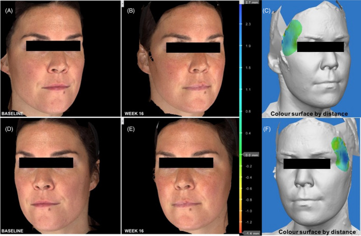
The mean unstratified volume increase after the treatment was [mean (standard deviation)] 1.89 cc (2.0; Figure 4), with 1.96 cc (2.0) for the SIT and with 1.83 cc (2.1) for the MIT with P = .898 for differences between injection techniques.
Figure 4.

Pre‐ and postinjection outcomes in temporal fossa volume with green demonstrating moderate (0.2‐0.4 mm) increase in volume, and blue demonstrating major (1‐2.7 mm) increase in volume
4. DISCUSSION
This study objectively measured the volumizing effects in the temporal fossa following the treatment of the masseter muscle with incobotulinumtoxin Type A for masseteric hypertrophy in a small sample size (n = 9). The results revealed that independent of the neuromodulator injection technique (single‐injection vs. multi‐injection technique), a reduction in masseteric hypertrophy occurred represented by a decrease in the masseter prominence scale with P = .005. The treatment additionally resulted in a significant improvement of the temporal volume scale with P = .003 and in an increase in the measured volume of the temporal fossa by 1.89 cc. None of the presented measurements were statistically significant different between the two utilized injection techniques with all P > .05.
A strength of the study is that the treatment outcome assessments were independently rated by two experienced blinded observers with high intra‐ and inter‐rater agreement coefficients. Additionally, the measurements of the volume of the temporal fossa were objectively analyzed via 3D imaging, which provides increased objectivity in the analyses of the effects of the neuromodulator injections. 5 , 10 , 11 , 12 The objective 3D measurements were in line with the clinically assessed improvement, which underlines the clinical effect of the masseter injections in the evaluated cases.
One central limitation of the study is the small sample size. A larger sample size allows more robust interpretation of results from occurring events, as clusters of observation can be built to guide conclusions. However, the objective of the initial study trial was the comparison of the two injection techniques with regard to efficacy and safety following masseteric hypertrophy treatment with incobotulinumtoxin Type A and did not include temporal volume assessment, at first. 6 The current study included the objective measurements only toward the end of the initial trial, to document the effects observed in the previously treated study participants. Having utilized the temporal measurements at the beginning of the initial trial would have allowed increasing the sample size of the current report. Future studies will need to design new clinical studies to objectively investigate and confirm the effects presented in this report. Another limitation is the generalizability of the presented results, as only young females were investigated. It remains to be tested whether the effects are similar in outcome and magnitude if male patients and/or with a different age range are treated. It could be speculated that in males, a greater effect could be expected as the muscular response might be larger resulting a greater volume increase of the temporal fossa. On the other side, it can be speculated that age might result in a limited effect of temporal volumization, as with increasing age the soft tissue of the temple additionally contributes to temporal hollowing. 13 , 14 , 15 , 16 , 17 Future clinical trials should account for various gender‐ and age‐related factors to best elaborate on the results and discussions presented herein.
The results revealed that neuromodulator treatment of the masseter muscle for masseteric hypertrophy resulted in an increase in the measured volume of the temporal fossa by 1.89 cc. This can be explained by the interaction of the muscles of mastication. Weakening the ability of the masseter muscle to contract after the neuromodulator injections resulted in this study in a potential increase in contractility of the remainder muscles of mastication. Of those, only the temporalis muscle is visible on the skin surface and the increase in volume of the temporal fossa could be interpreted as an increase in its function. Electromyographic investigations of all muscles of mastication should be performed to test whether all, or just the visible temporalis muscle, is affected by the neuromodulator treatment of the masseter muscle.
The observed effects on the temporal fossa emphasize the concept of a full‐face assessment when aesthetically treating a patient's face. The results show that by injecting the lower face with neuromodulators, a volumizing effect of the upper face was achieved. This full‐face concept can be best understood by the underlying anatomy if the treated components are seen as a unit of a full‐face spanning system. In this study, the muscles of mastication should be respected with its components: masseter, temporalis and medial and lateral pterygoids. Affecting one component (here, the masseter muscles) resulted in a potential increase in activity of the temporalis muscle, which could be assumed by the increase in volume of the temporal fossa.
Understanding the components that contribute to shaping the face can guide practitioners to more effective treatments. When viewed from the frontal view, the shape of each individual's face can be described according to various terms like: heart‐shaped, square, pear, rectangle, round, oval, diamond, and oblong, and can significantly drive the desire for aesthetic intervention. 3 , 4 In the present study, the prominence of the masseter muscle was reduced due to the effects of the injected neuromodulator and the volume of the temporal fossa was additionally increased. This concept can be utilized clinically to change the facial shape from square to oval or heart‐shaped.
Volumizing temporal hollows with soft tissue fillers can often require the use of up to 2.0 cc of product, per temporal fossa. 5 , 18 , 19 , 20 , 21 In the present study, an increase in temporal volume by 1.89 cc was measured without targeting the temporal fossa directly. This increase in volume is respectable if the surface volume coefficient of the temporal fossa for the different layered injections is considered. 20 The assessed improvements of the temple via the temporal volume scale was a two‐point improvement in one subject, whereas the remainder of the subjects displayed a one‐point improvement. Comparing the effects from the present study to other treatment algorithms, which utilized hyaluronic acid 22 or calcium hydroxylapatite 23 with improvements by two or three points on the temporal volume scale, reveal limitations in clinical effectiveness if this concept is thought to be utilized as a stand‐alone treatment for temporal volumization. The findings presented herein should not be considered as a new concept for treating the temporal fossa but rather an additional possibility for increasing the temporal volume.
5. CONCLUSIONS
The results revealed that, independent of the neuromodulator injection technique (single‐injection versus multi‐injection technique), a reduction in masseteric hypertrophy occurred. This was represented by a decrease in the masseter prominence scale with P = .005. The treatment additionally resulted in a significant improvement of the temporal volume scale with P = .003 and in an increase in the measured volume of the temporal fossa by 1.89 cc. The findings presented herein should not be considered as a new concept for treating the temporal fossa, but rather an additional possibility for increasing the temporal volume.
CONFLICT OF INTERESTS
Dr Andreas Nikolis is a consultant speaker and research collaborator for Merz Pharma. The remaining authors have no conflicts of interest to disclose. This research did not receive any specific funding from the public, commercial or not‐for‐profit sectors.
ACKNOWLEDGMENTS
We would like to thank Dr Konstantin Frank and Prof. Thilo Schencki for their contributions.
Nikolis A, Enright KM, Rudolph C, Cotofana S. Temporal volume increase after reduction of masseteric hypertrophy utilizing incobotulinumtoxin type A. J Cosmet Dermatol. 2020;19:1294–1300. 10.1111/jocd.13434
REFERENCES
- 1. American Society of Plastic Surgeons . 2018 National Plastic Surgery Statistics. 2018. https://www.plasticsurgery.org/documents/News/Statistics/2018/plastic‐surgery‐statistics‐report‐2018.pdf. Published 2019. Accessed April 14, 2019.
- 2. Patel J, Cardoso JA, Mehta S. A systematic review of botulinum toxin in the management of patients with temporomandibular disorders and bruxism. Br Dent J. 2019;226(9):667‐672. [DOI] [PubMed] [Google Scholar]
- 3. Goodman GJ. The oval female facial shape—a study in beauty. Dermatol Surg. 2015;41(12):1375‐1383. [DOI] [PubMed] [Google Scholar]
- 4. Samizadeh S. The ideals of facial beauty among Chinese aesthetic practitioners: results from a large National Survey. Aesthetic Plast Surg. 2019;43(1):102‐114. [DOI] [PMC free article] [PubMed] [Google Scholar]
- 5. Cotofana S, Koban K, Pavicic T, et al. Clinical validation of the surface volume coefficient for minimally invasive treatment of the temple. J Drugs Dermatol. 2019;18(6):533. [PubMed] [Google Scholar]
- 6. The World Medical Association . WMA Declaration of Helsinki – Ethical Principles for Medical Research Involving Human Subjects – WMA. https://www.wma.net/policies‐post/wma‐declaration‐of‐helsinki‐ethical‐principles‐for‐medical‐research‐involving‐human‐subjects/. Accessed August 5, 2018.
- 7. Nikolis A, Enright KM, Masouri S, Bernstein S, Antoniou C. Prospective evaluation of incobotulinumtoxina in the management of the masseter using two different injection techniques. Clin Cosmet Investig Dermatol. 2018;11:347‐356. [DOI] [PMC free article] [PubMed] [Google Scholar]
- 8. Carruthers J, Jones D, Hardas B, et al. Development and validation of a photonumeric scale for evaluation of volume deficit of the temple. Dermatol Surg. 2016;42(Suppl 1):S203‐S210. [DOI] [PMC free article] [PubMed] [Google Scholar]
- 9. Nelson KP, Edwards D. Measures of agreement between many raters for ordinal classifications. Stat Med. 2015;34(23):3116‐3132. [DOI] [PMC free article] [PubMed] [Google Scholar]
- 10. Koban KC, Cotofana S, Frank K, et al. Precision in 3‐dimensional surface imaging of the face: a handheld scanner comparison performed in a cadaveric model. Aesthetic Surg J. 2019;39(4):NP36‐NP44. [DOI] [PubMed] [Google Scholar]
- 11. Koban KC, Härtnagl F, Titze V, Schenck TL, Giunta RE. Chances and limitations of a low‐cost mobile 3D scanner for breast imaging in comparison to an established 3D photogrammetric system. J Plast Reconstr Aesthetic Surg. 2018;71(10):1417‐1423. [DOI] [PubMed] [Google Scholar]
- 12. Koban K, Schenck T, Metz P, et al. Auf dem Weg zur objektiven Evaluation von Form, Volumen und Symmetrie in der Plastischen Chirurgie mittels intraoperativer 3D Scans. Handchir Mikrochir Plast Chir. 2016;48(02):78‐84. [DOI] [PubMed] [Google Scholar]
- 13. Cotofana S, Gotkin RH, Ascher B, et al. Calvarial volume loss and facial aging: a computed tomographic (CT)‐Based Study. Aesthetic Surg J. 2018;38(10):1043‐1051. [DOI] [PubMed] [Google Scholar]
- 14. Cotofana S, Gotkin RH, Morozov SP, et al. The relationship between bone remodeling and the clockwise rotation of the facial skeleton: a computed tomographic imaging‐based evaluation. Plast Reconstr Surg. 2018;142(6):1447‐1454. [DOI] [PubMed] [Google Scholar]
- 15. Frank K, Gotkin RH, Pavicic T, et al. Age and gender differences of the frontal bone: a computed tomographic (CT)‐based study. Aesthetic Surg J. 2018;7:699‐710. 10.1093/asj/sjy270. [DOI] [PubMed] [Google Scholar]
- 16. Cotofana S, Mian A, Sykes JM, et al. An update on the anatomy of the forehead compartments. Plast Reconstr Surg. 2017;139(4):864e‐872e. [DOI] [PubMed] [Google Scholar]
- 17. Cotofana S, Gotkin RH, Frank K, et al. The functional anatomy of the deep facial fat compartments. Plast Reconstr Surg. 2019;143(1):53‐63. [DOI] [PubMed] [Google Scholar]
- 18. Lambros V. A technique for filling the temples with highly diluted hyaluronic acid: The “dilution solution”. Aesthetic Surg J. 2011;31(1):89‐94. [DOI] [PubMed] [Google Scholar]
- 19. Pavicic T, Mohmand HM, Yankova M, et al. Influence of needle size and injection angle on the distribution pattern of facial soft tissue fillers. J Cosmet Dermatol. 2019;18(5):1230–1236. [DOI] [PubMed] [Google Scholar]
- 20. Cotofana S, Koban CK, Frank K, et al. The surface‐volume‐coefficient of the superficial and deep facial fat compartments – a cadaveric 3D volumetric analysis. Plast Reconstr Surg. 2019;143(6):1605–1613. [DOI] [PubMed] [Google Scholar]
- 21. Schenck TL, Koban KC, Schlattau A, et al. The functional anatomy of the superficial fat compartments of the face: a detailed imaging study. Plast Reconstr Surg. 2018;141(6):1351‐1359. [DOI] [PubMed] [Google Scholar]
- 22. Juhász MLW, Marmur ES. Temporal fossa defects: techniques for injecting hyaluronic acid filler and complications after hyaluronic acid filler injection. J Cosmet Dermatol. 2015;14(3):254‐259. [DOI] [PubMed] [Google Scholar]
- 23. Juhász MLW, Levin MK, Marmur ES. Pilot study examining the safety and efficacy of calcium hydroxylapatite filler with integral lidocaine over a 12‐month period to correct temporal fossa volume loss. Dermatol Surg. 2018;44(1):93‐100. [DOI] [PubMed] [Google Scholar]


