Figure 1.

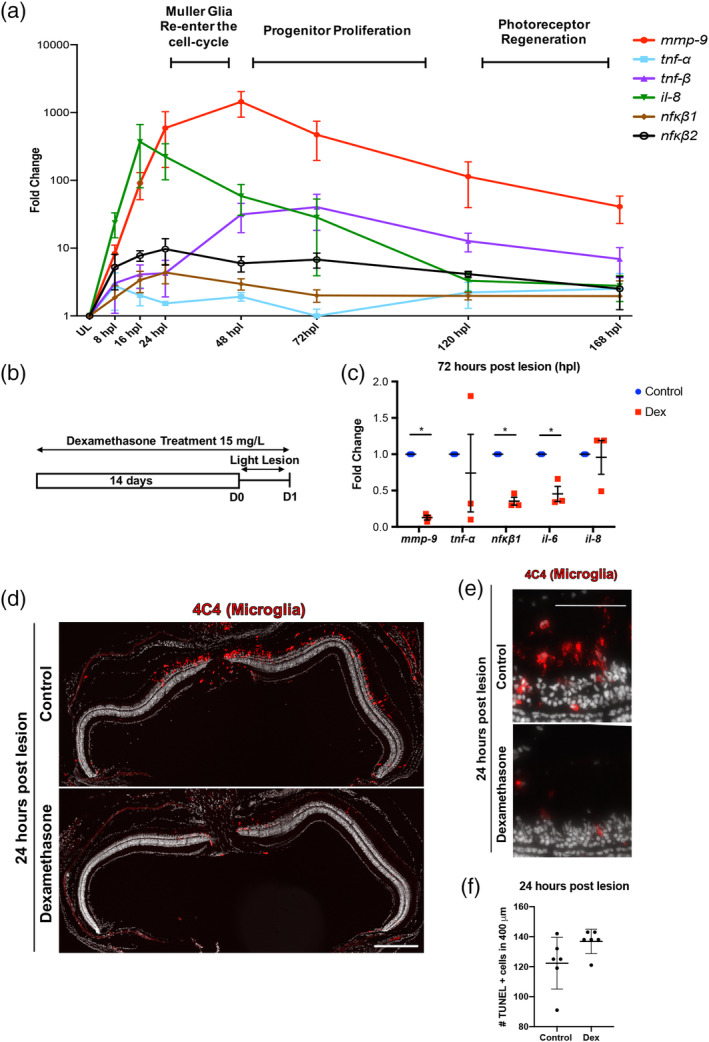
Inflammation is induced following photoreceptor ablation. Dexamethasone treatment suppresses microglia activation and cytokine production, but not photoreceptor death. (a) Time‐course for the expression of inflammatory genes, from 8 to 168 hr post lesion (hpl). Unlesioned retinas served as controls. Expression levels are represented as fold change calculated using DDC T method. (b) Experimental paradigm for the immune suppression. (c) qRT‐PCR for the inflammatory genes mmp‐9 (ANOVA F‐ratio = 22.237, p < .0001), tnf‐α (ANOVA F‐ratio = 1.5507, p = .2209), tnf‐β (ANOVA F‐ratio = 6.5120, p = .001), il‐8 (ANOVA F‐ratio = 8.2296, p = .0003), nfκb1 (ANOVA F‐ratio = 2.5207, p = .0596), and nfκb2 (ANOVA F‐ratio = 3.5591, p = .0168) from control and Dex‐treated retinas at 72 hpl. *p ≤ .05. (d) Immunostaining for microglia using the 4C4 antibody in control (top) and Dex‐treated retinas (bottom). (e) High magnification image of 4C4 staining at 1 dpl control (top) and Dex‐treated retinas (bottom). (f) The number of TUNEL positive cells at 1 dpl in control (122.27 ± 17.03 cells; n = 6) and Dex‐treated animals (136.93 ± 8.29 cells; n = 6). Scale bar equals 50 μm [Color figure can be viewed at wileyonlinelibrary.com]
