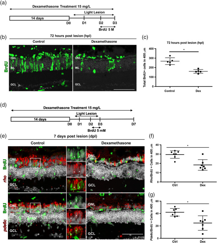
Figure 2.

Dexamethasone treatment suppresses proliferation of Müller glia and photoreceptor regeneration. (a) Experimental paradigm of anti‐inflammatory treatment to assay proliferation. (b) BrdU immunostained cells (green) in controls (left) and Dex‐treated animals (right) at 72 hpl. (c) Number of BrdU‐labeled cells in controls (268 ± 36.1 cells; n = 5) and Dex‐treated animals (160.2 ± 20.02 cells; n = 5) at 72 hpl; *p = .0079. (d) Experimental paradigm of anti‐inflammatory treatment. Regenerated photoreceptors were identified as BrdU‐labeled nuclei surrounded by in situ hybridization signal for either rho (rods) or pde6c (cones) at 7 dpl. (e) Double labeled, regenerated photoreceptors using in situ hybridization for rods (rho) and cones (pde6c; red signal) and BrdU (green). The high magnification insets show the colocalization of the two labels (asterisks). (f) Number of regenerated rod photoreceptors in control (29.52 ± 4.1 cells; n = 7) and experimental retinas (18.33 ± 5.71 cells; n = 7); *p = .0047. (g) Number of regenerated cone photoreceptors in control (42 ± 6.1 cells; n = 7) and Dex‐treated animals (25 ± 11.72 cells; n = 7). *p = .0012. Scale bar equals 50 μm. GCL, ganglion cell layer; INL, inner nuclear layer; ONL, outer nuclear layer [Color figure can be viewed at wileyonlinelibrary.com]
