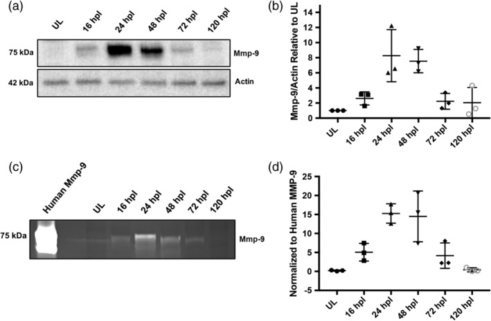
Figure 4.

Mmp‐9 is expressed and catalytically active following photoreceptor death. (a) Western blot of retinal proteins from unlesioned retinas (UL) and lesioned retinas at 16, 24, 48, 72, and 120 hpl, stained with anti‐Mmp‐9 and anti‐Actin (loading control) antibodies. (b) Densitometry of Mmp‐9 protein, plotted relative to unlesioned controls. (ANOVA F‐ratio = 8.377, p = .0013) (c) Zymogram of Mmp‐9 catalytic activity from unlesioned retinas (UL) and lesioned retinas at 16, 24, 48, 72, and 120 hpl. Lanes in zymogram correspond to those in the Western blot. Purified human recombinant protein serves as a positive control. (d) Densitometry of Mmp‐9 catalytic activity, plotted relative to the positive control. (ANOVA F‐ratio = 11.870, p = .0003)
