Abstract
Background
Induced pluripotent stem cells (iPSCs) have enormous potential in developmental biology studies and in cellular therapies. Although extensively studied and characterized in human and murine models, iPSCs from animals other than mice lack reproducible results.
Methods
Herein, we describe the generation of robust iPSCs from equine and bovine cells through lentiviral transduction of murine or human transcription factors Oct4, Sox2, Klf4, and c-Myc and from human and murine cells using similar protocols, even when different supplementations were used. The iPSCs were analyzed regarding morphology, gene and protein expression of pluripotency factors, alkaline phosphatase detection, and spontaneous and induced differentiation.
Results
Although embryonic-derived stem cells are yet not well characterized in domestic animals, generation of iPS cells from these species is possible through similar protocols used for mouse or human cells, enabling the use of pluripotent cells from large animals for basic or applied purposes. Herein, we also infer that bovine iPS (biPSCs) exhibit similarity to mouse iPSCs (miPSCs), whereas equine iPSs (eiPSCs) to human (hiPSCs).
Conclusions
The generation of reproducible protocols in different animal species will provide an informative tool for producing in vitro autologous pluripotent cells from domestic animals. These cells will create new opportunities in animal breeding through transgenic technology and will support a new era of translational medicine with large animal models.
Keywords: Bovine, Cellular reprogramming, Equine, Induced pluripotency, iPSCs, stem cells
Background
Induced pluripotent stem cells (iPSCs) have enormous potential in cellular therapy, reprogramming, and early development approaches as they can differentiate into numerous autologous cell lineages, including the three germ layers. Since the major breakthrough of iPSC generation in 2006 [1], several studies have reproduced the induction of cellular pluripotency through overexpression of specific transcription factors. Although some reports describe iPSC generation in dogs, pigs, horses, cattle, and some other species as reviewed elsewhere [2], the most thoroughly characterized iPSC cells are undoubtedly from humans and mice [3–16].
Interestingly, iPSCs can be generated from species that do not have well-characterized embryonic-derived pluripotent stem cells (ES cells) yet. For example, in bovine and equine models, the lack of characterization of pluripotent features leads to their denomination as embryonic “stem cell-like” cells [17, 18], although recent advances have been reported recently in cattle [19]. The clinical use of iPSCs is still limited by several factors, including the low efficiency of reprogramming and the lack of studies assuring the safety of transplantation procedures, a consequence of unstable karyotypes and genomic alterations due to viral integration. iPSC production, therefore, must be improved to be reproducible and safe for its therapeutic use to become possible [20].
Though mice are still the most studied and used model for basic and applied research, it has considerable limitations, in special, in early development, and in overall physiology when compared to humans [21]. Rabbits are also considered adequate models for translational research [22, 23]; however, its in vitro cell culture and pluripotency acquisition still lack further robust characterization [24]. In this context, domestic animal species such as swine, small ruminants, and even companion animals such as dogs may provide an important contribution to regenerative and translational medicine. iPSC technology suggests that although species-specific differences are evident, the basic mechanisms of pluripotency acquisition may follow similar patterns in mammals, and iPSCs have been reported from these animals [25–27]. Specifically, large farm animals are important models not only for pre-clinical stem cell therapies due to their physiological and morphological similarity to humans [28, 29], but they also may greatly benefit from pluripotency in vitro to the generation of genetically superior or modified organisms for agricultural and biomedical applications, for example, for reproductive sciences [30].
A deeper understanding of the process of acquisition and maintenance of pluripotency and reprogramming in large domestic models will allow the optimization of several reproductive biotechnologies, the development of genetically engineered herds that may be useful as pre-clinical models for gene and cellular therapies, enhanced animal breeding programs and bioreactors [30]. This study tested whether the mechanisms used to induce pluripotency in human and mouse models can generate and maintain pluripotency in bovine and equine cells. Similarities and differences between the species in which ES cells were or were not reported yet are described and discussed.
Methods
All procedures were performed in accordance with the Guide for the Care and Use of Laboratory Animals of the National Institutes of Health and The ARRIVE Guidelines, as well as with the rules issued by the National Council for Control of Animal Experimentation (CONCEA, Ministry of Science, Technology and Innovations and Communications, and in accordance with Law 11.794 of October 8, 2008, Decree 6899 of July 15, 2009). Protocols were then approved by the Ethics Committee on Animal Use of the School of Veterinary Medicine and Animal Science, University of São Paulo, Brazil (protocol number 2913/2013), and by the Ethics Committee on the Use of Animals of the Faculty of Animal Science and Food Engineering, University of São Paulo, Brazil (protocols number 3526250717 and 2192250918).
Primary cell isolation and culture
Fibroblasts and adipose tissue-derived mesenchymal cells (AdMsc) were used in this study. Bovine (Bos taurus × Bos indicus) fetal fibroblasts (bFF) were isolated from a 50-day gestation fetus, murine (Mus musculus) fetal fibroblasts (mFF) were obtained from a 13-day gestation pool of fetuses, equine (Equus caballus) fibroblasts were derived from adult females (eAF), and human (Homo sapiens) fibroblasts used were acquired commercially (hAF, HDFa, Thermo Scientific). Bovine, human, and equine mesenchymal cells (bAdMSCs, hAdMSC, eAdMSCs, respectively) were derived from approximately 2 cm3 of adipose tissue, minced and incubated for 3 h at 38.5 °C in 0.040 g/mL collagenase IV (Sigma Aldrich). Fibroblasts and AdMsc were maintained in Iscove’s Modified Dulbecco’s Media (IMDM, Life Technologies) supplemented with 10% fetal bovine serum (Hyclone) and antibiotics (Life Technologies).
Induction of pluripotency
A minimum of 3 independent replicates were performed for each cell lineage for pluripotency induction. All cell lineages used were under 10 cell passages.
Polycistronic lentiviral vectors (stem cell cassette (STEMCCA) [31]) containing human OCT4, SOX2, c-MYC, and KLF4 (hOSKM) or murine OSKM (mOSKM) were produced through 293FT lipofection (Lipofectamine 2000, Life Technologies) as described previously [32]; however, we used a ratio of 6:1:2 μg of OSKM; auxiliary vectors REV, TAT, and hgpm2; and packaging vector VSVG, respectively.
Transduction was performed overnight [32, 33], and 5 or 6 days after transduction, the cells were transferred to mitotically inactivated mouse embryonic fibroblasts (MEFs) and cultured for at least 14 days. iPSC medium consisted of DMEM/F12 KO (Life Technologies) supplemented with 20% KSR (Life Technologies), 1% glutamine (Life Technologies), 1:1000 β-mercaptoethanol (Life Technologies), 1% non-essential amino acids (Life Technologies), 10 ng/mL basic fibroblast growth factor (bFGF, Peprotech), antibiotics (pen/strep, Life Technologies), and, when specified, leukemia inhibitor factor (mouse or human -hLIF or hLIF, 1000 U/mL, Millipore, for mouse and human iPSCs, respectively) and 2i (two inhibitor - GSK3 inhibitor or iGSK3, 3.3 μM, Stemgent and MEK inhibitor or iMEK, 1 μM, Stemgent).
The first cell passage was performed manually in all experiments, and clonal lineages were then cultured in vitro. Human iPSCs (hiPSCs) did not respond well to enzymatic digestion with collagenase, trypsin, or dispase (data not shown). Murine, bovine, and equine (miPSCs, biPSCs, eiPSCs) iPSCs were dissociated from each other after a second passage with TrypLE Express (Life Technologies).
Induced pluripotent cell characterization
Cell cultures were visually assessed every 2 days for morphological changes. Alkaline phosphatase (AP) staining was performed with the Leukocyte Alkaline Phosphatase Kit (Sigma) following the manufacturer’s directions. At least 3 lines of biPSCs, eiPSCs, hiPSCs, and miPSCs were maintained for a minimum of 10 passages. At least one lineage of biPSCs was maintained for more than 50 passages, 1 lineage of eiPSCs and of hiPSCs for 30 passages, and 1 of miPSCs for more than 10 passages, and then cryopreserved.
Immunofluorescence of OCT4 protein was based on a protocol described in Oliveira et al. [34]. Briefly, iPSC cell cultures were fixed in 4% PFA for 20 min and maintained at 4 °C in PBS supplemented with 3% BSA and 0.5% Triton X-100 for a minimum of 12–16 h. Cell cultures were incubated in a blocking solution (PBS supplemented with 3% BSA and 0.2% Tween-20) for 1 h at room temperature. Cells were incubated with the primary antibody (OCT4 - rabbit anti OCT3/4, 1:50, Sigma) for 12–16 h at 4 °C, washed, and subsequently incubated with secondary antibodies (goat anti-rabbit 488, 1:100, Alexa Fluor, Invitrogen) for 2 h. Additionally, cells were incubated without primary antibodies for control to assess the immunofluorescence technique. Detection of NANOG was performed in biPSCs and miPSCs through immunofluorescence as described above (rabbit anti-NANOG, 1:100, Abcam 80892) and by RT-qPCR analysis in hiPSCs (Power SYBR Green, Life Technologies, F: 5′ CCAAAGGCAAACAACCCACTT 3′, R: 5′ CGGGACCTTGTCTTCCTTTTT 3′).
Embryoid bodies (EBs) were produced by seeding iPSC cells in dishes previously treated with 0.6% agarose and cultured in the absence of bFGF for 48–60 h. For in vitro induction of spontaneous differentiation, EBs were plated in dishes previously treated with 0.1% gelatin for a minimum of 6 days in DMEM/F12 KO supplemented with 20% FBS and antibiotics.
For in vivo differentiation assay, iPSC equine, bovine, and human as well as control cells (not reprogrammed) were injected subcutaneously in BALB/c nude female mice. One to four injections of approximately 1.5 × 10⁶ cells in 30% Matrigel (BD Biosciences) in PBS were injected per animal. Tumors, when present, were collected, fixed in PFA 4%, processed for histopathology, and stained with hematoxylin and eosin stain (H&E) for microscopic evaluation [35].
Results
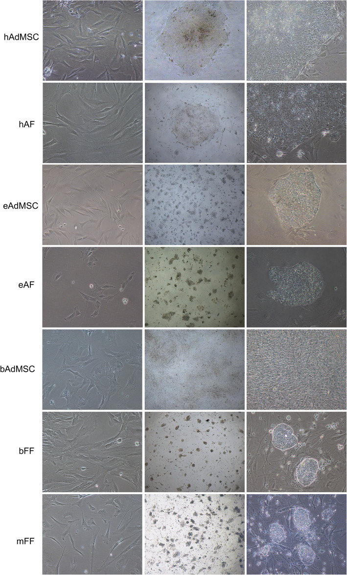
Human cells showed the first signs of morphological differentiation into pluripotent cells approximately 10 days after transduction. Human adult fibroblasts (hAF) and human adipose-derived mesenchymal cells (hAdMSC) displayed initial colony formation approximately 5 days after transduction, and colonies were picked approximately 20 days after transduction (Fig. 1).
Fig. 1.
hAdMSC, hAF, eAdMSC, eAF, bAdMSC, bFF, and mFF throughout in vitro cellular reprogramming: before transduction (× 200) and iPS colonies after replating (× 40 and × 200)
Enzymatic dissociation of hiPSCs did not allow culture viability after incubation with TrypLE Express or Dispase (BD Biosciences); therefore, hiPSCs were manually passaged throughout the experiments.
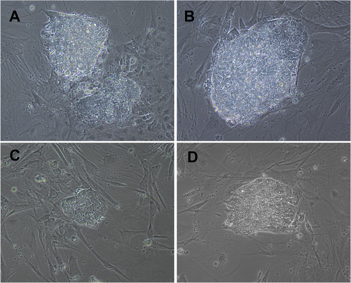
Equine adult fibroblasts (eAF) and adipose tissue mesenchymal cells (eAdMSCs) were transduced with murine or human OSKM, which resulted in induced colonies approximately 3 days after transduction only with human OSKM (Fig. 1) that were independent of hLIF/2i supplementation (Fig. 2). Cells were replated approximately 15 days after transduction.
Fig. 2.
a, b Bovine in vitro reprogrammed cells (p0) with murine OSKM non-supplemented and supplemented with 2i+LIF, respectively. c, d Equine in vitro reprogrammed cells (p0) with human OSKM non-supplemented and supplemented with 2i+LIF, respectively (× 200)
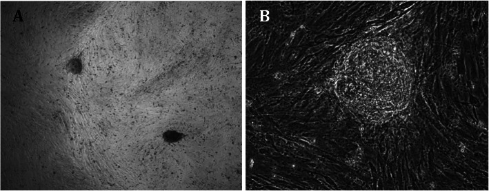
Bovine fetal fibroblasts (bFF) and adipose tissue mesenchymal cells (bAdMSCs) were transduced with murine or human OKSM, and each group was cultured in iPSC medium supplemented with 2i+LIF or not. After approximately 10 days, bFF cells transduced with murine OSKM developed colonies morphologically similar to mouse iPSCs or ES cells. bFF cells transduced with human OSKM developed non-replicative cell colonies approximately 30 days post-transduction (Fig. 3). The bAdMSC lineage used in this study did not develop colonies after transduction with either hOSKM or mOSKM.
Fig. 3.
Non-replicative cell colonies of bFF transduced with hOSKM. a × 40. b × 200
Murine fetal fibroblasts (mFF) were transduced with murine OSKM, and the first colonies developed approximately 3 days after transduction and were clonally replated after 15 days. Only colonies cultured with LIF maintained typical morphology after replating.
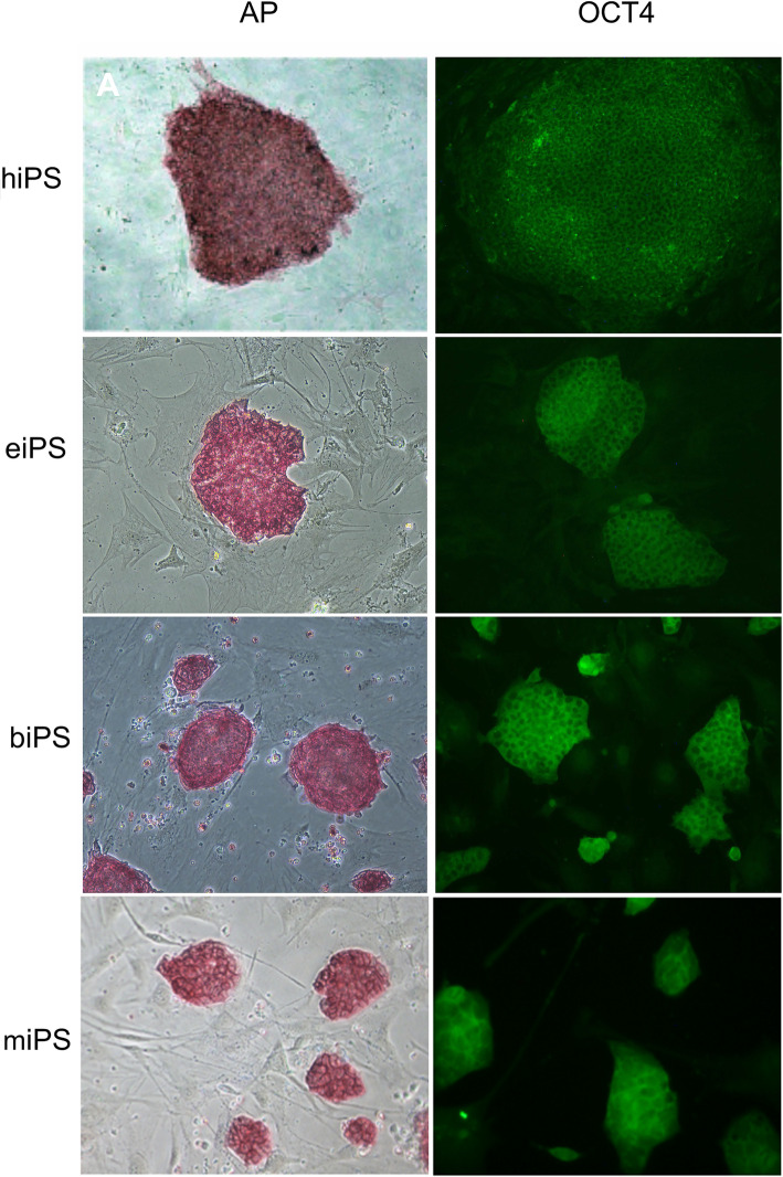
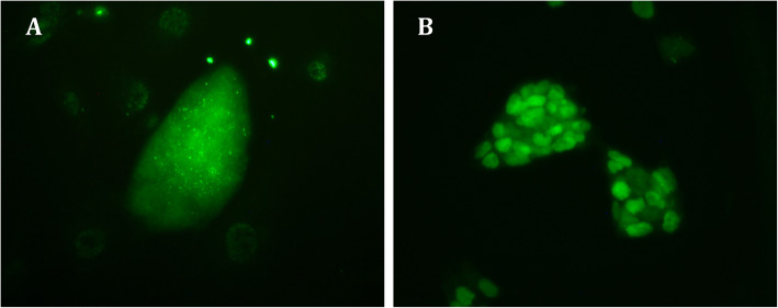
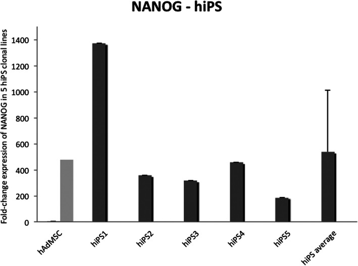
Alkaline phosphatase enzyme (AP) and OCT4 were detected in iPSC colonies from all four species (Fig. 4). NANOG expression was detected in biPS and miPS by immunofluorescence (Fig. 5) and in hiPS clonal lines by RT-qPCR (Fig. 6). eiPSCs were further characterized elsewhere (Pessôa et al. 2019).
Fig. 4.
AP detection and immunofluorescence of OCT4 in iPS colonies derived from human, equine, bovine, and mouse cells. × 200
Fig. 5.
Immunofluorescence of NANOG in iPS colonies derived from bovine (a) and mouse (b) cells. × 200
Fig. 6.
Detection of NANOG expression in hiPS colonies
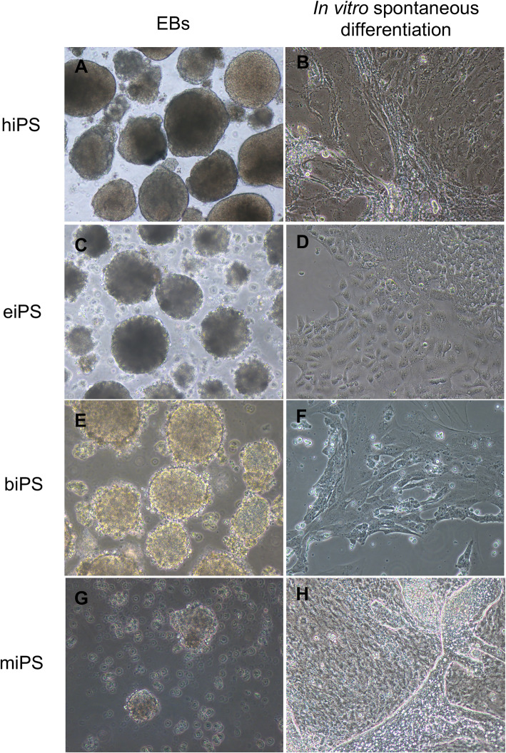
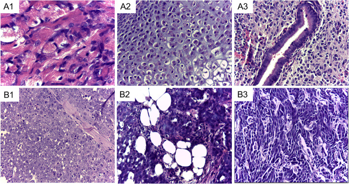
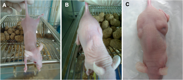
Embryoid bodies are presented in Fig. 7. No teratoma formation was observed when human or equine iPS cells were inoculated in nude mice after 6 months. Tumors were collected from animals injected with biPSCs or miPSCs (Fig. 8) after approximately 2 months. Histopathological evaluation revealed the growth of at least 3 different tissues per lineage, including well- and poorly differentiated specimens. Animals that received mesenchymal cells or fibroblasts did not present teratoma formation. The macroscopic identification of teratomas derived from biPSCs in different periods is presented in Fig. 9.
Fig. 7.
Embryoid bodies (a, c, e, g, × 100) and spontaneous in vitro differentiation (b, d, f, h; × 200) of hiPS, eiPS, biPS, and miPS
Fig. 8.
Photomicrograph of a teratoma derived from miPS showing three different tissues: (A1) striated muscle, (A2) cartilage, and (A3) pseudostratified ciliated epithelium. Photomicrograph of a tumor derived from biPS composed of (B1) undifferentiated tissue with high mitotic activity, (B2) adipose tissue, and (B3) fibroblast-like fusiform cells. Hematoxylin and eosin, objective × 40
Fig. 9.
Balb/c nude mice injected with bFF (a); biPS, 30 days (b); and biPS, 37 days (c). The same animal is presented in b and c
Discussion
Currently, the most common criteria for the characterization of human and murine embryonic stem cells include the presence of a typical phenotype, which is described by an increased nucleus/cytoplasm ratio; the expression of pluripotency markers; high telomerase activity; and in vitro and in vivo pluripotency [36]. In other species, however, these criteria were not yet reproducible until the advent of iPSC technology. In cattle, for example, isolation of embryonic cells results in colonies that may correspond to some of the criteria required for human and murine cells; however, in vitro culture of these stem cells revealed differences from the characteristics outlined above in several reports [37–41].
Possible causes for these inter-species differences are the difficulties of maintaining pluripotency conditions in vitro, as well as the lack of knowledge about factors that regulate stem cells isolated from embryos of domestic species. Therefore, in these species, they are called stem-cell-like [42].
Some studies have already reported bovine and equine potential iPSC cell lines with interspecific vectors (human, swine, or murine cDNAs), but obtained cells with divergent characteristics [8, 11, 43–45]. Huang et al. [46] reported the use of a non-integrative polycistronic vector containing bovine OCT4, SOX2, KLF4, and c-MYC driven by independent promoters. Media were supplemented with LIF, MEK1/2, and GSK3 inhibitors (2i); however, only quiescent non-proliferating cells were generated. Interestingly, this “iPS-like” state described by Huang and collaborators showed similarity to our non-proliferating bovine colonies obtained when human OKSM was used. Zhao et al. [47] reported the maintenance of biPSCs for more than 50 passages when reprogramming was achieved through piggyBac transposon integration of CAG-promoting OSKM, and Han et al. [10] were able to maintain biPSC cell lines for at least 16 passages when embryonic fibroblasts were used for reprogramming through the retroviral mechanism. Nonetheless, bovine reprogramming factors were also used elsewhere [47, 48], and the biPSCs presented showed characteristic pluripotency features, such as teratoma formation and pluripotency markers OCT4, SOX2, NANOG, and others (for extensive review, see Pessôa et al. 2019b).
eiPSCs have already been obtained by transposon-mediated or retroviral expression of human or murine OSKM or OSK (without c-Myc) in fetal or adult fibroblasts [49–51] [45, 52, 53], keratinocytes [54], blood and muscle cells [55], adipose tissue cells [45, 56], and umbilical cord tissue cells [45]. Overall, these cells presented different pluripotency features, such as teratoma and embryoid body formation, alongside the detection of pluripotency markers OCT4 and NANOG, as well as others, as recently reviewed [2]. Regarding medium supplementation, although there are reports of eiPS dependent of the combination of LIF and FGF [49–51, 53], and even only LIF [52, 54, 56], eiPSCs produced here were dependent exclusively of bFGF, as also seen in other reports [45, 55].
Herein, cellular reprogramming was performed in bovine and equine somatic cell cultures through the lentiviral transduction of interspecific OSKM (murine and human). We also generated murine and human iPSC cells, aiming to define a reproducible and controlled pluripotency induction protocol for domestic species. The iPS cells derived herein were positive for NANOG by immunofluorescence, which was used as a pluripotency reporter because no exogenous NANOG was used for reprogramming. The iPSC lineages were characterized after 10 passages and were maintained at least 30 passages in vitro. Exogenous expression was not silenced even after 30 passages, as expected and reported previously in other studies using viral vectors [57, 58]. Similarities and differences between species are summarized in Table 1.
Table 1.
Summary of pluripotency-related characteristics of hiPS, eiPS, and biPS derived from fibroblasts or mesenchymal cells and miPS derived from fibroblasts
| Exogenous transcription factors | hiPS | eiPS | miPS | biPS | ||
|---|---|---|---|---|---|---|
| human OSKM | Murine OSKM | Human OSKM | Murine OSKM | Murine OSKM | Human OSKM | |
| Morphology | Flat | n.a.* | Flat | Dome-shaped | Dome-shaped | Dome-shaped |
| 1st appearance of typical colonies | 5 days | n.a.* | 3 days | 3 days | 7 days | > 30 days |
| Replating | Manual | n.a.* | TrypLE | TrypLE | TrypLE | n.a.** |
| AP | + | n.a.* | + | + | + | – |
| EB | + | n.a.* | + | + | + | n.a.** |
| Tumor formation | – | n.a.* | – | + | + | n.a.** |
| LIF dependence for iPSCs generation | – | n.a.* | – | + | – | n.a.** |
n.a.* not assessed due to no colony formation, n.a.** not assessed due to quiescent-like colonies
In this study, interspecific transcription factors were able to reprogram somatic cells into a pluripotent state. In fact, evidence that regulatory interactions are conserved among organisms has already been reported [59, 60]. When OCT4, SOX2, c-Myc, and KLF4 cDNA and protein are compared between species, both equine and bovine OSKM transcription factors are more similar to human than murine homologs [Table 2, mRNA similarity % (protein similarity %)]*. The reason why bovine fibroblasts were successfully reprogrammed with murine but not human OSKM is still unclear. However, our results stress the need for further research into a possible new mechanism of transcriptional networks.
Table 2.
Homology of mRNA and protein of transcription factors OCT4, SOX2, c-Myc, and KLF4 between species
| Equus caballus | Bos taurus | |||||||
|---|---|---|---|---|---|---|---|---|
| OCT4 | SOX2 | c-MYC | KLF4 | OCT4 | SOX2 | c-MYC | KLF4 | |
| Homo sapiens | 92% (96%) | 92% (91%) | 88% (92%) | 90% (92%) | 90% (94%) | 95% (99%) | 86% (92%) | 91% (94%) |
| Mus musculus | 85% (88%) | 88% (88%) | 83% (90%) | 87% (91%) | 83% (88%) | 88% (98%) | 85% (90%) | 88% (92%) |
mRNA and protein (ncbi.nlm.nih.gov), respectively:
Equus caballus OCT4: XM_001490108.4, XP_001490158.1; SOX2: XM_003363345.2, XP_003363393.1; c-Myc: XM_001497991.3, XP_001498041.2; KLF4: XM_005605684.1, XP_005605741.1
Bos taurus OCT4: NM_174580.2, NP_777005.1, SOX2: NM_001105463.2, NP_001098933.1, c-Myc: NM_001046074.2, NP_001039539.1; KLF4: NM_001105385.1, NP_001098855.1
Homo sapiens OCT4: NM_001173531.2, NP_001167002.1; SOX2: NM_003106.3, NP_003097.1, c-Myc: NM_002467.4, NP_002458.2; KLF4: NM_004235.4, NP_004226.3
Mus musculus OCT4: NM_001252452.1, NP_001239381.1; SOX2: NM_011443.3, NP_035573.3; c-Myc: NM_001177353.1, NP_001170824.1; KLF4: NM_010637.3, NP_034767.2
Conclusions
Herein, we showed the generation of equine and bovine stem cells in vitro through the expression of exogenous and interspecific transcription factors. Although a similar protocol was able to reprogram these cells, it was observed that they differ regarding requirements and characterization. The acquaintance and maintenance of pluripotency in vitro provide a powerful tool to improve the understanding of early development in these species and may also facilitate the production of genetically improved organisms if combined with other reproductive technologies.
Abbreviations
- iPSCs
Induced pluripotent stem cells
- OSKM
Oct4, Sox2, KFL4, and c-Myc
- biPSCs
Bovine induced pluripotent stem cells
- miPSCs
Mouse induced pluripotent stem cells
- eiPSCs
Equine induced pluripotent stem cells
- hiPSCs
Human induced pluripotent stem cells
- ES cells
Embryonic stem cells
- AdMsc
Adipose tissue-derived mesenchymal cells
- bFF
Bovine fetal fibroblasts
- mFF
Murine fetal fibroblasts
- eAF
Equine adult fibroblasts
- hAF
Human adult fibroblasts
- bADMscs
Bovine adipose tissue-derived mesenchymal cells
- hADMSC
Human adipose tissue-derived mesenchymal cells
- eADMSCs
Equine tissue-derived mesenchymal cells
- IMDM
Iscove’s Modified Dulbecco’s Media
- STEMCCA
Stem cell cassette
- MEFs
Mitotically inactivated mouse embryonic fibroblasts
- DMEM/F12 KO
Dulbecco’s modified Eagle’s medium/Ham’s nutrient mixture F12 knockout
- KSR
Knockout serum replacement
- bFGF
Basic fibroblast growth factor
- pen/strep
Penicillin and streptomycin
- LIF
Leukemia inhibitor factor
- hLIF
Human leukemia inhibitor factor
- mLIF
Mouse inhibitor factor
- 2i
iGSK3+iMEK
- iGSK3
Glycogen synthase kinase 3 inhibitor
- iMEK
Mitogen-activated protein kinase inhibitor
- AP
Alkaline phosphatase
- PFA
Paraformaldehyde
- PBS
Phosphate-buffered saline buffer
- BSA
Bovine serum albumin
- RT-qPCR
Reverse transcription quantitative polymerase chain reaction (real-time PCR)
- EBs
Embryoid bodies
- H&E
Hematoxylin and eosin stain
Authors’ contributions
FFB carried out the design of the study, its execution, and writing. VB, CSB, TMM, PFN, RS, and LVFP participated in its execution and helped to draft the manuscript. SKH, JEK, and DTC participated in the design of the study. FVM participated in its design and coordination. The authors read and approved the final manuscript.
Funding
Funding support: FAPESP 2009/11631-6, 2011/08376-4, 2013/13686-8, 2013/08135-2, 2015/26818-5, CNPq 82163/2013-5, and CAPES Financial code 001.
Availability of data and materials
Not applicable.
Ethics approval and consent to participate
All procedures were performed in accordance with the Guide for the Care and Use of Laboratory Animals of the National Institutes of Health and The ARRIVE Guidelines, as well as with the rules issued by the National Council for Control of Animal Experimentation (CONCEA, Ministry of Science, Technology and Innovations and Communications, and in accordance with Law 11.794 of October 8, 2008, Decree 6899 of July 15, 2009). Protocols were then approved by the Ethics Committee on Animal Use of the School of Veterinary Medicine and Animal Science, University of São Paulo, Brazil (protocol number 2913/2013), and by the Ethics Committee on the Use of Animals of the Faculty of Animal Science and Food Engineering, University of São Paulo, Brazil (protocols number 3526250717 and 2192250918).
Consent for publication
Not applicable.
Competing interests
The authors declare that they have no competing interests.
Footnotes
Publisher’s Note
Springer Nature remains neutral with regard to jurisdictional claims in published maps and institutional affiliations.
Contributor Information
Fabiana Fernandes Bressan, Email: fabianabressan@usp.br.
Vinícius Bassanezze, Email: vinicius_bassaneze@harvard.edu.
Laís Vicari de Figueiredo Pessôa, Email: laisvpessoa@usp.br.
Chester Bittencourt Sacramento, Email: bittencourtsacrament@medimmune.com.
Tathiane Maistro Malta, Email: tathimalta@hemocentro.fmrp.com.br.
Simone Kashima, Email: skashima@hemocentro.fmrp.usp.br.
Paulo Fantinato Neto, Email: fantinato@usp.br.
Ricardo De Francisco Strefezzi, Email: rstrefezzi@usp.br.
Naira Caroline Godoy Pieri, Email: nairagodoy@usp.br.
José Eduardo Krieger, Email: krieger@incor.usp.br.
Dimas Tadeu Covas, Email: dimas@fmrp.usp.br.
Flávio Vieira Meirelles, Email: meirellf@usp.br.
References
- 1.Takahashi K, Yamanaka S. Induction of pluripotent stem cells from mouse embryonic and adult fibroblast cultures by defined factors. Cell. 2006;126(4):663–676. [cited 2017 Aug 23] Available from: http://www.ncbi.nlm.nih.gov/pubmed/16904174. [DOI] [PubMed]
- 2.Pessôa LV de F, Bressan FF, Freude KK. Induced pluripotent stem cells throughout the animal kingdom: availability and applications. World J Stem Cells. 2019;11(8):491–505. [cited 2020 Feb 20]Available from: http://www.ncbi.nlm.nih.gov/pubmed/31523369. [DOI] [PMC free article] [PubMed]
- 3.Okita K, Ichisaka T, Yamanaka S. Generation of germline-competent induced pluripotent stem cells. Nature. 2007;448(7151):313–317. doi: 10.1038/nature05934. [DOI] [PubMed] [Google Scholar]
- 4.Takahashi K, Tanabe K, Ohnuki M, Narita M, Ichisaka T, Tomoda K, et al. Induction of pluripotent stem cells from adult human fibroblasts by defined factors. Cell. 2007;131(5):861–872. doi: 10.1016/j.cell.2007.11.019. [DOI] [PubMed] [Google Scholar]
- 5.Honda A, Hirose M, Hatori M, Matoba S, Miyoshi H, Inoue K, et al. Generation of induced pluripotent stem cells in rabbits: potential experimental models for human regenerative medicine. J Biol Chem. 2010;285(41):31362–31369. doi: 10.1074/jbc.M110.150540. [DOI] [PMC free article] [PubMed] [Google Scholar]
- 6.Bao L, He L, Chen J, Wu Z, Liao J, Rao L, et al. Reprogramming of ovine adult fibroblasts to pluripotency via drug-inducible expression of defined factors. Cell Res. 2011;21(4):600–608. doi: 10.1038/cr.2011.6. [DOI] [PMC free article] [PubMed] [Google Scholar]
- 7.Picanço-Castro V, Russo-Carbolante E, Reis LCJ, Fraga AM, de Magalhães DAR, Orellana MD, et al. Pluripotent reprogramming of fibroblasts by lentiviral mediated insertion of SOX2, C-MYC, and TCL-1A. Stem Cells Dev. 2011;20(1):169–180. doi: 10.1089/scd.2009.0424. [DOI] [PubMed] [Google Scholar]
- 8.Sumer H, Liu J, Malaver-Ortega LF, Lim ML, Khodadadi K, Verma PJ. NANOG is a key factor for induction of pluripotency in bovine adult fibroblasts. J Anim Sci. 2011;89(9):2708–2716. doi: 10.2527/jas.2010-3666. [DOI] [PubMed] [Google Scholar]
- 9.Wernig M, Meissner A, Foreman R, Brambrink T, Ku M, Hochedlinger K, et al. In vitro reprogramming of fibroblasts into a pluripotent ES-cell-like state. Nature. 2007;448(7151):318–324. doi: 10.1038/nature05944. [DOI] [PubMed] [Google Scholar]
- 10.Han X, Han J, Ding F, Cao S, Lim SS, Dai Y, et al. Generation of induced pluripotent stem cells from bovine embryonic fibroblast cells. Cell Res. 2011;21(10):1509–1512. doi: 10.1038/cr.2011.125. [DOI] [PMC free article] [PubMed] [Google Scholar]
- 11.Cao H, Yang P, Pu Y, Sun X, Yin H, Zhang Y, et al. Characterization of bovine induced pluripotent stem cells by lentiviral transduction of reprogramming factor fusion proteins. Int J Biol Sci. 2012;8(4):498–511. doi: 10.7150/ijbs.3723. [DOI] [PMC free article] [PubMed] [Google Scholar]
- 12.Huangfu D, Maehr R, Guo W, Eijkelenboom A, Snitow M, Chen AE, et al. Induction of pluripotent stem cells by defined factors is greatly improved by small-molecule compounds. Nat Biotechnol. 2008;26(7):795–797. doi: 10.1038/nbt1418. [DOI] [PMC free article] [PubMed] [Google Scholar]
- 13.Liu J, Balehosur D, Murray B, Kelly JM, Sumer H, Verma PJ. Generation and characterization of reprogrammed sheep induced pluripotent stem cells. Theriogenology. 2012;77(2):338–346. doi: 10.1016/j.theriogenology.2011.08.006. [DOI] [PubMed] [Google Scholar]
- 14.Nakagawa M, Koyanagi M, Tanabe K, Takahashi K, Ichisaka T, Aoi T, et al. Generation of induced pluripotent stem cells without Myc from mouse and human fibroblasts. Nat Biotechnol. 2008;26(1):101–106. doi: 10.1038/nbt1374. [DOI] [PubMed] [Google Scholar]
- 15.Esteban MA, Xu J, Yang J, Peng M, Qin D, Li W, et al. Generation of induced pluripotent stem cell lines from Tibetan miniature pig. J Biol Chem. 2009;(26):284, 17634–17240 [cited 2018 Sep 27] Available from: http://www.jbc.org/lookup/doi/10.1074/jbc.M109.008938. [DOI] [PMC free article] [PubMed]
- 16.Wu Z, Chen J, Ren J, Bao L, Liao J, Cui C, et al. Generation of pig induced pluripotent stem cells with a drug-inducible system. J Mol Cell Biol., Available from. 2009;1(1):46–54 http://www.ncbi.nlm.nih.gov/pubmed/19502222. [DOI] [PubMed]
- 17.Telugu BPVL, Ezashi T, Roberts RM. The promise of stem cell research in pigs and other ungulate species. Stem Cell Rev. 2010;6(1):31–41. doi: 10.1007/s12015-009-9101-1. [DOI] [PubMed] [Google Scholar]
- 18.Gandolfi F, Pennarossa G, Maffei S, Brevini TAL. Why is it so difficult to derive pluripotent stem cells in domestic ungulates? Reprod Domest Anim. 2012;47:11–17. doi: 10.1111/j.1439-0531.2012.02106.x. [DOI] [PubMed] [Google Scholar]
- 19.Bogliotti YS, Wu J, Vilarino M, Okamura D, Soto DA, Zhong C, et al. Efficient derivation of stable primed pluripotent embryonic stem cells from bovine blastocysts. Proc Natl Acad Sci. 2018;115(9):2090–5. [cited 2018 Sep 27] Available from: http://www.ncbi.nlm.nih.gov/pubmed/29440377. [DOI] [PMC free article] [PubMed]
- 20.Liu N, Lu M, Tian X, Han Z. Molecular mechanisms involved in self-renewal and pluripotency of embryonic stem cells. J Cell Physiol. 2007;211(2):279–286. doi: 10.1002/jcp.20978. [DOI] [PubMed] [Google Scholar]
- 21.Simmet K, Zakhartchenko V, Philippou-Massier J, Blum H, Klymiuk N, Wolf E. OCT4/POU5F1 is required for NANOG expression in bovine blastocysts. Proc Natl Acad Sci. 2018;115(11):2770–2775. doi: 10.1073/pnas.1718833115. [DOI] [PMC free article] [PubMed] [Google Scholar]
- 22.Casals JB, Pieri NCG, Feitosa MLT, Ercolin ACM, Roballo KCS, Barreto RSN, et al. The use of animal models for stroke research: a review. Comp Med. 2011; Available from: http://gateway.webofknowledge.com/gateway/Gateway.cgi?GWVersion=2&SrcAuth=ORCID&SrcApp=OrcidOrg&DestLinkType=FullRecord&DestApp=WOS_CPL&KeyUT=WOS:000302043200002&KeyUID=WOS:000302043200002. [PMC free article] [PubMed]
- 23.Mazeto Ercolin AC, Santos Roballo KC, Casals JB, Godoy Pieri NC, Souza AF, Nunes Barreto R da S, et al. Rabbit olfactory stem cells. Isolation protocol and characterization. Acta Cir Bras. 2016; Available from: http://gateway.webofknowledge.com/gateway/Gateway.cgi?GWVersion=2&SrcAuth=ORCID&SrcApp=OrcidOrg&DestLinkType=FullRecord&DestApp=WOS_CPL&KeyUT=WOS:000369657900009&KeyUID=WOS:000369657900009. [DOI] [PubMed]
- 24.Zomer HD, Roballo KCS, Lessa TB, Bressan FF, Gonçalves NN, Meirelles FV, et al. Distinct features of rabbit and human adipose-derived mesenchymal stem cells: implications for biotechnology and translational research. Stem Cells Cloning Adv Appl. 2018;11. [DOI] [PMC free article] [PubMed]
- 25.Gonçalves NJN, Bressan FF, Roballo KCS, Meirelles FV, Xavier PLP, Fukumasu H, et al. Generation of LIF-independent induced pluripotent stem cells from canine fetal fibroblasts. Theriogenology. 2017;92:75–82. [cited 2018 Jan 15] Available from: http://www.ncbi.nlm.nih.gov/pubmed/28237347. [DOI] [PubMed]
- 26.Li Y, Cang M, Lee AS, Zhang K, Liu D. Reprogramming of sheep fibroblasts into pluripotency under a drug-inducible expression of mouse-derived defined factors. Kaufman D, editor. PLoS One. 2011;6(1):e15947. [cited 2018 Jan 15] Available from: http://dx.plos.org/10.1371/journal.pone.0015947. [DOI] [PMC free article] [PubMed]
- 27.Li D, Secher J, Hyttel P, Ivask M, Kolko M, Hall VJ, et al. Generation of transgene-free porcine intermediate type induced pluripotent stem cells. Cell Cycle. 2018;17(23):2547–2563. doi: 10.1080/15384101.2018.1548790. [DOI] [PMC free article] [PubMed] [Google Scholar]
- 28.Kues WA, Niemann H. The contribution of farm animals to human health. Trends Biotechnol. 2004;22(6):286–294. doi: 10.1016/j.tibtech.2004.04.003. [DOI] [PubMed] [Google Scholar]
- 29.Brevini T, Antonini S, Pennarossa G, Gandolfi F. Recent progress in embryonic stem cell research and its application in domestic species. Reprod Domest Anim. 2008;43:193–199. doi: 10.1111/j.1439-0531.2008.01161.x. [DOI] [PubMed] [Google Scholar]
- 30.Pieri NCG, de Souza AF, Botigelli RC, Machado LS, Ambrosio CE, dos Santos MD, et al. Stem cells on regenerative and reproductive science in domestic animals. Vet Res Commun. 2019;43(1). [DOI] [PubMed]
- 31.Sommer CA, Stadtfeld M, Murphy GJ, Hochedlinger K, Kotton DN, Mostoslavsky G. Induced pluripotent stem cell generation using a single lentiviral stem cell cassette. Stem Cells. 2009;27(3):543–549. doi: 10.1634/stemcells.2008-1075. [DOI] [PMC free article] [PubMed] [Google Scholar]
- 32.Bressan FF, dos Santos Miranda M, Perecin F, De Bem TH, Verechia Pereira FT, Russo-Carbolante EM, et al. Improved production of genetically modified fetuses with homogeneous transgene expression after transgene integration site analysis and recloning in cattle. Cell Reprogram. 2011; Available from: http://gateway.webofknowledge.com/gateway/Gateway.cgi?GWVersion=2&SrcAuth=ORCID&SrcApp=OrcidOrg&DestLinkType=FullRecord&DestApp=WOS_CPL&KeyUT=WOS:000287318300004&KeyUID=WOS:000287318300004. [DOI] [PubMed]
- 33.Bressan FF, Miranda MS, Bajgelman MC, Perecin F, Mesquita LG, Fantinato-Neto P, et al. Effects of long-term in vitro culturing of transgenic bovine donor fibroblasts on cell viability and in vitro developmental potential after nuclear transfer. 2013; Available from: http://gateway.webofknowledge.com/gateway/Gateway.cgi?GWVersion=2&SrcAuth=ORCID&SrcApp=OrcidOrg&DestLinkType=FullRecord&DestApp=WOS_CPL&KeyUT=WOS:000318170800002&KeyUID=WOS:000318170800002. [DOI] [PubMed]
- 34.Oliveira CS, de Souza MM, Saraiva NZ, Tetzner TAD, Lima MR, Lopes FL, et al. In vitro culture of bovine embryos in murine ES cell conditioned media negatively affects expression of pluripotency-related markers OCT4, SOX2 and SSEA1. Reprod Domest Anim. 2012;47(3):428–435. doi: 10.1111/j.1439-0531.2011.01896.x. [DOI] [PubMed] [Google Scholar]
- 35.Prophet EB, U.S. AFI of P. Laboratory methods in histotechnology. 1992. Available from: https://books.google.com.br/books/about/Laboratory_methods_in_histotechnology.html?id=R1xrAAAAMAAJ&pgis=1.
- 36.Sidhu KS, Tuch BE. Derivation of three clones from human embryonic stem cell lines by FACS sorting and their characterization. Stem Cells Dev. 2006;15(1):61–9. Available from: http://www.ncbi.nlm.nih.gov/pubmed/16522163. [DOI] [PubMed]
- 37.Gjørret JO, Maddox-Hyttel P. Attempts towards derivation and establishment of bovine embryonic stem cell-like cultures. Reprod Fertil Dev. 2005;17(1–2):113–24. Available from: http://www.ncbi.nlm.nih.gov/pubmed/15745636. [DOI] [PubMed]
- 38.Wang L, Duan E, Sung L, Jeong B-S, Yang X, Tian XC. Generation and characterization of pluripotent stem cells from cloned bovine embryos. Biol Reprod. 2005;73(1):149–155. doi: 10.1095/biolreprod.104.037150. [DOI] [PubMed] [Google Scholar]
- 39.Roach M, Wang L, Yang X, Tian XC. Bovine embryonic stem cells. Methods Enzymol. 2006;418:21–37. doi: 10.1016/S0076-6879(06)18002-7. [DOI] [PubMed] [Google Scholar]
- 40.Muñoz M, Díez C, Caamaño JN, Jouneau A, Hue I, Gómez E. Embryonic stem cells in cattle. Reprod Domest Anim. 2008;43(Suppl 4):32–37. doi: 10.1111/j.1439-0531.2008.01229.x. [DOI] [PubMed] [Google Scholar]
- 41.Pant D, Keefer CL. Expression of pluripotency-related genes during bovine inner cell mass explant culture. Cloning Stem Cells. 2009;11(3):355–365. doi: 10.1089/clo.2008.0078. [DOI] [PubMed] [Google Scholar]
- 42.Koh S, Piedrahita JA. From “ES-like” cells to induced pluripotent stem cells: a historical perspective in domestic animals. Theriogenology. 2014;81(1):103–111. doi: 10.1016/j.theriogenology.2013.09.009. [DOI] [PMC free article] [PubMed] [Google Scholar]
- 43.Talluri TR, Kumar D, Glage S, Garrels W, Ivics Z, Debowski K, et al. Derivation and characterization of bovine induced pluripotent stem cells by transposon-mediated reprogramming. Cell Reprogram. 2015;17(2):131–40. [cited 2020 Feb 20] Available from: http://www.ncbi.nlm.nih.gov/pubmed/25826726. [DOI] [PMC free article] [PubMed]
- 44.Cravero D, Martignani E, Miretti S, Accornero P, Pauciullo A, Sharma R, et al. Generation of induced pluripotent stem cells from bovine epithelial cells and partial redirection toward a mammary phenotype in vitro. Cell Reprogram. 2015 ;17(3):211–20. [cited 2020 Feb 20] Available from: http://www.ncbi.nlm.nih.gov/pubmed/26053520. [DOI] [PubMed]
- 45.Pessôa LV de F, Pires PRL, del Collado M, Pieri NCG, Recchia K, Souza AF, et al. Generation and miRNA characterization of equine induced pluripotent stem cells derived from fetal and adult multipotent tissues. Stem Cells Int. 2019;2019:1–15. [cited 2020 Feb 20] Available from: https://www.hindawi.com/journals/sci/2019/1393791/. [DOI] [PMC free article] [PubMed]
- 46.Huang B, Li T, Alonso-Gonzalez L, Gorre R, Keatley S, Green A, et al. A virus-free poly-promoter vector induces pluripotency in quiescent bovine cells under chemically defined conditions of dual kinase inhibition. PLoS One. 2011;6(9):e24501. Available from: http://www.pubmedcentral.nih.gov/articlerender.fcgi?artid=3166309&tool=pmcentrez&rendertype=abstract. [DOI] [PMC free article] [PubMed]
- 47.Zhao L, Wang Z, Zhang J, Yang J, Gao X, Wu B, et al. Characterization of the single-cell derived bovine induced pluripotent stem cells. Tissue Cell. 2017;49(5):521–7. [cited 2020 Feb 20] Available from: https://linkinghub.elsevier.com/retrieve/pii/S004081661730023X. [DOI] [PubMed]
- 48.Bai C, Li X, Gao Y, Yuan Z, Hu P, Wang H, et al. Melatonin improves reprogramming efficiency and proliferation of bovine-induced pluripotent stem cells. J Pineal Res. 2016;61(2):154–67. [cited 2020 Feb 20] Available from: http://doi.wiley.com/10.1111/jpi.12334. [DOI] [PubMed]
- 49.Nagy K, Sung H-K, Zhang P, Laflamme S, Vincent P, Agha-Mohammadi S, et al. Induced pluripotent stem cell lines derived from equine fibroblasts. Stem Cell Rev Reports. 2011;7(3):693–702. [cited 2018 Jan 15] Available from: http://www.ncbi.nlm.nih.gov/pubmed/21347602. [DOI] [PMC free article] [PubMed]
- 50.Breton A, Sharma R, Diaz AC, Parham AG, Graham A, Neil C, et al. Derivation and characterization of induced pluripotent stem cells from equine fibroblasts. Stem Cells Dev. 2013;22(4):611–621. doi: 10.1089/scd.2012.0052. [DOI] [PMC free article] [PubMed] [Google Scholar]
- 51.Khodadadi K, Sumer H, Pashaiasl M, Lim S, Williamson M, Verma PJ. Induction of pluripotency in adult equine fibroblasts without c-MYC. Stem Cells Int. 2012;2012:429160. Available from: http://www.pubmedcentral.nih.gov/articlerender.fcgi?artid=3328202&tool=pmcentrez&rendertype=abstract. [DOI] [PMC free article] [PubMed]
- 52.Whitworth DJ, Ovchinnikov DA, Sun J, Fortuna PRJ, Wolvetang EJ. Generation and characterization of leukemia inhibitory factor-dependent equine induced pluripotent stem cells from adult dermal fibroblasts. Stem Cells Dev. 2014;23(13):1515–23. [cited 2018 Jan 15] Available from: https://www.liebertpub.com/doi/10.1089/scd.2013.0461. [DOI] [PMC free article] [PubMed]
- 53.Moro LN, Amin G, Furmento V, Waisman A, Garate X, Neiman G, et al. MicroRNA characterization in equine induced pluripotent stem cells. Pereira L V., editor. PLoS One. 2018;13(12):e0207074. [cited 2020 Feb 20] Available from: http://dx.plos.org/10.1371/journal.pone.0207074. [DOI] [PMC free article] [PubMed]
- 54.Sharma R, Livesey MR, Wyllie DJA, Proudfoot C, Whitelaw CBA, Hay DC, et al. Generation of functional neurons from feeder-free, keratinocyte-derived equine induced pluripotent stem cells. Stem Cells Dev. 2014;23(13):1524–1534. doi: 10.1089/scd.2013.0565. [DOI] [PubMed] [Google Scholar]
- 55.Quattrocelli M, Giacomazzi G, Broeckx SY, Ceelen L, Bolca S, Spaas JH, et al. Equine-induced pluripotent stem cells retain lineage commitment toward myogenic and chondrogenic fates. Stem Cell Reports. 2016;6(1):55–63. doi: 10.1016/j.stemcr.2015.12.005. [DOI] [PMC free article] [PubMed] [Google Scholar]
- 56.Lee E-M, Kim A-Y, Lee E-J, Park J-K, Park S-I, Cho S-G, et al. Generation of equine-induced pluripotent stem cells and analysis of their therapeutic potential for muscle injuries. Cell Transplant. 2016;25(11):2003–2016. doi: 10.3727/096368916X691691. [DOI] [PubMed] [Google Scholar]
- 57.Pessôa LVF, Bressan FF, Freude KK. Induced pluripotent stem cells throughout the animal kingdom: availability and applications. World J Stem Cells. 2019;11(8):491–505. https://www.wjgnet.com/1948-0210/full/v11/i8/491.htm. [DOI] [PMC free article] [PubMed]
- 58.Ezashi T, Yuan Y, Roberts RM. Pluripotent stem cells from domesticated mammals. Annu Rev Anim Biosci. 2016;4(1):223–253. doi: 10.1146/annurev-animal-021815-111202. [DOI] [PubMed] [Google Scholar]
- 59.Babu MM, Luscombe NM, Aravind L, Gerstein M, Teichmann SA. Structure and evolution of transcriptional regulatory networks. Curr Opin Struct Biol. 2004;14(3):283–291. doi: 10.1016/j.sbi.2004.05.004. [DOI] [PubMed] [Google Scholar]
- 60.Yu H, Luscombe NM, Lu H, Zhu X, Xia Y. Annotation transfer for genomics: assessing the transferability of protein-protein and protein-DNA interactions between organisms. Genome …. 2004; Available from: http://archive.gersteinlab.org/papers/e-print/interolog/preprint.pdf.
Associated Data
This section collects any data citations, data availability statements, or supplementary materials included in this article.
Data Availability Statement
Not applicable.


