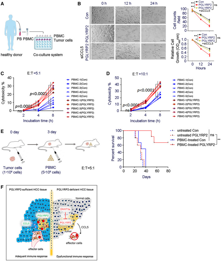
Figure 7.

PGLYRP2 elevates antitumor activity of PBMC. (A) Model of PBMC–tumor cell coculture system. (B) PBMCs were incubated with stable Huh7/Con, Huh7/PGLYRP2, or Huh7/PGLYRP2/siCCL5 cells at an E:T ratio of 5:1 for the indicated incubation times. Scale bar, 300 μm. After 12 or 24 hours of incubation, PBMCs were removed from the cell culture plates, and the antitumor activity of PBMCs on tumor cells was determined using cell counts and MTT assay. **P < 0.001. (C,D) The effect of PGLYRP2 expression on the antitumor activity of PBMCs was analyzed by incubating target Huh7/Con or Huh7/PGLYRP2 cells with five independent PBMC donors for 2, 4, and 8 hours at an E:T ratio of 5:1 (C) or 10:1 (D). (E) Left, Evaluation model of antitumor activity of PBMCs in Huh7 xenografted BALB/c‐nu/nu mice. Right, Survival of mice in untreated or PBMC‐treated Huh7/Con or Huh7/PGLYRP2 group (n = 5) is shown. (F) Working model of PGLYRP2‐mediated antitumor immune response against HCC. All results are expressed as mean ± SEM. *P < 0.05, **P < 0.001. Abbreviations: ns, not significant; OD, optical density; PB, peripheral blood.
