Abstract
Amebiasis caused by Entamoeba histolytica is nowadays a serious public health problem worldwide, especially in developing countries. Annually, up to 100,000 deaths occur across the world. Due to the resistance that pathogenic protozoa exhibit against commercial antiprotozoal drugs, a growing emphasis has been placed on plants used in traditional medicine to discover new antiparasitics. Previously, we reported the in vitro antiamoebic activity of a methanolic extract of Lippia graveolens Kunth (Mexican oregano). In this study, we outline the isolation and structure elucidation of antiamoebic compounds occurring in this plant. The subsequent work-up of this methanol extract by bioguided isolation using several chromatographic techniques yielded the flavonoids pinocembrin (1), sakuranetin (2), cirsimaritin (3), and naringenin (4). Structural elucidation of the isolated compounds was achieved by spectroscopic/spectrometric analyses and comparing literature data. These compounds revealed significant antiprotozoal activity against E. histolytica trophozoites using in vitro tests, showing a 50% inhibitory concentration (IC50) ranging from 28 to 154 µg/mL. Amebicide activity of sakuranetin and cirsimaritin is reported for the first time in this study. These research data may help to corroborate the use of this plant in traditional Mexican medicine for the treatment of dyspepsia.
Keywords: infectious diseases, amoebiasis, Mexican oregano, bioguided isolation, flavonoids, antiprotozoal agents
1. Introduction
Amoebiasis is caused by Entamoeba histolytica, which is a protozoan of the family Endomoebidae [1]. It is related to elevated morbidity and mortality worldwide, and has become a serious public health problem in developing countries [2]. Traveling to endemic countries is a risk factor for acquiring an E. histolytica infection [3]. After malaria, amoebiasis is the second cause of death due to parasitic diseases [4,5]. The symptoms vary from mild diarrhea to dysentery, but, occasionally, E. histolytica can invade the intestinal mucosal barrier and trigger liver abscesses [6]. Asymptomatic infections occur in 90% of individuals, whereas the remaining 10% contract symptomatic infections [7]. Around 50 million people suffer from severe amoebiasis, and 40,000–100,000 deaths occur annually due to this parasitosis [8,9].
Currently, metronidazole is the most used commercial drug for the treatment of amoebiasis, however, since drug resistance by E. histolytica is increasing, the use of higher doses to overcome the infection is needed, thus causing unpleasant side effects [10,11]. Considering these undesired side effects as well as the development of resistant strains of E. histolytica against metronidazole, more efficient and safer antiamoebic agents are required [12,13,14].
Natural products occurring in medicinal plants have proved to be an important source of leading compounds for the design of new drugs [15]. Mexican oregano (Lippia graveolens Kunth) has been used in traditional Mexican medicine for curing inflammation-related diseases, such as respiratory and digestive disorders, headaches, and rheumatism, among others [16,17]. Oregano’s essential oil, regardless of the species, shows a broad range of effects on bacteria, with some of them being resistant to antibiotics of clinical use, as well as on fungi and parasites [18,19,20,21,22].
Recently, we reported the in vitro antiamoebic activity of a methanolic extract of Lippia graveolens Kunth and the bioguided isolation of carvacrol, as one of the bioactive compounds with antiprotozoal activity [23]. This work aims to isolate and achieve the structure elucidation of additional antiamoebic compounds present in this plant.
2. Results
2.1. Bioguided Isolation of Flavonoids from Lippia graveolens Kunth
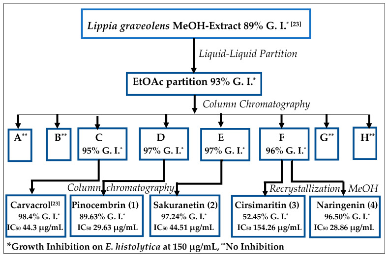
As previously reported, the partition of a methanolic extract of Lippia graveolens by extraction with n-hexane and fractionation of the hexane phase led to the isolation of carvacrol with excellent antiamoebic activity [23]. The subsequent handling of the remaining methanol (MeOH) by partition between methanol-water and ethyl acetate (EtOAc), carried out in this research, yielded an EtOAc residue with 93.3% growth inhibition of E. histolytica. After column chromatography (silica gel, Sephadex), this residue afforded the known flavonoids pinocembrin (1), sakuranetin (2), cirsimaritin (3), and naringenin (4), with significant antiamoebic activity (Figure 1).
Figure 1.
General scheme for the bioguided isolation of compounds with antiamoebic activity from Lippia graveolens Kunth (Mexican oregano).
The isolated flavonoids were identified by comparing their physical and spectral data with those reported in the literature.
The electron ionization (EI) mass spectrum showed a molecular ion with m/z = 256 for pinocembrin 1 (calcd. for C15H12O4, 256.253), m/z = 286 for sakuranetin 2 (calcd. for C16H14O5, 286.283), m/z = 314 for cirsimaritin 3 (calcd. for C17H14O6, 314.28), and m/z = 272 for naringenin 4 (calcd. for C15H12O5, 272.252).
The infrared (IR) spectrum of all isolated flavonoids (see Supplementary material) contained absorption bands at 1600–1650 cm−1 (medium) and 1100–1250 cm−1 (strong), consistent with a C=O bond in the molecules [24,25,26,27].
One- and two-dimensional nuclear magnetic resonance (NMR) spectra were recorded for the isolated compounds using deuterated dimethyl sulfoxide (DMSO-d6). 1H- and 13C-NMR chemical shifts (see Supplementary Material) were in accordance with those reported for pinocembrin 1 [28,29,30], sakuranetin 2 [31,32,33,34], cirsimaritin 3 [26,35,36,37], and naringenin 4 [27,28,38,39,40,41]. An unambiguous assignment of the 13C-NMR spectrum of these compounds was deduced from 1H-1H COSY, NOESY, HSQC, and HMBC spectra (see Supplementary Material).
The four isolated flavonoids have the common characteristic that the rotameric hydroxy group at C-5 forms an intramolecular H-bond with the carbonyl group [42]. That explains the shift of this proton absorption to the range of about δ 13.0–12.0 as a sharp singlet in DMSO-d6 [43].
2.2. Entamoeba Histolytica Growth Parameters
After collecting data from E. histolytica growth kinetics experiments, it was estimated that the generation time is 14.76 h and the duplication time is 21.30 h.
The ideal growth time of E. histolytica for evaluating the amebicide activity of the compounds was set at 72 h because, at this point, protozoa are still in the exponential and sustained growth stage, thus decreasing the number of false positives.
2.3. In Vitro Assay for Entamoeba histolytica
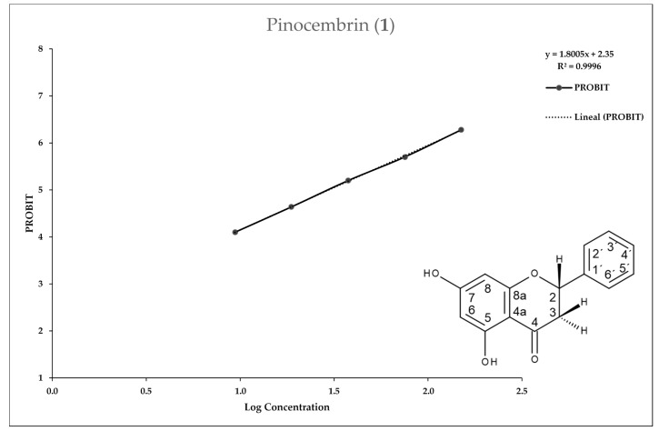
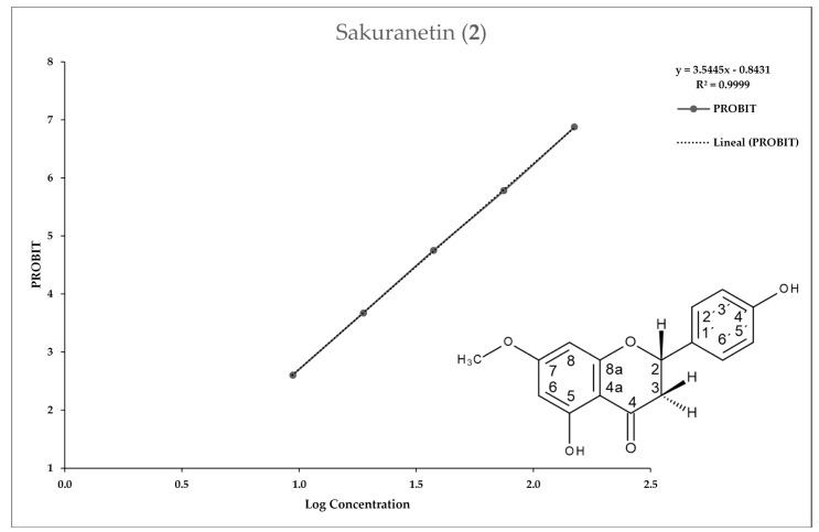
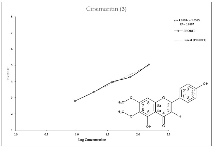
The pure compounds were dissolved in DMSO to a concentration of 150 µg/mL in a suspension of E. histolytica trophozoites in a logarithmic phase in peptone, pancreas, and liver extract plus 10% bovine serum (PEHPS medium). They showed significant growth inhibition of E. histolytica at this concentration. The 50% inhibitory concentration (IC50) values of these compounds ranged from 28.86 to 154.26 µg/mL (metronidazole IC50 0.205 µg/mL).
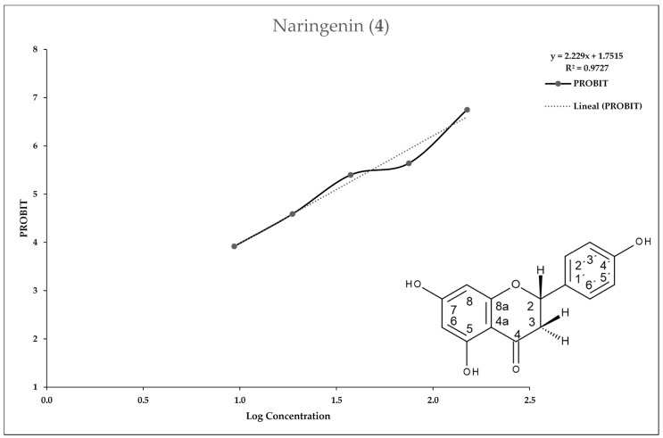
Figure 2, Figure 3, Figure 4 and Figure 5 show the 50% inhibitory concentration of each compound calculated by using a Probit analysis, considering a 95% confidence level.
Figure 2.
Antiprotozoal activity of Pinocembrin 1 against Entamoeba histolytica. Growth inhibition of 89.63% at 150 µg/mL, 50% inhibitory concentration value (IC50) = 29.63 µg/mL.
Figure 3.
Antiprotozoal activity of Sakuranetin 2 against Entamoeba histolytica. Growth inhibition of 97.24% at 150 µg/mL, IC50 = 44.51 µg/mL.
Figure 4.
Antiprotozoal activity of Cirsimaritin 3 against Entamoeba histolytica. Growth inhibition of 52.45% at 150 µg/mL, IC50 = 154.26 µg/mL.
Figure 5.
Antiprotozoal activity of Naringenin 4 against Entamoeba histolytica. Growth inhibition of 96.50% at 150 µg/mL, IC50 = 28.86 µg/mL.
3. Discussion
It is estimated that about 6000 flavonoids are present in different plants worldwide [44,45], and many of them are common ingredients of our daily food. Flavonoids exhibit a variety of biological properties, such as antioxidant, anticancer, antibacterial, antifungal, antiparasitic [46,47,48,49,50,51,52,53], as well as those for treating other kinds of illness [54,55,56,57].
More than 20 flavonoids have been identified in the leaves of Lippia graveolens by high pressure liquid chromatography (HPLC), according to previous reports [58]. Pinocembrin, sakuranetin, naringenin, and cirsimaritin have already been isolated from this plant [58,59,60,61,62], but, in the respective studies, no antiamoebic activity is reported. Nevertheless, some research groups have reported antiprotozoal activity of these flavonoids isolated from sources other than Lippia graveolens.
A weak antiparasitic activity of pinocembrin has been shown against trypomastigotes of Trypanosoma cruzi, with inhibition values in the range of 40.13% (at 250 µg/mL) to 43.68% (at 500 µg/mL), according to Grael et al. [63]. Weak inhibition was also observed against Giardia lamblia trophozoites, with an IC50 of 174.4 µg/mL reported by Alday-Provencio et al. [64] and an IC50 of 57.39 µg/mL reported by Calzada et al. [65], respectively.
Sakuranetin presented activity against Leishmania amazonensis, Leishmania brazilians, Leishmania major, and Leishmania chagasi, with a range of 43–52 µg/mL, as well as against T. cruzi trypomastigotes, with an IC50 of 20.17 µg/mL, according to Grecco et al. [39].
Regarding parasitic diseases, cirsimaritin showed potent inhibition against Plasmodium falciparum resistant to chloroquine, with an IC50 of 16.9 µM [66], and similar activity against Leishmania donovani (IC50 = 3.9 µg/mL), Trypanosoma brucei rhodesiense (IC50 = 3.3 µg/mL), and T. cruzi (IC50 = 19.7 µg/mL), according to Tasdemir et al. [49].
Antiparasitic research has found giardicidal activity in naringenin (4), with an IC50 of 125.7 µg/mL [64] and 47.84 µg/mL [65], according to Alday-Provencio et al. and Calzada et al., respectively, but no damage against Trypanosoma cruzi and Leishmania spp. was observed, according to Grecco et al. [39].
Some results of pinocembrin and naringenin tested against the strain E. histolytica HM1:IMSS have been published; pinocembrin isolated from Teloxys graveolens and naringenin from a commercial source exhibited an IC50 of 80.76 and 98.24 µg/mL, respectively [65,67]. This indicates a lower effectiveness compared with our results (IC50 of 29.51 µg/mL for pinocembrin and 28.85 µg/mL for naringenin, respectively), which could be explained by the difference between the methodology reported by Calzada [65] and that used by our group. The principal difference was the time that the trophozoites were exposed to the chemical compound. In our methodology, we incubated the trophozoites with the flavonoids for 72 h, while Calzada [65] incubated them for 48 h, which could represent a factor in the certainty of the biological activity.
In our view, this is the first report on the amebicide activity of sakuranetin and cirsimaritin.
The antiprotozoal activity of pinocembrin and naringenin (IC50 of 29.63 and 28.86 µg/mL, respectively) was higher compared with sakuranetin (44.51 µg/mL), and the most remarkable comparison was with cirsimaritin (154.26 µg/mL), revealing that a 5,7-dihydroxylated A ring is essential for antiprotozoal activity, as remarked by Calzada [65]. The structure–effect correlations also showed that a 2,3-double bond in ring B (as in cirsimaritin) reduces the antiprotozoal activity.
Currently, there are no available studies on the mechanism of action against E. histolytica of the flavonoids isolated from L. graveolens, although there are reports on some structurally related flavonoids [68,69]. The ultrastructural changes in the morphology of Entamoeba histolytica when it was assayed with (−)-epicatechin, a flavan-3-ol flavonoid, have previously been demonstrated, showing an IC50 of 1.9 µg/mL. The results indicated programmed cell death activation with nuclear alterations (small clumps around the nuclear membrane), in addition to cytoplasmatic modifications, such as an increase of glycogen deposits and a reduction of the size and number of vacuoles [70]. Recently, Bolaños et al. [68] have also demonstrated that the flavonoids (−)-epicatechin and kaempferol affect cytoskeleton proteins and functions in E. histolytica [68,71], leading to changes in essential cellular mechanisms, such as adhesion, migration, phagocytosis, and cytolysis. These findings lead us to have an idea of the possible targets and mechanisms of action of the flavonoids isolated from L. graveolens. None of the flavonoids isolated from L. graveolens present a hydroxyl group at position 3, as in the case of epicatechin and kaempferol, so there must be subtle differences in the mechanism of action of pinocembrin, sakuranetin, naringenin, and cirsimaritin, and part of the future work of our group will be devoted to this issue.
The significant inhibitory effect against E. histolytica observed for the methanolic extract of Lippia graveolens (IC50 = 59.15 µg/mL) can be attributed to the presence of the flavonoids isolated from the ethyl acetate partition as well as the carvacrol mainly obtained from the hexane partition [23]. Compounds with a higher polarity occurring in L. graveolens, soluble in methanol or water, are not involved in the antiprotozoal activity of this plant against E. histolytica.
4. Materials and Methods
4.1. General
An Electrothermal 9100 apparatus (Electrothermal Engineering Ltd., Southend-on-Sea, UK) was used for melting point acquisition. IR spectra were measured on a Frontier Fourier transform infrared (FT-IR) spectrometer (PerkinElmer, Waltham, MA, USA) with an ATR accessory. NMR spectra were recorded on an Avance DPX 400 spectrometer (Bruker, Billerica, MA, USA) running at 400.13 MHz for 1H and 100.61 MHz for 13C. EI-MS were obtained on a MAT 95 spectrometer (70 eV, Finnigan, San Jose, CA, USA). Thin layer chromatography (TLC) was realized on precoated silica gel glass plates (5 × 10 cm, Merck silica gel 60 F254, Darmstadt, Germany). Column chromatography was carried out on silica gel (60–200 mesh) purchased from J. T. Baker (Phillipsburg, NJ, USA). Size-exclusion chromatography was performed on Sephadex LH-20 (Lipophilic Sephadex, Amersham Biosciences Ltd., purchased from Sigma-Aldrich Chemie, Steinheim, Germany).
4.2. Plant Material
Aerial parts of Lippia graveolens were collected near the town General Cepeda (Mexican State Coahuila) in March 2011 and identified by Maria del Consuelo González. A voucher specimen (No. 025554) was deposited at the Herbario de la Facultad de Ciencias Biológicas (UANL), Nuevo León, México. The plant name has been checked with http://www.theplantlist.org. The vegetal material was dried and ground to powder.
4.3. Plant Extraction and Bioguided Isolation of Antiamoebic Compounds from Lippia graveolens Kunth
In total, 600 g of dried and ground Lippia graveolens leaves were extracted in a Soxhlet apparatus for 40 h with MeOH. After filtration, the solvent was removed in a rotatory evaporator to yield 260 g of crude extract. This extract was analyzed for its amebicide activity on trophozoites of E. histolytica (HM1:IMSS strain), showing a significant inhibition percentage (89%; IC50 59.15 µg/mL) in terms of the standard concentration of 150 µg/mL. Afterward, the extract was redissolved in 2 L methanol and divided into four portions of 500 mL each for conducting liquid-liquid partition with n-hexane. After solvent evaporation, 19.9 g of a residue with high amebicidal activity (90.9% growth inhibition) was obtained. Bioguided fractionation of this hexane partition, using column chromatography on silica gel, provided 2.2 g of carvacrol (98.4% growth inhibition; IC50 44.30 µg/mL), as previously reported [23].
The methanolic phase was concentrated under reduced pressure up to a volume of ca. 500 mL, and 1.5 L distilled water was then added, gradually and under constant stirring. Afterward, the methanol/water mixture was divided into four portions of 500 mL each and submitted to liquid–liquid partition with ethyl acetate to yield, after solvent evaporation, 67.3 g of a combined residue with 93.3% growth inhibition against E. histolytica.
The EtOAc partition was suspended in 400 mL of chloroform (CHCl3), and, after stirring, filtration, and solvent evaporation, 25.8 g of CHCl3-soluble residue was obtained. The material recovered from the filter was then suspended in 400 mL of EtOAc, and, after stirring, filtration, and solvent evaporation, 8.8 g of EtOAc-soluble residue was obtained. This second material recovered from the filter was then suspended in 100 mL MeOH, and the same procedure was applied to yield 8.8 g of MeOH-soluble residue and ca. 24 g of an insoluble powder from the final filtration. Each residue was analyzed for its anti-Entamoeba histolytica activity, and remarkable results were obtained, mainly for the CHCl3-residue, with 90.9% growth inhibition, followed by the EtOAc residue, with 61.6% growth inhibition. The MeOH residue and insoluble powder did not show any activity.
The CHCl3 fraction was divided into five portions of ca. 5 g, and each of them was chromatographed on a silica gel (100 g) column (60 × 2.6 cm) and eluted with stepwise gradients of chloroform-ethyl acetate, and finally with methanol. For each column, a total of 110 subfractions (50 mL) were obtained and collected, and considering their TLC (CHCl3–EtOAc, 9:1) profiles, split into eight main fractions (A-H). These main fractions, containing the nonpolar to the more polar compounds, were used for amebicide assays. Out of the eight fractions, only fractions C, D, E, and F showed amebicide activity, with 95.34%, 96.89%, 97.24%, and 95.85% growth inhibition, respectively.
Fraction C (showing the main compound, according to TLC, with Rf = 0.77; CHCl3-Ethyl acetate, 9:1) was divided into five portions of approx. 1 g, and each of them was chromatographed on a silica gel (20 g) column (39 × 2 cm) and eluted with a stepwise gradient solvent system of chloroform and ethyl acetate, and finally with methanol. Sixty subfractions (10 mL) were collected for each column and combined, based on their TLC (CHCl3-Ethyl acetate, 9:1) profiles, into five main fractions (CA-CE). Fraction CB presented the main compound, with Rf = 0.77, so it was subjected to subsequent purification with three columns packed with silica gel as stationary phase (data not shown). Afterward, 315 mg of a viscous liquid with a high amebicide activity (96.76% growth inhibition; IC50 44.1 µg/mL) was recovered. The spectroscopic results indicated that this compound is carvacrol, previously isolated from hexane partition [23].
Fraction D (showing the main compound, according to TLC, with Rf = 0.51; CHCl3-Ethyl acetate, 9:1) was divided into four portions of ca. 1 g and each of them was submitted to chromatography on a silica gel (20 g) column (39 × 2 cm) and eluted with a stepwise gradient solvent system of chloroform and ethyl acetate, and finally with methanol. Sixty subfractions (10 mL) were collected for each column and combined, based on their TLC (CHCl3-Ethyl acetate, 9:1) profiles, into five main fractions (DA-DE). Fraction DB presented the main compound, with Rf = 0.51, so it was subjected to subsequent purification with several columns packed with Sephadex as stationary phase (data not shown). Afterward, 32 mg of a solid with a high amebicide activity (89.63% growth inhibition; IC50 29.63 µg/mL) was recovered. The spectroscopic results indicated that this compound is pinocembrin 1 (C15H12O4; M.p. 210 °C: Lit. 223–236 °C [72], 191–193 °C [24]).
Fraction E (showing the main compound, according to TLC, with Rf = 0.33; CHCl3-Ethyl acetate, 9:1) was divided into three portions of approx. 1 g and each of them was chromatographed on a silica gel (20 g) column (39 × 2 cm) and eluted with a stepwise gradient solvent system of chloroform and ethyl acetate, and finally with methanol. Sixty subfractions (10 mL) were collected for each column and combined, based on their TLC (CHCl3-Ethyl acetate, 9:1) profiles, into five main fractions (EA-EE). Fractions EB and EC presented the main compound, with Rf = 0.33, so they were subjected to subsequent purification with several columns packed with Sephadex as stationary phase (data not shown). Afterward, 102 mg of a solid with a high amebicide activity (97.24% growth inhibition; IC50 44.51 µg/mL) was recovered. The spectroscopic results indicated that this compound is sakuranetin 2 (C16H14O5; M.p. 160 °C: Lit. 151–153 °C [73], 143–144 °C [74]).
Fraction F showed only a main compound, according to TLC, with Rf = 0.10 (CHCl3-Ethyl acetate, 9:1). Using an eluent of a higher polarity (CHCl3-Ethyl acetate, 1:1), this compound was revealed to be a mixture of two compounds, with an Rf of 0.70 and 0.53, respectively. This mixture was no longer soluble in chloroform and it was therefore not possible to use column chromatography with silica gel of a normal phase to try to separate the two compounds. Then, this fraction was suspended in 30 mL of chloroform and heated to reflux for 15 min, noting that a large quantity of the material remained insoluble. After cooling and filtering, 645 mg of an insoluble solid consisting of the two compounds of the mixture (TLC) was recovered, while in the chloroform solution, a mixture of the same compounds was also observed, but with additional impurities, mainly of a lower polarity. The insoluble solid (645 mg) recovered from the filter was suspended in 30 mL methanol and heated to reflux until complete solubility was observed. After cooling, the MeOH solution was kept refrigerated for 72 h with hermetic closure, and during that time, 50.6 mg of a greenish powder containing only the compound with Rf = 0.53 (CHCl3-Ethyl acetate, 1:1) was separated. This solid presented no significant amebicide activity (52.45% growth inhibition; IC50 154.26 µg/mL). The spectroscopic results indicated that this compound is cirsimaritin 3 (C17H14O6; M.p. 268 °C: Lit. 256–258 °C [26], 267–268 °C [75]).
The filtered MeOH solution was concentrated and chromatographed on a Sephadex (50 g) column (160 × 1.5 cm) and eluted with methanol (300 mL). A total of 60 subfractions (5 mL) were collected and combined, based on their TLC (CHCl3-Ethyl acetate, 1:1) profiles, into seven main fractions (FA-FG). From fraction FC, additional cirsimaritin with Rf = 0.53 was recovered. Fraction FF showed only the pure compound, with Rf = 0.70. This solid also presented high amebicide activity (96.50% growth inhibition; IC50 28.86 µg/mL). The spectroscopic results indicated that this compound is naringenin 4 (C15H12O5; M.p. 254 °C: Lit. 250–252 °C [27], 248–250 °C [73]).
4.4. Antiprotozoal Assay
4.4.1. Test Microorganisms
Strain HM-1:IMSS of Entamoeba histolytica was obtained from the microorganism culture collection of the Centro de Investigación Biomédica del Noreste (CIBIN) in Monterrey, Mexico. The trophozoites were grown axenically and maintained in peptone, pancreas, and liver extract plus bovine serum and employed at the log phase of growth (2 × 10⁴ cells/mL) by all of the bioassays performed [76,77].
The procedure for determining the growth curve for E. histolytica was performed in 13 × 100 mm screw cap test tubes, by inoculating 20,000 trophozoites of E. histolytica in 5 mL of PEHPS medium, to which 10% bovine serum was added. Subsequently, they were incubated at 36.5 °C for 120 h, and every 24 h, the number of trophozoites was determined and the growth parameters in the medium were evaluated. The process was conducted in three separate experiments per triplicate.
4.4.2. In Vitro Assay for Entamoeba histolytica
Each compound was dissolved in DMSO and adjusted to a concentration of 150 µg/mL in a suspension of E. histolytica trophozoites at a logarithmic phase in PEHPS medium with 10% bovine serum. Vials were incubated for 72 h, and then chilled in iced water for 20 min, and, by using a hemocytometer, the number of dead trophozoites per milliliter was calculated. Each extract assay was carried out in triplicate. Metronidazole was used as a positive control, and as a negative control, an E. histolytica suspension in PEHPS medium with no extract added was used. The percentage of inhibition was estimated as the number of dead trophozoites compared to the negative controls.
4.4.3. In Vitro IC50 Determination
Each compound was dissolved in dimethyl sulfoxide and adjusted to 150, 75, 37.5, 18.75, and 9.375 µg/mL by adding a suspension of E. histolytica trophozoites at a logarithmic phase in PEHPS medium with 10% bovine serum. Vials were incubated for 72 h, and then chilled in cold water for 20 min, and the number of dead trophozoites per milliliter was evaluated using a hemocytometer. All assays were performed in triplicate. Metronidazole was used as a positive control, and as a negative control, an E. histolytica suspension in PEHPS medium with no extract added was used. The percentage of inhibition was estimated as the number of dead trophozoites compared to the negative controls. The 50% inhibitory concentration of each compound was calculated by using a Probit analysis, considering a 95% confidence level.
5. Conclusions
The isolated and pure flavonoids from L. graveolens showed significant growth inhibition against E. histolytica (52% to 97% at a concentration of 150 µg/mL). The IC50 values of these compounds ranged from 28.86 to 154.26 µg/mL, so they were not as effective as metronidazole (IC50 0.205 µg/mL), but these IC50 values can be used as a guideline for further research on this plant as a source of potential antiamoebic agents.
The main contribution of this research work lies in the fact that it has shown that the presence of the flavonoids described herein in Lippia graveolens has a direct relationship with the antiprotozoal activity of extracts of this plant against Entamoeba histolytica. These flavonoids could be used as biomarkers [78] for the quality control of phytotherapeutics developed based on this work.
The results of our research may also form the basis for directly incorporating the use of Lippia graveolens extracts into conventional and complementary medicine for the treatment of amebiasis, as well as other infectious diseases.
Acknowledgments
For the spectroscopic measurements, we thank Noemi Waksman-Minsky of the College of Medicine from Universidad Autónoma de Nuevo León.
Supplementary Materials
The following are available online, Figures S1–S30: supplementary spectroscopic data.
Author Contributions
Conceptualization, R.Q.-L.; investigation, R.Q.-L., J.V.-V., M.J.V.-S., and V.M.R.-G.; writing—review and editing, R.Q.-L. and Á.D.T.-H.; R.Q.-L. conceived and designed the experiments; R.Q.-L. and M.J.V.-S. performed the extraction and isolation; V.M.R.-G. performed the spectral analysis and structure determination; J.V.-V. conceived, designed, and performed the antiprotozoal assay; R.Q.-L. wrote the paper. All authors read and approved the final manuscript.
Funding
We thank the Universidad Autónoma de Nuevo León (Mexico) for PAICYT grants CN-422-10, and CN-662-11.
Conflicts of Interest
The authors declare no conflicts of interest.
Footnotes
Sample Availability: Not available.
References
- 1.Ávila-García R., Valdés J., Jáuregui-Wade J.M., Ayala-Sumuano J.T., Cerbón-Solórzano J. The metabolic pathway of sphingolipids biosynthesis and signaling in Entamoeba histolytica. Biochem. Biophys. Res. Commun. 2020;522:574–579. doi: 10.1016/j.bbrc.2019.11.116. [DOI] [PubMed] [Google Scholar]
- 2.Carrero J.C., Reyes-López M., Serrano-Luna J., Shibayama M., Unzueta J., León-Sicairos N., De La Garza M. Intestinal amoebiasis: 160 years of its first detection and still remains as a health problem in developing countries. Int. J. Med. Microbiol. 2020;310:151358. doi: 10.1016/j.ijmm.2019.151358. [DOI] [PubMed] [Google Scholar]
- 3.Broucke S.V.D., Verschueren J., Van Esbroeck M., Bottieau E., Ende J.V.D. Clinical and microscopic predictors of Entamoeba histolytica intestinal infection in travelers and migrants diagnosed with Entamoeba histolytica/dispar infection. PLoS Negl. Trop. Dis. 2018;12:e0006892. doi: 10.1371/journal.pntd.0006892. [DOI] [PMC free article] [PubMed] [Google Scholar]
- 4.Aguilar-Rojas A., Olivo-Marin J.-C., Guillen N. The motility of Entamoeba histolytica: Finding ways to understand intestinal amoebiasis. Curr. Opin. Microbiol. 2016;34:24–30. doi: 10.1016/j.mib.2016.07.016. [DOI] [PubMed] [Google Scholar]
- 5.Muñoz P.L.A., Minchaca A.Z., Mares R.E., Ramos-Ibarra M.A. Activity, stability and folding analysis of the chitinase from Entamoeba histolytica. Parasitol. Int. 2016;65:70–77. doi: 10.1016/j.parint.2015.10.006. [DOI] [PubMed] [Google Scholar]
- 6.Guo F., Forde M., Werre S.R., Krecek R.C., Zhu G. Seroprevalence of five parasitic pathogens in pregnant women in ten Caribbean countries. Parasitol. Res. 2016;116:347–358. doi: 10.1007/s00436-016-5297-6. [DOI] [PubMed] [Google Scholar]
- 7.Singh R.S., Walia A.K., Kanwar J.R., Kennedy J.F. Amoebiasis vaccine development: A snapshot on E. histolytica with emphasis on perspectives of Gal/GalNAc lectin. Int. J. Boil. Macromol. 2016;91:258–268. doi: 10.1016/j.ijbiomac.2016.05.043. [DOI] [PubMed] [Google Scholar]
- 8.Iyer L.R., Verma A.K., Paul J., Bhattacharya A. Phagocytosis of Gut Bacteria by Entamoeba histolytica. Front. Microbiol. 2019;9:34. doi: 10.3389/fcimb.2019.00034. [DOI] [PMC free article] [PubMed] [Google Scholar]
- 9.Institut-Pasteur Amoebiasis. [(accessed on 19 April 2020)]; Available online: https://www.pasteur.fr/en/medical-center/disease-sheets/amoebiasis-0.
- 10.Gómez-García C., Marchat L.A., López-Cánovas L., Pérez-Ishiwara D.G., Rodriguez M.A., Orozco E. Drug Resistance Mechanisms in Entamoeba histolytica, Giardia lamblia, Trichomonas vaginalis, and Opportunistic Anaerobic Protozoa. In: Mayers D.L., Sobel J.D., Oullette M., Kaye K.S., Marchaim D., editors. Antimicrobial Drug Resistance. Mechanisms of Drug Resistance. Springer International Publishing AG; Cham, Switzerland: 2017. pp. 613–628. [Google Scholar]
- 11.Luevano J.H.E., Castro-Ríos R., Sánchez-García E., Hernández-García M.E., Villarreal J.V., Rodríguez-Luis O.E., Chávez-Montes A. In Vitro Study of Antiamoebic Activity of Methanol Extracts of Argemone mexicana on Trophozoites of Entamoeba histolytica HM1-IMSS. Can. J. Infect. Dis. Med. Microbiol. 2018;2018:1–8. doi: 10.1155/2018/7453787. [DOI] [PMC free article] [PubMed] [Google Scholar]
- 12.Singh B., Singh J.P., Kaur A., Singh N. Bioactive compounds in banana and their associated health benefits—A review. Food Chem. 2016;206:1–11. doi: 10.1016/j.foodchem.2016.03.033. [DOI] [PubMed] [Google Scholar]
- 13.Hayat F., Azam A., Shin D. Recent progress on the discovery of antiamoebic agents. Bioorganic Med. Chem. Lett. 2016;26:5149–5159. doi: 10.1016/j.bmcl.2016.09.040. [DOI] [PubMed] [Google Scholar]
- 14.Varghese S.S., Ghosh S.K. Stress-responsive Entamoeba topoisomerase II: A potential antiamoebic target. FEBS Lett. 2020;594:1005–1020. doi: 10.1002/1873-3468.13677. [DOI] [PubMed] [Google Scholar]
- 15.Newman D.J., Cragg G.M. Natural Products as Sources of New Drugs from 1981 to 2014. J. Nat. Prod. 2016;79:629–661. doi: 10.1021/acs.jnatprod.5b01055. [DOI] [PubMed] [Google Scholar]
- 16.Leyva-López N., Nair V., Bang W.Y., Cisneros-Zevallos L., Heredia J.B. Protective role of terpenes and polyphenols from three species of Oregano (Lippia graveolens, Lippia palmeri and Hedeoma patens) on the suppression of lipopolysaccharide-induced inflammation in RAW 264.7 macrophage cells. J. Ethnopharmacol. 2016;187:302–312. doi: 10.1016/j.jep.2016.04.051. [DOI] [PubMed] [Google Scholar]
- 17.Ramirez-Moreno E., Soto-Sanchez J., Rivera G., Marchat L.A. Mexican Medicinal Plants as an Alternative for the Development of New Compounds Against Protozoan Parasites. In: Hanem K., Govindarajan M., Benelli G., editors. Natural Remedies in the Fight Against Parasites. INTECH; London, UK: 2017. pp. 61–91. [Google Scholar]
- 18.Martínez-Rocha A., Puga R., Hernández-Sandoval L., Loarca-Piña G., Mendoza S. Antioxidant and Antimutagenic Activities of Mexican Oregano (Lippia graveolens Kunth) Plant Foods Hum. Nutr. 2007;63:1–5. doi: 10.1007/s11130-007-0061-9. [DOI] [PubMed] [Google Scholar]
- 19.Arana-Sánchez A., Estarrón-Espinosa M., Obledo-Vázquez E., Padilla-Camberos E., Silva-Vázquez R., Lugo-Cervantes E. Antimicrobial and antioxidant activities of Mexican oregano essential oils (Lippia graveolens H. B. K.) with different composition when microencapsulated inβ-cyclodextrin. Lett. Appl. Microbiol. 2010;50:585–590. doi: 10.1111/j.1472-765X.2010.02837.x. [DOI] [PubMed] [Google Scholar]
- 20.Rivero-Cruz I., Duarte G., Navarrete A., Bye R., Linares E., Mata R. Chemical composition and antimicrobial and spasmolytic properties of Poliomintha longiflora and Lippia graveolens essential oils. J. Food Sci. 2011;76:C309–C317. doi: 10.1111/j.1750-3841.2010.02022.x. [DOI] [PubMed] [Google Scholar]
- 21.Adame-Gallegos J., Ochoa S.A., Nevárez-Moorillón G.V. Potential Use of Mexican Oregano Essential Oil against Parasite, Fungal and Bacterial Pathogens. J. Essent. Oil Bear. Plants. 2016;19:553–567. doi: 10.1080/0972060X.2015.1116413. [DOI] [Google Scholar]
- 22.García-Heredia A., Garcia S., Merino-Mascorro J.Á., Feng P., Heredia N. Natural plant products inhibits growth and alters the swarming motility, biofilm formation, and expression of virulence genes in enteroaggregative and enterohemorrhagic Escherichia coli. Food Microbiol. 2016;59:124–132. doi: 10.1016/j.fm.2016.06.001. [DOI] [PubMed] [Google Scholar]
- 23.Quintanilla-Licea R., Mata-Cárdenas B.D., Villarreal J.V., Bazaldúa-Rodríguez A.F., Kavimngeles-Hernández I., Garza-González J.N., Hernández-García M.E., Ángeles-Hernández I.K. Antiprotozoal Activity against Entamoeba histolytica of Plants Used in Northeast Mexican Traditional Medicine. Bioactive Compounds from Lippia graveolens and Ruta chalepensis. Molecules. 2014;19:21044–21065. doi: 10.3390/molecules191221044. [DOI] [PMC free article] [PubMed] [Google Scholar]
- 24.Ching A.Y.L., Wah T.S., Sukari M.A., Lian G.E.C., Rahmani M., Khalid K. Characterization of Flavonoid Derivatives From Boesenbergia Rotunda (L.) Malays. J. Anal. Sci. 2007;11:154–159. [Google Scholar]
- 25.Jerz G., Waibel R., Achenbach H. Cyclohexanoid protoflavanones from the stem-bark and roots of Ongokea gore. Phytochemistry. 2005;66:1698–1706. doi: 10.1016/j.phytochem.2005.04.031. [DOI] [PubMed] [Google Scholar]
- 26.Suleimen Y., Raldugin V.A., Adekenov S.M. Cirsimaritin from Stizolophus balsamita. Chem. Nat. Compd. 2008;44:398. doi: 10.1007/s10600-008-9077-0. [DOI] [Google Scholar]
- 27.Jain R., Mittal M. Naringenin, a flavonone from the stem of Nyctanthes arbortristis Linn. Int. J. Biol. Pharm. Allied Sci. 2012;1:964–972. [Google Scholar]
- 28.Bertelli D., Papotti G., Bortolotti L., Marcazzan G.L., Plessi M. 1H-NMR Simultaneous Identification of Health-Relevant Compounds in Propolis Extracts. Phytochem. Anal. 2011;23:260–266. doi: 10.1002/pca.1352. [DOI] [PubMed] [Google Scholar]
- 29.Katerere D.R., Gray A.I., Nash R.J., Waigh R.D. Phytochemical and antimicrobial investigations of stilbenoids and flavonoids isolated from three species of Combretaceae. Fitoterapia. 2012;83:932–940. doi: 10.1016/j.fitote.2012.04.011. [DOI] [PubMed] [Google Scholar]
- 30.Ramírez J., Cartuche L., Morocho V., Aguilar S., Malagón O. Antifungal activity of raw extract and flavanons isolated from Piper ecuadorense from Ecuador. Rev. Bras. Farm. 2013;23:370–373. doi: 10.1590/S0102-695X2013005000012. [DOI] [Google Scholar]
- 31.Guzmán-Avendaño A.J., Torrenegra-Guerrero R.D., Rodríguez-Aguirre O.E. Flavonoids from Chromolaena subscandens (Hieron.) R.M. King & H. Rob. Rev. Prod. Nat. 2008;2:1–5. [Google Scholar]
- 32.McNulty J., Nair J.J., Bollareddy E., Keskar K., Thorat A., Crankshaw D., Holloway A.C., Khan G., Wright G.D., Ejim L. Isolation of flavonoids from the heartwood and resin of Prunus avium and some preliminary biological investigations. Phytochemistry. 2009;70:2040–2046. doi: 10.1016/j.phytochem.2009.08.018. [DOI] [PubMed] [Google Scholar]
- 33.Grecco S.D.S., Dorigueto A.C., Landre I.M., Soares M.G., Martho K., Lima R., Pascon R.C., Vallim M.A., Capello T.M., Romoff P., et al. Structural Crystalline Characterization of Sakuranetin—An Antimicrobial Flavanone from Twigs of Baccharis retusa (Asteraceae) Molecules. 2014;19:7528–7542. doi: 10.3390/molecules19067528. [DOI] [PMC free article] [PubMed] [Google Scholar]
- 34.Yamashita Y., Hanaya K., Shoji M., Sugai T. Simple Synthesis of Sakuranetin and Selinone via a Common Intermediate, Utilizing Complementary Regioselectivity in the Deacetylation of Naringenin Triacetate. Chem. Pharm. Bull. 2016;64:961–965. doi: 10.1248/cpb.c16-00190. [DOI] [PubMed] [Google Scholar]
- 35.Alwahsh M.A.A., Khairuddean M., Chong W.K. Chemical constituents and antioxidant activity of Teucrium barbeyanum Aschers. Rec. Nat. Prod. 2015;9:159–163. [Google Scholar]
- 36.Exarchou V., Kanetis L., Charalambous Z., Apers S., Pieters L., Gekas V., Goulas V. HPLC-SPE-NMR Characterization of Major Metabolites in Salvia fruticosa Mill. Extract with Antifungal Potential: Relevance of Carnosic Acid, Carnosol, and Hispidulin. J. Agric. Food Chem. 2015;63:457–463. doi: 10.1021/jf5050734. [DOI] [PubMed] [Google Scholar]
- 37.Lee S.-J., Jang H.-J., Kim Y., Oh H.-M., Lee S., Jung K., Kim Y.-H., Lee W.-S., Lee S.W., Rho M.-C. Inhibitory effects of IL-6-induced STAT3 activation of bio-active compounds derived from Salvia plebeia R.Br. Process. Biochem. 2016;51:2222–2229. doi: 10.1016/j.procbio.2016.09.003. [DOI] [Google Scholar]
- 38.Gaggeri R., Rossi D., Christodoulou M.S., Passarella D., Leoni F., Azzolina O., Collina S. Chiral Flavanones from Amygdalus lycioides Spach: Structural Elucidation and Identification of TNFalpha Inhibitors by Bioactivity-guided Fractionation. Molecules. 2012;17:1665–1674. doi: 10.3390/molecules17021665. [DOI] [PMC free article] [PubMed] [Google Scholar]
- 39.Grecco S.D.S., Reimão J.Q., Tempone A.G., Sartorelli P., Cunha R.L.O.R., Romoff P., Ferreira M.J.P., Fávero O.A., Lago J. In vitro antileishmanial and antitrypanosomal activities of flavanones from Baccharis retusa DC. (Asteraceae) Exp. Parasitol. 2012;130:141–145. doi: 10.1016/j.exppara.2011.11.002. [DOI] [PubMed] [Google Scholar]
- 40.Kyriakou E., Primikyri A., Charisiadis P., Katsoura M., Stamatis H., Gerothanassis I.P., Tzakos A. Unexpected enzyme-catalyzed regioselective acylation of flavonoid aglycones and rapid product screening. Org. Biomol. Chem. 2012;10:1739. doi: 10.1039/c2ob06784f. [DOI] [PubMed] [Google Scholar]
- 41.Ashour M.A.-G. New diacyl flavonoid derivatives from the Egyptian plant Blepharis edulis (Forssk.) Pers. Bull. Fac. Pharm. Cairo Univ. 2015;53:11–17. doi: 10.1016/j.bfopcu.2015.02.001. [DOI] [Google Scholar]
- 42.Joseph-Nathan P., Gordillo-Román B. Vibrational Circular Dichroism Absolute Configuration Determination of Natural Products. In: Kinghorn A.D., Falk H., Kobayashi J., editors. Progress in the Chemistry of Organic Natural Products. Volume 100. Springer International Publishing Switzerland; Cham, Switzerland: 2015. pp. 311–452. [DOI] [PubMed] [Google Scholar]
- 43.Silverstein R.M., Bassler G.C. Spectrometric identification of organic compounds. J. Chem. Educ. 1962;39:546. doi: 10.1021/ed039p546. [DOI] [Google Scholar]
- 44.Grotewold E. The Science of Flavonoids. Springer Science+Business Media, Inc.; New York, NY, USA: 2006. [Google Scholar]
- 45.Pinheiro P.F., Justino G.C. Structural analysis of flavonoids and related compounds—A review of spectroscopic applications. In: Rao V., editor. Phytochemicals–A Global Perspective of Their Role in Nutrition and Health. InTech Europe; Rijeka, Croatia: 2012. pp. 33–56. [Google Scholar]
- 46.Ren W., Qiao Z., Wang H., Zhu L., Zhang L. Flavonoids: Promising anticancer agents. Med. Res. Rev. 2003;23:519–534. doi: 10.1002/med.10033. [DOI] [PubMed] [Google Scholar]
- 47.Cai Y., Luo Q., Sun M., Corke H. Antioxidant activity and phenolic compounds of 112 traditional Chinese medicinal plants associated with anticancer. Life Sci. 2004;74:2157–2184. doi: 10.1016/j.lfs.2003.09.047. [DOI] [PMC free article] [PubMed] [Google Scholar]
- 48.Cushnie T.T., Lamb A.J. Antimicrobial activity of flavonoids. Int. J. Antimicrob. Agents. 2005;26:343–356. doi: 10.1016/j.ijantimicag.2005.09.002. [DOI] [PMC free article] [PubMed] [Google Scholar]
- 49.Tasdemir D., Kaiser M., Brun R., Yardley V., Schmidt T.J., Tosun F., Ruedi P. Antitrypanosomal and Antileishmanial Activities of Flavonoids and Their Analogues: In Vitro, In Vivo, Structure-Activity Relationship, and Quantitative Structure-Activity Relationship Studies. Antimicrob. Agents Chemother. 2006;50:1352–1364. doi: 10.1128/AAC.50.4.1352-1364.2006. [DOI] [PMC free article] [PubMed] [Google Scholar]
- 50.Plochmann K., Korte G., Koutsilieri E., Richling E., Riederer P., Rethwilm A., Schreier P., Scheller C. Structure–activity relationships of flavonoid-induced cytotoxicity on human leukemia cells. Arch. Biochem. Biophys. 2007;460:1–9. doi: 10.1016/j.abb.2007.02.003. [DOI] [PubMed] [Google Scholar]
- 51.Delgado L., Fernandes I., González-Manzano S., De Freitas V., Mateus N., Santos-Buelga C. Anti-proliferative effects of quercetin and catechin metabolites. Food Funct. 2014;5:797. doi: 10.1039/c3fo60441a. [DOI] [PubMed] [Google Scholar]
- 52.Resende F.A., Almeida C.P.D.S., Vilegas W., Varanda E.A. Differences in the hydroxylation pattern of flavonoids alter their chemoprotective effect against direct- and indirect-acting mutagens. Food Chem. 2014;155:251–255. doi: 10.1016/j.foodchem.2014.01.071. [DOI] [PubMed] [Google Scholar]
- 53.Martínez-Castillo M., Pacheco-Yépez J., Flores N., Guzman P.G., Jarillo-Luna R.A., Cárdenas-Jaramillo L.M., Campos-Rodríguez R., Shibayama M. Flavonoids as a Natural Treatment Against Entamoeba histolytica. Front. Microbiol. 2018;8:1–14. doi: 10.3389/fcimb.2018.00209. [DOI] [PMC free article] [PubMed] [Google Scholar]
- 54.Vaya J., Tamir S. The relation between the chemical structure of flavonoids and their estrogen-like activities. Curr. Med. Chem. 2004;11:1333–1343. doi: 10.2174/0929867043365251. [DOI] [PubMed] [Google Scholar]
- 55.Agrawal A.D. Pharmacological activities of flavonoids: A review. Int. J. Pharm. Sci. Nanotechnol. 2011;4:1394–1398. [Google Scholar]
- 56.Beekmann K., Actis-Goretta L., Van Bladeren P.J., Dionisi F., Destaillats F., Rietjens I.M.C.M. A state-of-the-art overview of the effect of metabolic conjugation on the biological activity of flavonoids. Food Funct. 2012;3:1008. doi: 10.1039/c2fo30065f. [DOI] [PubMed] [Google Scholar]
- 57.Kumar S., Pandey A.K. Chemistry and Biological Activities of Flavonoids: An Overview. Sci. World J. 2013;2013:1–16. doi: 10.1155/2013/162750. [DOI] [PMC free article] [PubMed] [Google Scholar]
- 58.Lin L.-Z., Mukhopadhyay S., Robbins R.J., Harnly J.M. Identification and quantification of flavonoids of Mexican oregano (Lippia graveolens) by LC-DAD-ESI/MS analysis. J. Food Compos. Anal. 2007;20:361–369. doi: 10.1016/j.jfca.2006.09.005. [DOI] [PMC free article] [PubMed] [Google Scholar]
- 59.Dominguez S., Sánchez H., Suárez M., Baldas J., González M.D.R. Chemical Constituents of Lippia graveolens. Planta Med. 1989;55:208–209. doi: 10.1055/s-2006-961937. [DOI] [Google Scholar]
- 60.González-Güereca M.C., Soto-Hernández M., Kite G., Martínez-VÁzquez M. Antioxidant activity of flavonoids from the stem of the Mexican oregano (Lippia graveolens HBK var. berlandieri Schauer) Rev. Fitotec. Mex. 2007;30:43–49. [Google Scholar]
- 61.Rufino-González Y., Ponce-Macotela M., González-Maciel A., Reynoso-Robles R., Jiménez-Estrada M., Sanchez-Contreras A., Martínez-Gordillo M.N. In vitro activity of the F-6 fraction of oregano against Giardia intestinalis. Parasitology. 2012;139:434–440. doi: 10.1017/S0031182011002162. [DOI] [PubMed] [Google Scholar]
- 62.Moreno-Rodríguez A., Vázquez-Medrano J., Hernández-Portilla L.B., Peñalosa-Castro I., Canales-Martínez M., Orozco-Segovia A., Jiménez-Estrada M., Colville L., Pritchard H.W., Flores-Ortiz C.M. The effect of light and soil moisture on the accumulation of three flavonoids in the leaves of Mexican oregano (Lippia graveolens Kunth) J. Food, Agric. Environ. 2014;12:1272–1279. [Google Scholar]
- 63.Grael C., De Albuquerque S., Lopes J.L. Chemical constituents of Lychnophora pohlii and trypanocidal activity of crude plant extracts and of isolated compounds. Fitoterapia. 2005;76:73–82. doi: 10.1016/j.fitote.2004.10.013. [DOI] [PubMed] [Google Scholar]
- 64.Alday-Provencio S., Diaz G., Rascon L., Quintero J., Alday E., Robles-Zepeda R., Garibay-Escobar A., Astiazaran H., Hernández J., Velázquez C. Sonoran Propolis and Some of its Chemical Constituents Inhibit in vitro Growth of Giardia lamblia Trophozoites. Planta Med. 2015;81:742–747. doi: 10.1055/s-0035-1545982. [DOI] [PubMed] [Google Scholar]
- 65.Calzada F., Meckes M., Cedillo-Rivera R. Antiamoebic and Antigiardial Activity of Plant Flavonoids. Planta Med. 1999;65:78–80. doi: 10.1055/s-2006-960445. [DOI] [PubMed] [Google Scholar]
- 66.Tasdemir D., Lack G., Brun R., Rüedi P., Scapozza L., Perozzo R. Inhibition of Plasmodium falciparum fatty acid biosynthesis: Evaluation of FabG, FabZ, and FabI as drug targets for flavonoids. J. Med. Chem. 2006;49:3345–3353. doi: 10.1021/jm0600545. [DOI] [PubMed] [Google Scholar]
- 67.Calzada F., Velazquez C., Cedillo-Rivera R., Esquivel B. Antiprotozoal activity of the constituents ofTeloxys graveolens. Phytother. Res. 2003;17:731–732. doi: 10.1002/ptr.1192. [DOI] [PubMed] [Google Scholar]
- 68.Bolaños V., Díaz-Martínez A., Soto J., Marchat L.A., Sánchez-Monroy V., Ramírez-Moreno E. Kaempferol inhibits Entamoeba histolytica growth by altering cytoskeletal functions. Mol. Biochem. Parasitol. 2015;204:16–25. doi: 10.1016/j.molbiopara.2015.11.004. [DOI] [PubMed] [Google Scholar]
- 69.Fatima S., Gupta P., Agarwal S.M. Insight into structural requirements of antiamoebic flavonoids: 3D-QSAR and G-QSAR studies. Chem. Boil. Drug Des. 2018;92:1743–1749. doi: 10.1111/cbdd.13343. [DOI] [PubMed] [Google Scholar]
- 70.Soto J., Gómez C., Calzada F., Ramírez M.E. Ultrastructural Changes on Entamoeba histolytica HM1-IMSS Caused by the Flavan-3-Ol, (-)-Epicatechin. Planta Med. 2009;76:611–612. doi: 10.1055/s-0029-1240599. [DOI] [PubMed] [Google Scholar]
- 71.Bolaños V., Díaz-Martínez A., Soto J., Rodríguez M.A., López-Camarillo C., Marchat L.A., Ramírez-Moreno E. The flavonoid (-)-epicatechin affects cytoskeleton proteins and functions in Entamoeba histolytica. J. Proteom. 2014;111:74–85. doi: 10.1016/j.jprot.2014.05.017. [DOI] [PubMed] [Google Scholar]
- 72.Yi W., Wei Q., Di G., Jing-Yu L., Luo Y.-L. Phenols and flavonoids from the aerial part of Euphorbia hirta. Chin. J. Nat. Med. 2012;10:40–42. doi: 10.1016/S1875-5364(12)60009-0. [DOI] [PubMed] [Google Scholar]
- 73.Vasconcelos J.M., Silva A.M.S., Cavaleiro J.A.S. Chromones and flavanones from artemisia campestris subsp. maritima. Phytochemistry. 1998;49:1421–1424. doi: 10.1016/S0031-9422(98)00180-0. [DOI] [Google Scholar]
- 74.Oyama K.-I., Kondo T. Total Synthesis of Flavocommelin, a Component of the Blue Supramolecular Pigment fromCommelinacommunis, on the Basis of Direct 6-C-Glycosylation of Flavan. J. Org. Chem. 2004;69:5240–5246. doi: 10.1021/jo0494681. [DOI] [PubMed] [Google Scholar]
- 75.Isobe T., Doe M., Morimoto Y., Nagata K., Ohsaki A. The anti-Helicobacter pylori flavones in a Brazilian plant, Hyptis fasciculata, and the activity of methoxyflavones. Boil. Pharm. Bull. 2006;29:1039–1041. doi: 10.1248/bpb.29.1039. [DOI] [PubMed] [Google Scholar]
- 76.Mata-Cárdenas B.D., Vargas-Villarreal J., González-Salazar F., Palacios-Corona R., Salvador S.-F. A new vial microassay to screen antiprotozoal drugs. Pharmacologyonline. 2008;1:529–537. [Google Scholar]
- 77.González-Salazar F., Mata-Cárdenas B.D., Vargas-Villarreal J. Sensibilidad de trofozoitos de Entamoeba histolytica a Ivermectina. Medicina (Buenos Aires) 2009;69:318–320. [PubMed] [Google Scholar]
- 78.Hirudkar J.R., Parmar K.M., Prasad R.S., Sinha S.K., Jogi M.S., Itankar P.R., Prasad S.K. Quercetin a major biomarker of Psidium guajava L. inhibits SepA protease activity of Shigella flexneri in treatment of infectious diarrhoea. Microb. Pathog. 2019;138:103807. doi: 10.1016/j.micpath.2019.103807. [DOI] [PubMed] [Google Scholar]
Associated Data
This section collects any data citations, data availability statements, or supplementary materials included in this article.


