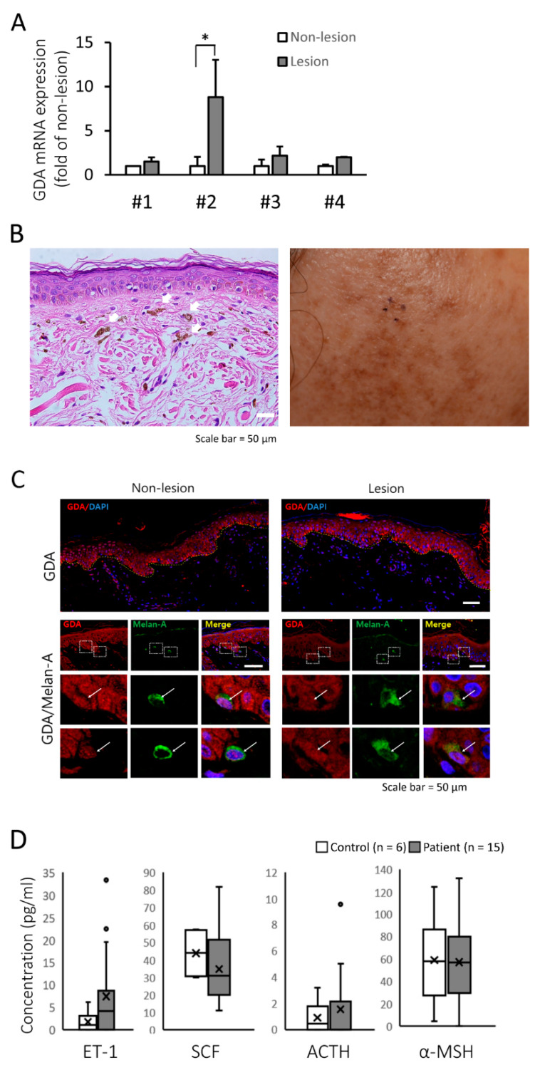
Figure 1.
Guanine deaminase (GDA) expression in Riehl’s melanosis (RM) skin tissue. (A) Relative mRNA expression of GDA in lesional skin compared with that in non-lesional skin tissues from each patient with RM (n = 4). (B) Histopathologic (left panel, hematoxylin and eosin staining, original magnification 400×, dermal melanophages are indicated by white arrows) and clinical features (right panel) of a patient with RM. (C) Immunofluorescence and immunohistochemical staining for GDA in non-lesional and lesional skin from a patient with RM. The images demonstrated GDA (red) with 4′,6-diamidino-2-phenylindole (DAPI) (blue) as a nuclear counterstain (upper panel). Double immunofluorescence images for GDA (red), Melan-A (MART-1; melanocyte marker; green). Merged images with DAPI (blue) showed GDA and Melan-A double positive cells (white arrow; lower panel, scale bar = 50 μm). (D) Box plots displaying the minimum, the maximum, the median, and the first and third quartiles for serum endothelin-1 (ET-1), stem cell factor (SCF), adrenocorticotropic hormone (ACTH), and alpha-melanocyte-stimulating hormone (α-MSH) levels in patients with RM (n = 15) and age-matched healthy volunteers (n = 6). * p < 0.05.

