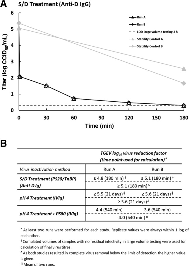
Fig 2.

(A) Susceptibility of the coronavirus TGEV to S/D treatment (1.7% wt/wt PS20, 1.0% wt/wt TnBP, 30°C) in an anti‐D IgG intermediate. Results of two independent replicates are shown (Run A and Run B). Stability controls (virus spiked into anti‐D IgG intermediate and held at process temperature) are shown in light gray. Open symbols indicate when the virus titer has reached the detection limit (no residual infectivity detected). Limit of detection (LOD) is shown for 180‐minute test samples (based on cumulated volume of samples with no residual infectivity). (B) Reduction factors for inactivation of TGEV by manufacturing steps of different plasma‐derived products. Individual as well as final reduction factors are given.
