Abstract
Rationale: MicroRNAs are potent regulators of biologic systems that are critical to tissue homeostasis. Individual microRNAs have been identified in airway samples. However, a systems analysis of the microRNA–mRNA networks present in the sputum that contribute to airway inflammation in asthma has not been published.
Objectives: Identify microRNA and mRNA networks in the sputum of patients with asthma.
Methods: We conducted a genome-wide analysis of microRNA and mRNA in the sputum from patients with asthma and correlated expression with clinical phenotypes. Weighted gene correlation network analysis was implemented to identify microRNA networks (modules) that significantly correlate with clinical features of asthma and mRNA expression networks. MicroRNA expression in peripheral blood neutrophils and lymphocytes and in situ hybridization of the sputum were used to identify the cellular sources of microRNAs. MicroRNA expression obtained before and after ozone exposure was also used to identify changes associated with neutrophil counts in the airway.
Measurements and Main Results: Six microRNA modules were associated with clinical features of asthma. A single module (nely) was associated with a history of hospitalizations, lung function impairment, and numbers of neutrophils and lymphocytes in the sputum. Of the 12 microRNAs in the nely module, hsa-miR-223-3p was the highest expressed microRNA in neutrophils and was associated with increased neutrophil counts in the sputum in response to ozone exposure. Multiple microRNAs in the nely module correlated with two mRNA modules enriched for TLR (Toll-like receptor) and T-helper cell type 17 (Th17) signaling and endoplasmic reticulum stress. hsa-miR-223-3p was a key regulator of the TLR and Th17 pathways in the sputum of subjects with asthma.
Conclusions: This study of sputum microRNA and mRNA expression from patients with asthma demonstrates the existence of microRNA networks and genes that are associated with features of asthma severity. Among these, hsa-miR-223-3p, a neutrophil-derived microRNA, regulates TLR/Th17 signaling and endoplasmic reticulum stress.
Keywords: microRNAs, severe asthma, hsa-miR-223-3p, TH17, gene networks
At a Glance Commentary
Scientific Knowledge on the Subject
MicroRNAs are involved in the regulation of normal cell function and are also dysregulated in asthma. Existing studies have demonstrated a role in type 2 inflammation; however, our understanding of microRNA roles in neutrophilic asthma is limited.
What This Study Adds to the Field
We demonstrate that a network of microRNAs in the sputum is associated with increased airflow obstruction and neutrophilic airway inflammation in asthma. These microRNAs may play a role in the development and maintenance of neutrophilic airway inflammation and may represent potential therapeutic targets.
MicroRNAs are small non–protein-coding RNA molecules that are key regulators of gene expression. MicroRNAs regulate approximately 30% of the human genome (1), and genes targeted by a specific microRNA are frequently functionally related and organ-specific. As a result, microRNAs control networks of genes responsible for normal cell function and inflammatory responses. Not surprisingly, the evidence that microRNAs are associated with airway inflammation in asthma continues to emerge. However, a genome-wide, quantitative assessment of sputum microRNA expression in asthma has never been conducted.
Several microRNA families and corresponding networks have been shown to regulate pathways that are associated with asthma pathogenesis. The let-7 family of microRNAs, miR-1, mir-19, miR-126, miR-155, and miR-221, have been shown to regulate T-helper cell type 2 (Th2) inflammatory responses by downregulating IL-13 and VEGF as well as modulating T-cell, macrophage, and mast cell function (2–10). MicroRNAs have also been shown to regulate Th1 cell polarization (11) and smooth muscle function and proliferation (12–16). In severe asthma, miR-221 regulates smooth muscle proliferation (16) and miR-28-5p, and miR-146a/b activate circulating CD8+ (cluster of differentiation 8–positive) T cells (17). MicroRNA expression has also been shown to be influenced by inhaled steroids (18). Combined, these studies indicate that microRNAs are important mediators of asthma pathogenesis and heterogeneity.
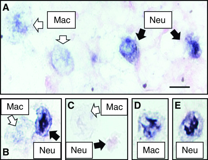
Despite the evolving knowledge on the role of microRNAs in asthma, our understanding of their association with disease severity and regulatory effects on gene networks in the human airway remains largely unknown. We hypothesized that microRNA expression levels in inflammatory cells isolated from sputum would correlate with the phenotypic expression of asthma and the expression of their associated gene regulatory networks. To test this hypothesis, we performed a genome-wide expression analysis of microRNA levels in cells isolated from induced sputum from patients with asthma. MicroRNA networks were identified and correlated with clinical, physiological, and inflammatory phenotypes of disease. In situ hybridization and microRNA expression of the sputum obtained before and after ozone exposure were used to identify hsa-miR-223-3p expression in neutrophils and changes associated with neutrophil counts in the airway. MicroRNA expression was correlated with mRNA expression in the sputum to identify downstream targets and microRNA–mRNA networks using systems biology analyses. These studies reveal that hsa-miR-223-3p is a neutrophil-derived microRNA with a prominent regulatory effect on Th17 signaling and endoplasmic reticulum (ER) stress in severe asthma.
Methods
Study Subjects
We performed a cross-sectional analysis of individuals with asthma recruited from the Yale Center for Asthma and Airways Disease based in New Haven, Connecticut. Data from 71 subjects, those with asthma (n = 62) and controls (n = 9), were available for analysis. The asthma cohort data analysis included additional data from 11 follow-up visits in 10 subjects and 5 technical replicates. The inclusion/exclusion criteria and phenotyping protocol have been previously described (19–21). Severe asthma was defined on the basis of the Expert Panel Report 3 guidelines (22). Features of severe asthma were consistent with those derived from the Severe Asthma Research Program (23) and include healthcare use and airflow limitation. The Yale Human Investigation Committee approved this protocol. All participants provided informed consent.
Sputum Induction, MicroRNA, and mRNA Expression Measurements
Sputum induction was performed using inhaled hypertonic saline, as previously described (24). Sputum cells were isolated using the plug selection technique, followed by dithiothreitol and phosphate-buffered saline incubation and filtration using a 60-micron mesh. Viability was measured manually using the trypan blue exclusion method. Absolute cell counts were the product of differential cell counts (%) and total number of cells in sputum per microliter. Total RNA was isolated from sputum cell pellets using the All-in-One purification kit (Norgen Biotek) and checked on an Agilent Bioanalyzer (Agilent 2100 Bioanalyzer; Agilent Technologies, Inc.). Purified total RNA from the sputum was processed for microRNA expression using the Nanostring nCounter v3.0a array following the manufacturer’s protocols (Nanostring). Only samples with RNA integrity numbers >4 were analyzed. Nanostring array experiments are available under the gene expression omnibus accession number GSE146306. For mRNA profiling, paired-end sample preparation with Illumina prep kit was performed following the manufacturer's protocols (Illumina). Libraries were sequenced using the Illumina HiSeq2000 platform, with 75 nucleotides paired-end reads.
Peripheral Blood Cell Isolation and RNA Extraction
Whole blood was collected from two healthy donors and one patient with asthma that were not part of the sputum induction protocol and processed within 1 hour of collection. Neutrophils and lymphocytes were isolated with the EasySep Direct Human Neutrophil Isolation Kit and the EasySep Direct Human Total Lymphocyte Isolation Kit (STEMCELL Technologies), following the manufacturer’s instructions. A total of 1–2 × 10⁶ isolated cells were resuspended in RPMI-1640 with and without LPS (final concentration, 500 ng/mL) and incubated at 37°C and 5% CO2 for 1 hour. Total RNA was extracted using Norgen Total RNA Purification Kit, the All-in-One purification kit (Norgen Biotek). The protocol was modified to include a Shredder (Qiagen) and genomic DNA column and adjusted centrifugation times and speeds to maximize purity and yield of total RNA. RNA was quantified by Nanodrop (Thermo Scientific) and assessed for quality by capillary gel electrophoresis (Agilent 2100 Bioanalyzer; Agilent Technologies, Inc.).
MicroRNA in Healthy Volunteers before and after Ozone Exposure
Twenty healthy adults without asthma and with no history of smoking in the past 10 years participated in the study, as previously described (25). All subjects underwent sputum induction with hypertonic saline (3%, 4%, and 5%, 7 min/inhalation) before and after ozone exposure. Ozone exposures (2 h, 0.4 ppm, alternating 15 min exercise [Vemin = 30–40 L/min] and rest) were conducted in an exposure chamber at the U.S. Environmental Protection Agency Human Studies Facility on the campus of the University of North Carolina (Chapel Hill, NC). Preexposure sputum was collected at least 48 hours before the collection of the postexposure samples. Postexposure samples were obtained 6 hours after exposure. Sputum underwent quality control and processing to isolate small RNAs from sputum cell pellets. RNA was labeled and hybridized to the Agilent Human microRNA (miRNA) Microarray v1.0 (Agilent Technologies). Microarray experiments are available under the Gene expression omnibus accession number GSE47977. Further details can be found in an article by Fry and colleagues (25).
In Situ Hybridization hsa-miR-223-3p
Cytospins from sputum from asthmatics were mounted onto Superfrost Plus slides (Fisher Scientific) at an approximate density of 1 × 10⁶ cells/mL (50 μL sample and 50 μL phosphate-buffered saline) and were centrifuged at 1,000 × g for 8 minutes. Slides were air-dried for 10 minutes, immersed in paraformaldehyde for 10 minutes, and air-dried overnight. The Qiagen miRCURY locked nucleic acid miRNA in situ hybridization Buffer and Control kit, including the scramble miRNA negative control probe and the U6 small nuclear RNA positive control probe, was used in combination with the hsa-miR-223-3p detection probe (Qiagen). The 1-day miRNA in situ hybridization manufacturer’s protocol was used. Slides were mounted using 50 μL of Eukitt mounting medium and imaged using light microscopy.
Statistical Analysis
All statistical analyses were performed using R software version 3.3.1 (https://cran.r-project.org/). Results are reported as medians and interquartile ranges (25–75%) unless otherwise specified. Continuous variables were tested using nonparametric tests, including the Wilcoxon test to compare two groups and the Kruskal-Wallis test to compare more than two groups. Categorical variables were analyzed with chi-square tests. P values of less than 0.05 were considered significant.
Nanostring nCounter v3.0a arrays were normalized using the top 100 genes without background subtraction using the geometric mean in the nSolver Analysis Software.v3. (Nanostring). The normalized raw counts were used to identify expressed microRNAs. MicroRNAs were considered to be expressed if their overall mean signal was above the background mean plus two standard deviations of the negative ligation controls in the array. Subsequently, the log2 count normalized matrix of the expressed microRNAs was batch-adjusted using the ComBat function in the sva package (version 3.22.0) and used for all analyses (26). Pairwise analyses were considered significant with an false discovery rate (FDR)-adjusted P value <0.05. RNA sequencing data were aligned to the HG19 reference using the STAR software version 2.5 (27). The number of reads mapped to each gene was counted (raw counts), and these raw counts were further processed and normalized by edgeR (28). Cufflinks software version 2.2.1 was used to estimate the fragments per kilobase of transcript per million mapped reads (29). Fragments per kilobase of transcript per million was used for subsequent analyses. R software version 3.3.1 was used for all analyses.
Weighted gene correlation network analysis (WGCNA) version 1.42, a method that identifies comodulated genes, which when aggregated and reduced can then be correlated against external features, was used in two instances. First, WGCNA identified correlations between sputum microRNA expression and clinical, physiologic, and inflammatory characteristics of asthma (30, 31). A second WGCNA identified correlations between microRNAs and mRNAs expressed in the nely module found in the first WGCNA analysis.
Canonical pathway and upstream regulator (an analysis to identify molecules regulating gene expression) analyses in the RNA modules were performed with Ingenuity Pathway Analysis (IPA) software (Qiagen). Enrichment analyses based off the Fischer’s Exact test with an FDR-adjusted P value < 0.05 were considered significant.
Results
Study Population
The characteristics of the subjects are shown in Table 1. Subjects in this study were predominantly female (69%) and white (66%). Patients with asthma had a lower baseline and post-bronchodilator FEV1% predicted than controls (P < 0.05 for both comparisons), and 52% of the subjects had severe asthma.
Table 1.
Demographic, Clinical, and Inflammatory Characteristics of Study Participants
| Control | Mild | Moderate | Severe | |
|---|---|---|---|---|
| Prevalence, n | 9 | 12 | 18 | 32 |
| Age at visit, yr | 39 ± 13 | 51 ± 14 | 48 ± 12 | 46 ± 13 |
| Sex, F, n (%) | 5 (56) | 8 (67) | 12 (67) | 23 (81) |
| Race | ||||
| White, n (%) | 8 (89) | 9 (75) | 15 (83) | 15 (47) |
| Black, n (%) | — | 2 (17) | 1 (6) | 9 (28) |
| Other, n (%) | 1 (11) | 2 (11) | 8 (25) | |
| NA, n (%) | — | 1 (8) | — | — |
| Hispanic origin, n (%) | 1 (11) | 1 (8) | 2 (11) | 6 (19) |
| BMI, kg/m2 | 26.6 ± 3.8 | 29.4 ± 8.5 | 29.0 ± 6.8 | 36.9 ± 8.4 |
| History of atopy, n (%) | 4 (44) | 11 (92) | 15 (83) | 31 (97) |
| Age of symptom onset, yr | — | 26 ± 18 | 22 ± 19 | 16 ± 15 |
| Disease duration, yr | — | 22 ± 16 | 29 ± 21 | 29 ± 16 |
| History of hospitalization past year, n (%) | — | 0 | 2 (11) | 15 (47) |
| History of intubations, n (%) | — | 0 | 1 (6) | 7 (22) |
| ACT score | — | 20 ± 6 | 19 ± 5 | 15 ± 5 |
| ICS, n (%) | — | 2 (17) | 3 (17) | 3 (9) |
| ICS + LABA, n (%) | — | 2 (17) | 13 (72) | 31 (97) |
| Antihistamine, n (%) | — | 5 (42) | 6 (33) | 10 (31) |
| LTRA, n (%) | — | 4 (33) | 9 (50) | 22 (69) |
| Omalizumab, n (%) | — | 0 | 0 | 6 (19) |
| LAMA, n (%) | — | 0 | 1 (6) | 11 (34) |
| OCS, n (%) | — | 0 | 1 (6) | 6 (19) |
| Theophylline, n (%) | — | 0 | 0 | 2 (6) |
| ICS dose, mg/d | — | 42 ± 94 | 372 ± 276 | 649 ± 500 |
| ICS use, yes, n (%) | — | 4 (33) | 14 (78) | 32 (100) |
| Chronic OCS use, n (%) | — | 0 | 1 (6) | 6 (19) |
| FEV1% predicted value | ||||
| Pre–β2-agonist use | 98 ± 5 | 89 ± 20 | 79 ± 19 | 65 ± 21 |
| Post–β2-agonist use | 100 ± 6 | 97 ± 16 | 85 ± 17 | 72 ± 20 |
| FVC% predicted value | ||||
| Pre–β2-agonist use | 100 ± 10 | 95 ± 18 | 87 ± 14 | 79 ± 16 |
| Post–β2-agonist use | 99 ± 11 | 99 ± 14 | 90 ± 15 | 85 ± 14 |
| FEV1/FVC | ||||
| Pre–β2-agonist use | 0.80 ± 0.04 | 0.74 ± 0.08 | 0.72 ± 0.14 | 0.65 ± 0.12 |
| Post–β2-agonist use | 0.83 ± 0.05 | 0.78 ± 0.07 | 0.74 ± 0.13 | 0.68 ± 0.12 |
| BDR, % | 3 ± 3 | 11 ± 12 | 9 ± 11 | 13 ± 14 |
| FeNO, ppb | 21 ± 8 | 46 ± 35 | 28 ± 26 | 34 ± 29 |
| Sputum cell concentration* | 69.5 ± 33.8 | 118.2 ± 70.8 | 121.3 ± 92.2 | 243.4 ± 309.5 |
| Squamous, % | 8.2 ± 6.1 | 7.5 ± 7.3 | 8.6 ± 10.5 | 7.2 ± 6.9 |
| Viability, % | 69.0 ± 17.3 | 64.2 ± 16.8 | 55.5 ± 17.8 | 57.9 ± 22.1 |
| ACC neutrophils | 26.95 ± 23.04 | 26.22 ± 34.01 | 62.56 ± 78.12 | 81.730 ± 117.74 |
| ACC eosinophils | 4.89 ± 8.86 | 5.89 ± 7.41 | 4.40 ± 10.97 | 43.52 ± 218.89 |
| ACC macrophages | 34.78 ± 8.94 | 77.66 ± 64.76 | 54.77 ± 38.13 | 74.43 ± 62.55 |
| ACC lymphocytes | 2.42 ± 3.82 | 3.57 ± 3.68 | 4.74 ± 6.07 | 5.78 ± 7.50 |
| ACC bronchial epithelial cells | 0.49 ± 1.07 | 0.71 ± 0.96 | 0.33 ± 0.54 | 1.11 ± 2.17 |
| Sputum inflammatory profile asthma | ||||
| Eosinophilic, n (%) | — | 3 (25) | 4 (22) | 9 (28) |
| Neutrophilic, n (%) | — | 1 (8) | 7 (39) | 10 (31) |
| Mixed granulocytic, n (%) | — | 3 (25) | 1 (6) | 4 (13) |
| Paucigranulocytic, n (%) | — | 5 (42) | 6 (33) | 9 (28) |
| RIN | 6.6 ± 2.6 | 7.4 ± 2.0 | 7.4 ± 1.7 | 6.8 ± 2.1 |
Definition of abbreviations: ACC = absolute cell count; ACT = asthma control test; BDR = bronchodilator response; BMI = body mass index; FeNO = fractional exhaled nitric oxide; ICS = inhaled corticosteroids; LABA = long-acting β-agonist; LAMA = long-acting muscarinic antagonist; LTRA = leukotriene receptor antagonist; NA = not available; OCS = oral corticosteroid; RIN = RNA integrity number.
Data are shown as mean ± SD unless otherwise specified.
Cells per microliter × 104.
Sputum MicroRNAs in Asthma versus Controls
Expression levels of 800 microRNAs in the sputum were measured using the Nanostring nCounter array v3.0a. Two hundred and twenty-one microRNAs were expressed above the background level for the platform and were used for subsequent analyses. The pairwise comparison between asthma and control subjects demonstrated that 12 microRNAs had nominal P values < 0.05. None of these microRNAs were above the predetermined FDR significance threshold (summarized in Table E1 in the online supplement).
WGCNA Analysis of the Sputum in Asthma
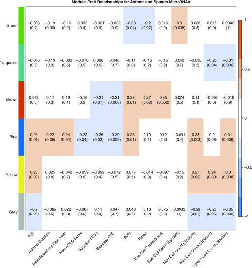
WGCNA analysis was conducted to identify associations between microRNA expression in the sputum and clinical features of asthma. This method uses the correlation between genes across samples to identify gene modules that are coexpressed. Each module can be used to identify correlations with disease traits in the cohort of subjects being examined (30). For this analysis, the 221 expressed microRNAs (Table E2) were correlated with clinical, physiologic, and inflammatory features of asthma. Six microRNA modules were identified, four of which correlated significantly with clinical features of asthma: the green, brown, blue, and yellow modules (Figure 1). None of the modules was correlated with inhaled corticosteroids (ICS) dose, suggesting the lack of ICS dose effect on these correlations (Figure E1). These disease feature–microRNA module associations suggest that the microRNAs in a given module have similar regulatory effects on the clinical feature correlated with the specific module.
Figure 1.
Weighted gene correlation network analysis of sputum microRNA expression and asthma features (n = 78 samples). Using expressed microRNAs (n = 221) in the sputum, six modules were identified. These modules had correlations with multiple demographic, clinical, physiologic, and sputum characteristics. Positive correlations are red, and negative correlations are blue. The microRNAs in the blue (nely) module were positively associated with age, asthma duration, hospitalizations in the previous year, bronchodilator response, and neutrophil and lymphocyte cell counts in the sputum; and negatively correlated with asthma quality of life (AQLQ), FEV1% predicted, and FVC% predicted at baseline. Values are correlation coefficient (nominal P value). BDR = bronchodilator response; Eos = eosinophil; FeNO = fractional exhaled nitric oxide; Lymph = lymphocyte; Mac = macrophage; miRNA = microRNA; Neu = neutrophil.
The green microRNA module, formed by hsa-miR-149-5p, hsa-miR-520f-3p, hsa-miR-574-5p, and hsa-miR-2682-5p, correlated positively with eosinophil counts in the sputum (r = 0.30; P = 0.008) and negatively with bronchodilator response (BDR) (r = −0.23; P = 0.04). The yellow microRNA module formed by hsa-let-7b-5p, hsa-let-7c-5p, hsa-miR-423-5p, and hsa-miR-4443 correlated positively with age (r = 0.25; P = 0.03), sputum macrophage (r = 0.24; P = 0.03), and lymphocyte cell counts (r = 0.30; P = 0.008). The brown microRNA module was formed by hsa-miR-15a-5p, hsa-miR-16-5p, hsa-miR-21-5p, hsa-miR-29b-3p, hsa-miR-142-3p, hsa-miR-148a-3p, hsa-miR-374a-5p, and hsa-miR-1246. Expression in the brown module correlated positively with BDR (r = 0.28; P = 0.01), fractional exhaled nitric oxide (FeNO) (r = 0.27; P = 0.02), and absolute blood eosinophil counts (r = 0.35; P = 0.002). The turquoise module was large, composed of 135 microRNAs (Table E3) and correlated positively with macrophage and lymphocyte cell counts (cells per microliter). The gray module was formed by microRNAs that did not meet membership criteria for all other modules (n = 57; Table E3). Despite the significant correlations between these modules and clinical features of asthma, there was no clear association with specific asthma subtypes (e.g., eosinophilic, type 2 inflammation–high, and neutrophilic) or severity. In contrast to these modules, the blue module correlated with multiple clinical, physiological, and inflammatory features of severe asthma.
The Blue Module Has Multiple Associations with Severe Asthma
The blue module was formed by 12 microRNAs: hsa-let-7a-5p, hsa-let-7d-5p, hsa-let-7 g-5p, hsa-let-7i-5p, hsa-miR-15b-5p, hsa-miR-23a-3p, hsa-miR-25-3p, hsa-miR-150-5p, hsa-miR-181a-5p, hsa-miR-191-5p, hsa-miR-223-3p, and hsa-miR-342. This module correlated positively with age (r = 0.23; P = 0.04), asthma duration (r = 0.22; P = 0.05), and multiple features of severe asthma (Figure 1). Specifically, the blue module correlated positively with hospitalizations in the previous year (r = 0.24; P = 0.04) and BDR (r = 0.28; P = 0.01) and correlated negatively with quality of life, Mini Asthma Quality of Life (32) (r = −0.23; P = 0.04), and baseline FEV1% predicted (r = −0.25; P = 0.02). It also correlated positively with the absolute neutrophil (r = 0.33; P = 0.003) and lymphocyte (r = 0.31; P = 0.006) cell counts in the sputum. Given these positive associations with neutrophil and lymphocyte counts in the sputum (henceforth, nely module), therefore, this microRNA module showed enrichment for phenotypic characteristics associated with severe asthma (23).
In contrast to the other modules, all the microRNAs composing the nely module correlated positively with each other (Table E4), several correlated with multiple phenotypes of asthma (Table 2), and all the microRNAs correlated were in the same direction with a given feature (e.g., all positive or all negative), suggesting a similar effect on the regulatory networks and association with features of severe asthma (23). Ten of the nely module microRNAs correlated with sputum neutrophils, longer duration of asthma, decreased quality of life, impaired lung function, and/or increased BDR (Table 2). Higher sputum expression of hsa-miR-15b and hsa-miR-223-3p was present in subjects hospitalized for asthma in the previous year (6.4 vs. 5.2 [log2 normalized microRNA expression], P = 0.02 and 9.5 vs. 6.9 [log2 normalized microRNA expression], P = 0.03, respectively.) In addition to these correlations, two microRNAs, hsa-let-7d-5p and hsa-miR-181a-5p, correlated positively with lymphocyte counts in the sputum.
Table 2.
Correlations between nely Module MicroRNAs and Asthma Features
| MicroRNA | Age (yr) | Asthma Duration (yr) | Mini-AQLQ Score | Baseline FEV1 (% Predicted) | BDR | Neutrophil Cell Count | Lymphocyte Cell Count |
|---|---|---|---|---|---|---|---|
| hsa-let-7a-5p | 0.22 (0.08) | 0.12 (0.37) | −0.14 (0.28) | −0.26 (0.05) | 0.25 (0.05) | 0.32 (0.05) | 0.25 (0.05) |
| hsa-let-7d-5p | 0.17 (0.18) | 0.20 (0.11) | −0.10 (0.45) | −0.19 (0.16) | 0.18 (0.17) | 0.25 (0.05) | 0.31 (0.01) |
| hsa-let-7 g-5p | 0.26 (0.05) | 0.26 (0.04) | −0.25 (0.05) | −0.23 (0.07) | 0.27 (0.04) | 0.17 (0.18) | 0.19 (0.14) |
| hsa-let-7i-5p | 0.24 (0.06) | 0.20 (0.13) | −0.27 (0.04) | −0.09 (0.49) | 0.10 (0.47) | −0.03 (0.79) | 0.07 (0.58) |
| hsa-miR-15b-5p | 0.28 (0.02) | 0.28 (0.03) | −0.09 (0.50) | −0.32 (0.01) | 0.26 (0.05) | 0.57 (<0.01) | 0.14 (0.29) |
| hsa-miR-23a-3p | 0.20 (0.12) | 0.27 (0.03) | −0.35 (0.01) | −0.34 (0.01) | 0.35 (0.01) | 0.25 (0.05) | 0.11 (0.39) |
| hsa-miR-25-3p | 0.23 (0.07) | 0.31 (0.02) | −0.23 (0.07) | −0.34 (0.01) | 0.31 (0.02) | 0.54 (<0.01) | −0.08 (0.56) |
| hsa-miR-150-5p | 0.17 (0.20) | 0.19 (0.14) | −0.20 (0.14) | −0.25 (0.06) | 0.19 (0.15) | 0.40 (<0.01) | 0.17 (0.19) |
| hsa-miR-181a-5p | 0.36 (<0.01) | 0.40 (<0.01) | −0.14 (0.29) | −0.33 (0.01) | 0.23 (0.08) | 0.36 (<0.01) | 0.29 (0.02) |
| hsa-miR-191-5p | 0.27 (0.03) | 0.24 (0.03) | −0.22 (0.10) | −0.35 (0.01) | 0.38 (<0.01) | 0.46 (<0.01) | 0.05 (0.68) |
| hsa-miR-223-3p | 0.27 (0.04) | 0.30 (0.05) | −0.35 (0.01) | −0.42 (<0.01) | 0.36 (<0.01) | 0.46 (<0.01) | 0.03 (0.81) |
| hsa-miR-342-3p | 0.12 (0.34) | 0.15 (0.25) | −0.35 (0.01) | 0.09 (0.50) | 0.15 (0.27) | −0.06 (0.64) | 0.20 (0.12) |
Definition of abbreviations: AQLQ = asthma quality of life; BDR = bronchodilator response.
Values are correlation coefficients (P value). Bold indicates nominal P values less than 0.05.
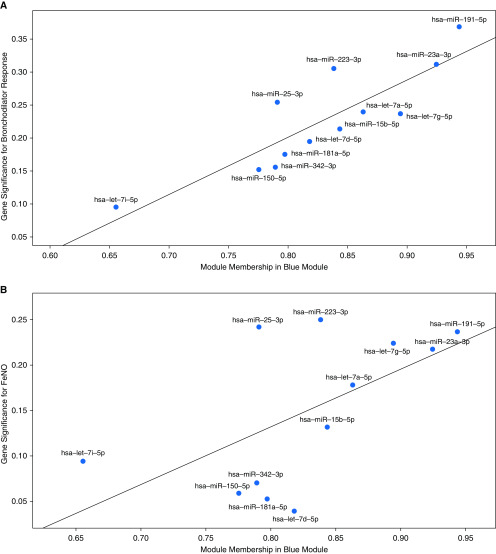
Two additional metrics derived from WGCNA analyses are module membership (MM) and gene significance (GS). MM is a measurement also known as eigengene-based connectivity. This is used to determine the significance of a microRNA in the module. Values close to −1 or 1 have the highest connection to the microRNAs in that module. GS is a measure that reflects the biological significance of a specific external trait that correlates with the module. To characterize the relationship between MM and GS within the nely module, we examined multiple asthma features and found a significant correlation between GS and MM with BDR (r = 0.86, 95% confidence interval: 0.57–0.96, P < 0.01) and FeNO (0.59, 95% confidence interval: 0.02–0.88, P = 0.045) with hsa-miR-23a-3p, hsa-miR-191-5p and hsa-miR-223-3p having the most significant correlation with BDR and FeNO (right upper quadrant, Figures 2A and 2B). These findings suggest that these three nely module microRNAs have significant pathophysiologic effects on the regulation of BDR and FeNO levels and potentially other clinical phenotypes of asthma.
Figure 2.
(A and B) Module membership and gene significance correlation with bronchodilator response (A) and fractional exhaled nitric oxide (FeNO) (B) for nely module microRNAs. The module membership and gene significance analyses demonstrate that hsa-miR-191-5p, hsa-miR-23a-3p, and hsa-miR-223-3p play a key role in the nely module and two traits of interest (A, bronchodilator response and B, FeNO).
Combined, these results showed that mature microRNAs are expressed and detectable in the sputum and correlate with clinical features of the disease; specifically, the nely module is formed by a network of coexpressed microRNAs that modulate the clinical expression of asthma and severe asthma. These microRNAs are likely produced by and/or affect the levels of neutrophils and lymphocytes in the sputum (Figure 1). Three microRNAs in particular, hsa-miR-23a-3p, hsa-miR-191-5p, and hsa-miR-223-3p, had the strongest associations with features of severe asthma. These findings suggest that this network of sputum microRNAs is associated with neutrophilic airway inflammation in severe asthma.
High Expression of nely Module MicroRNAs Is Associated with Severe Asthma Features
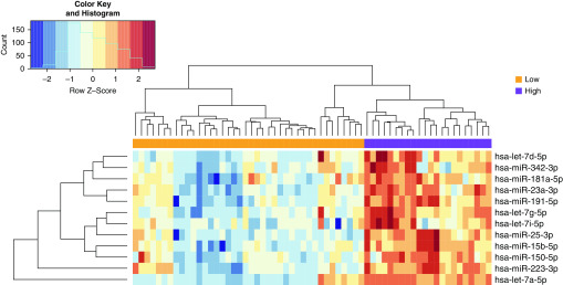
We performed unsupervised hierarchical clustering to determine if the expression levels of the 12 microRNAs in the nely module can discriminate subgroups of patients. This analysis identified two clusters of individuals with distinctly different levels of microRNAs and clinical features of the disease (Figure 3). The cluster with high microRNA expression had a longer duration of illness, lower lung function, higher sputum inflammatory cell levels, and higher sputum neutrophil and lymphocyte counts (Table 3). No differences were noted in serum IgE level or blood eosinophil counts between high and low blue module clusters. There were no differences in age, the age of onset, or doses of ICS between the two clusters. Other microRNAs in the sputum were also differentially expressed between the two clusters (Table E5). Therefore, high nely module microRNA expression was associated with a subgroup of asthma characterized by neutrophilic inflammation, low lung function, and low T2 biomarkers.
Figure 3.
nely module microRNA expression identifies two asthma clusters (n = 62). Hierarchical clustering of patients using nely module microRNA expression was used to identify two groups with high and low expression of sputum microRNA. These two groups were characterized by differences in lung function and neutrophil and lymphocyte counts (Table 3).
Table 3.
Clinical Characteristics of the High and Low nely Module MicroRNA Expression Patient Clusters
| Feature | Low MicroRNA (n = 40) | High MicroRNA (n = 22) | P Value |
|---|---|---|---|
| Age, yr | 45 ± 13 | 51 ± 12 | 0.10 |
| Sex, F, n (%) | 29 (73) | 17 (43) | 0.91 |
| Age of onset | 21 ± 16 | 17 ± 19 | 0.25 |
| Asthma duration | 23 ± 16 | 37 ± 19 | 0.05 |
| BMI, kg/m2 | 30.7 ± 7.9 | 32.3 ± 8.4 | 0.37 |
| Atopy, n (%) | 37 (94) | 20 (91) | 1 |
| Baseline FEV1% pred | 79 ± 21 | 63 ± 22 | 0.02 |
| Post-BD FEV1% pred | 86 ± 20 | 72 ± 19 | 0.02 |
| Post-BD FEV1/FVC ratio | 0.74 ± 0.12 | 0.68 ± 0.12 | 0.06 |
| ICS total | 492 ± 432 | 377 ± 492 | 0.20 |
| Cell concentration in sputum* | 138.80 ± 227.75 | 214.00 ± 136.89 | <0.01 |
| ACC neutrophil* | 30.70 ± 34.42 | 128.50 ± 136.49 | <0.01 |
| ACC lymphocytes* | 4.42 ± 6.2 | 6.20 ± 7.04 | 0.03 |
| ACC macrophages* | 68.36 ± 57.61 | 71.14 ± 56.94 | 0.72 |
| ACC eosinophils* | 34.55 ± 195.96 | 7.29 ± 11.53 | 0.51 |
| ACC bronchial epithelial cells* | 0.83 ± 1.68 | 0.78 ± 1.70 | 0.69 |
| Sputum inflammatory profiles | 0.07 | ||
| Eosinophilic, n (%) | 12 (30) | 4 (18) | |
| Neutrophilic, n (%) | 9 (22) | 9 (41) | |
| Mixed granulocytic, n (%) | 3 (8) | 5 (23) | |
| Paucigranulocytic, n (%) | 16 (40) | 4 (18) |
Definition of abbreviations: ACC = absolute cell count; BD = bronchodilator; BMI = body mass index; ICS = inhaled corticosteroids; pred = predicted.
Data are shown as mean ± SD unless otherwise specified.
Cells per microliter × 104.
hsa-miR-223-3p Expression in Neutrophils
Because the nely module microRNAs correlated with neutrophils and lymphocytes in the sputum, and previous studies demonstrated expression of hsa-mir-223-3p from myeloid-derived cells including neutrophils (33, 34), we sought to determine the cellular source of the nely module microRNAs (Figure E2). Neutrophils and lymphocytes were isolated from peripheral blood in three subjects (two controls and one with asthma). All microRNAs in the nely module were expressed in these two cell types (Figure E2). hsa-miR-23a-3p, hsa-miR-191-5p, and hsa-miR-223-3p were highly expressed in neutrophils and correlated with each other (Figure E2 and Table E6).
In contrast with the correlations seen in sputum (Table E4), the other eight microRNAs in the nely module correlated negatively with hsa-miR-223-3p expression in neutrophils isolated from the blood (Table E6). hsa-let-7 g-5p, hsa-let-7i-5p, hsa-miR-150-5p, and hsa-miR-342-3p were highly expressed in lymphocytes and correlated negatively with the microRNAs expressed in neutrophils (Figure E2 and Table E6). These findings suggest possible tissue-specific differences in microRNA expression.
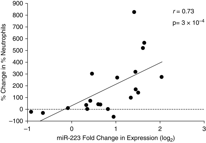
We also evaluated the expression of blue module microRNAs in the sputum from a cohort of healthy subjects exposed to ozone (25) to test the responsivity of the microRNAs to a known stimulus like ozone that induces increased neutrophils in sputum. This analysis demonstrated that increases in sputum neutrophil counts in the airway following ozone exposure positively correlated with fold changes in hsa-miR-223-3p (r = 0.73, P < 0.01) (Figure 4). This analysis suggests that neutrophils are strongly associated with changes in hsa-miR-223-3p in the sputum. Together, these findings suggest that blue module microRNAs are part of a regulatory network that is expressed in neutrophils and lymphocytes in patients with asthma and with neutrophilic inflammation following exposure to ozone in nonasthmatic individuals. To confirm neutrophil-specific hsa-miR-223 expression in the sputum, we performed in situ hybridization (Figure 5).
Figure 4.
hsa-miR-223-3p expression changes are correlated with sputum neutrophil counts following ozone exposure in healthy individuals. In healthy individuals exposed to ozone, changes in sputum neutrophil counts were positively correlated with fold change in expression of hsa-miR-223-3p utilizing standard Pearson’s correlation. miR = microRNA.
Figure 5.
In situ hybridization for hsa-miR-223-3p on sputum cytospins. (A and B) Samples incubated with a locked nucleic acid probe targeting hsa-miR-223-3p (blue color). hsa-miR-223-3p was expressed in neutrophils (Neu; black arrows) and not expressed in macrophages (Mac; white arrows). Scale bar, 10 μm. (C) Negative control staining. (D and E) Positive control staining using locked nucleic acid targeting U6 RNA in Mac (D) and Neu (E), respectively.
Integrated Analysis of mRNA and Blue Module MicroRNA Expression Defines a Link to Th17 Pathways
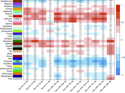
On the basis of the observation that nely module microRNAs were associated with multiple phenotypes of severe asthma, we performed a second WGCNA analysis to evaluate the potential regulatory effect of nely module microRNAs on gene expression in the airway. RNA sequencing was performed on total RNA isolated from 21 sputum samples, as described above. Applying WGCNA analysis, gene expression was correlated with the 12 microRNAs composing the nely module. Thirty-two mRNA modules were identified, of which 14 had at least one significant correlation with a nely module microRNA (Figure 6). Genes in each of these modules are presented in Table E7. Nine of the mRNA modules correlated with multiple microRNAs in the same direction, suggesting that the microRNAs associated with those specific mRNA modules had a similar regulatory effect on gene expression.
Figure 6.
Weighted gene correlation network analysis of microRNA (miR) and mRNA expression in matched sputum samples (n = 21). Weighted gene correlation network analysis heatmap of the correlation between genes expressed in the sputum (n = 16,966). This analysis identified 32 gene modules correlated with the 12 microRNAs included in the nely microRNA module in paired mRNA and microRNA samples obtained from the same individuals. Positive correlations are red, and negative correlations are blue. Two modules had the most positive and negative correlations. The lightcyan mRNA module had positive correlations with 10 nely module microRNAs, and the turquoise module had negative correlations with 8 nely module microRNAs. Gene enrichment analysis of the lightcyan mRNA module revealed enrichment for TLR (Toll-like receptor) pathways and T-helper cell type 17 pathways, and the turquoise mRNA module was enriched for the unfolded protein response (UPR). These putative biological functions for the two modules are highlighted in the module. Values are correlation coefficient (nominal P value).
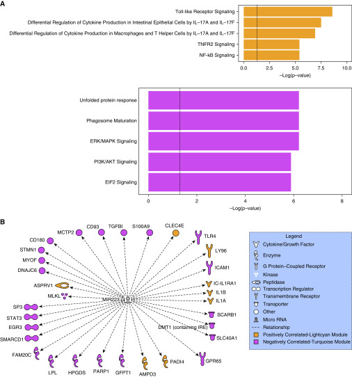
IPA software was used to perform canonical pathway enrichment of each mRNA module that correlated most significantly with microRNA nely module member microRNAs, the mRNA lightcyan, and mRNA turquoise mRNA modules. This analysis identified multiple pathways at an FDR <0.05. The top five pathways in the mRNA lightcyan and mRNA turquoise modules are illustrated in Figure 7A, and Tables E8 and E9 show all the pathways enriched in these two modules, respectively. The TLR (Toll-like receptor)–IL-17 signaling pathway was the top pathway enriched in the lightcyan module (henceforth TLR–IL-17 module). Despite the positive correlation between nely module microRNAs and the TLR signaling pathway, TLR4, a key molecule in the TLR pathway, had a negative correlation with both hsa-miR-223 and let-7, suggesting that the TLR pathway may have a complex regulatory interaction with nely module microRNAs potentially through the regulation of downstream molecules associated with these TLR pathways. This finding is consistent with the known complex regulation of TLR responses by microRNAs (35). Also, both IL-17A and IL-17F pathways were among the top three enriched pathways in the TLR-IL17 module. The association of nely module microRNAs with IL-17 expression and neutrophil counts may be mediated through the effect of IL-17 in neutrophil recruitment to the airway (36). The top negatively correlated pathway enriched in the turquoise mRNA module was the unfolded protein response (UPR) in the ER (henceforth UPR module), suggesting a role in the regulation of ER stress, a novel pathway in asthma (37, 38).
Figure 7.
(A) Pathways enriched in lightcyan (Toll-like receptor–IL-17) and turquoise (unfolded protein response) weighted gene correlation network analysis modules of microRNA–mRNA expression. This figure summarizes the top five pathways enriched in the lightcyan and turquoise mRNA modules. (B) hsa-miR-223-3p gene regulation network. Upstream regulation enrichment analysis of the lightcyan and turquoise mRNA modules identified miR-223 as the only nely module microRNA with known or predicted regulatory effects in multiple genes present in the two mRNA modules. EIF2 = eukaryotic initiation factor 2; ERK/MAPK = extracellular-signal-regulated kinase/mitogen-activated protein kinase; NF-κB = nuclear factor–kappa B; PI3K/AKT = phosphatidylinositol 3-kinase/protein kinase B; TLR = Toll-like receptors; TNFR2 = tumor necrosis factor receptor 2.
The upstream analysis feature of the IPA software was used to identify microRNAs involved in the TLR-IL17 and UPR mRNA module regulation. This analysis identified hsa-miR-223 as a regulator in both TLR-IL17 and UPR mRNA modules (Table E10). Figure 7B illustrates the network of genes regulated by hsa-miR-223 in the TLR-IL17 and UPR modules. These analyses suggest that although multiple nely module microRNAs are associated with TLR-IL17 and UPR mRNA modules, hsa-miR-223-3p is a prominent Th17 pathway regulator in the sputum.
Discussion
Systematic characterization of microRNA profiles in the airway of patients with asthma, particularly severe disease, has been hampered by the inherent limitations of invasive sampling associated with bronchoscopy. Here we show the feasibility of noninvasive assessment of microRNA expression in sputum inflammatory cells and the identification of a network of 12 microRNAs that is associated with severe asthma. Specifically, microRNA expression in the nely module is associated with age, decreased asthma quality of life, impaired lung function, increased bronchodilator response, increased sputum neutrophils, and lymphocytes. Further stratification of subjects with asthma using hierarchical clustering analysis based on the expression of the 12 microRNAs identified two distinct groups of patients with differential expression of microRNAs. Patients with high nely module microRNA expression had increased asthma-related morbidity, impaired lung function, and increased neutrophilic inflammation in the airway. Additional analyses of the potential effect of these microRNAs on the regulation of gene expression in the airway identified TLR function, IL-17 response, and the UPR as processes tied to nely module microRNAs, particularly hsa-miR-223-3p. These findings suggest that higher expression of these microRNAs contributes to increased disease activity and more neutrophilic inflammation that is not controlled by conventional asthma therapy and distinct transcriptomic networks.
We also found that blue module microRNAs that correlate positively with each other in the sputum have distinct cellular origins. First, the positive correlation of nely module microRNAs with sputum neutrophil and lymphocyte counts suggested these microRNAs originate from both cell types. We followed this observation with quantification of microRNAs in isolated neutrophils and lymphocytes, demonstrating that all 12 nely module microRNAs were expressed in both neutrophils and lymphocytes. Importantly, and consistent with other reports, hsa-miR-223-3p was the most abundant microRNA detected in neutrophils. To validate this observation in the airway, in situ hybridization of hsa-miR-223-3p in sputum from asthmatics demonstrated increased expression in neutrophils. Our findings are consistent with a previous study that identified hsa-miR-223-3p in the sputum supernatant of patients with severe neutrophilic asthma (39). However, in contrast with Maes and colleagues, we used cell-derived microRNAs rather than microRNAs isolated from the sputum supernatant, showing that neutrophils are the source of hsa-miR-223-3p, and we suggest this miRNA is released into the airway where it exerts its regulatory effects on the bronchial epithelium, dendritic cells, and other cells in the airway. Also, the studies herein demonstrate that the downstream effects of hsa-miR-223-3p likely influence multiple pathways in the airway, including TLR function, IL-17 response, and the UPR in the ER, a major regulator of inflammation (32, 33). The association between nely module microRNAs hsa-miR-25-3p and hsa-miR-223-3p and sputum neutrophils is further supported by an additional study of airway response to ozone exposure in healthy subjects (25). Further examination of the correlation between changes in neutrophil counts and hsa-miR-223-3p in this group of healthy subjects demonstrated a strong cell-specific association. Together, these data support an association between blue module microRNAs, especially hsa-miR-223-3p, and airway neutrophilia, in addition to IL-17 expression in the airway, suggesting a potential effect of IL-17 in neutrophil recruitment to the airway (36).
We also found several members of the let-7 microRNA family in the nely module. Previous experimental evidence showed an association between members of this family and IL-13 expression (3, 4). Consistent with those reports, we found increased expression of this family of microRNAs in lymphocytes. Other microRNAs in the nely module, including hsa-miR-15b-5p and hsa-miR-342, are associated with lung function in asthma (40); hsa-miR-150-5p is associated with severe asthma (41) and T-cell function (42). Therefore, our findings are in line with previous studies and support the presence of coregulated microRNA networks between neutrophils and lymphocytes in the airway and point to their interaction leading to specific associations with severe asthma features, namely, airflow obstruction.
In addition to the nely module microRNA, we found other microRNA modules associated with phenotypes of asthma. We found a correlation between the green module and absolute eosinophil counts in the sputum and a correlation between the brown module and absolute blood eosinophil counts. Of these, hsa-miR-15a, hsa-miR-21, and hsa-miR-574 have been studied in humans and animal models of asthma (11, 39, 43–46). Importantly, a study of circulating microRNAs in asthma stratified by blood eosinophil counts identified multiple microRNAs present in the brown module (47). Together, our analyses point to specific cellular sources of microRNAs in the airway, including not only neutrophils and lymphocytes in the nely module, but also eosinophils in the green module and macrophages in the yellow module. In this study of microRNA profiles in the sputum, we found an association between neutrophils and lymphocytes in the airway of patients with severe asthma and a network of microRNAs that includes a key role for hsa-miR-223-3p.
On the basis of the association between nely module microRNAs and multiple phenotypes of severe asthma, we performed a WGCNA analysis to identify gene networks that are likely to be regulated by this module. Two mRNA modules, TLR-IL17 and UPR, showed inverse correlations with hsa-let-7 g-5p, hsa-miR-15b-5p, hsa-miR-23a-3p, hsa-miR-25-3p, hsa-miR-150-5p, hsa-miR-181a-5p, hsa-miR-191-5p, and hsa-miR-223-3p in these analyses, indicating that nely module microRNAs have opposing regulatory effects in similar gene networks. Further pathway enrichment of these two modules identified the regulation of TLR signaling, IL-17 production, ER stress, and phagosome maturation. Additional analyses of upstream regulators of genes in the TLR-IL17 and UPR modules identified hsa-miR-223-3p. This upstream analysis demonstrates that hsa-miR-223-3p directly targets several genes in these gene modules. Multiple microRNAs in the blue module have been associated with Th17 cell differentiation in infectious and inflammatory diseases, including hsa-let-7a, hsa-let-7i, hsa-miR-15b, and hsa-miR-223 (48–51). These analyses and observations suggest that both neutrophils and lymphocytes are the cellular sources of the nely module microRNAs and that they are dysregulated in asthma and underlying association between Th17 responses and neutrophilic inflammation in the lung and the emerging role of ER stress in severe asthma (36–38, 52).
There are some strengths and limitations of this study. Future studies will need to be conducted to extend beyond the cross-sectional study design and results inferred by bioinformatics analyses. Validation of the observations will need to be confirmed in additional cohorts of patients. Also, the results could be influenced by the analytical platform used and may not reflect the wider dynamic range seen with other technologies like small RNA-seq. To overcome these limitations, we used a rigorous approach to determine the presence of expressed microRNAs to limit false-positive bias. Nanostring measures mature microRNA, which has an actual modulatory effect and is more likely to correlate with clinical phenotypes. Last, our results are consistent with existing literature and are complemented by the demonstration of a positive association between hsa-miR-223-3p expression and airway neutrophils in healthy individuals following exposure to ozone.
In conclusion, we identified multiple microRNA modules associated with distinct features of asthma, particularly severe disease. We found a dominant microRNA network associated with hsa-miR-223-3p that is associated with increased disease activity, morbidity, lung function impairment, and sputum neutrophil counts, suggesting an association with patients that have severe neutrophilic asthma. This microRNA network is associated with the expression of TLR, Th17, and ER stress pathways. hsa-miR-223-3p, a key member of the nely module, had the strongest associations with neutrophils and regulatory effects for the gene networks identified in this study. These findings highlight the regulatory effect of a network of microRNAs in neutrophilic airway inflammation and identify hsa-miR-223-3p as a critical marker and potential therapeutic target in asthma.
Supplementary Material
Footnotes
Supported by K01HL125474 and FAMRI Young Clinical Scientist Award 113393 (J.L.G.); R01 HL118346 (G.L.C.); and NIH grants ES-012796, ES-018417, ES-010126, AT-002620, U19-AI077437, ES019315, ES007018, and Environmental Protection Agency Cooperative Agreement CR83346301 (N.A.).
Author Contributions: Conception and design: J.L.G. and G.L.C. Data acquisition and analysis: J.L.G., A.C., M.P.D, N.Z., A.G., C.B., M.S., X.Y., E.S., K.S., N.G., Q.L., R.F., J.R., and N.A. Article drafting/revision: J.L.G., A.C., L.C., J.R., N.A., and G.L.C. Final approval: all authors.
This article has an online supplement, which is accessible from this issue's table of contents online at www.atsjournals.org.
Originally Published in Press as DOI: 10.1164/rccm.201912-2360OC on April 7, 2020
Author disclosures are available with the text of this article at www.atsjournals.org.
References
- 1.Lewis BP, Burge CB, Bartel DP. Conserved seed pairing, often flanked by adenosines, indicates that thousands of human genes are microRNA targets. Cell. 2005;120:15–20. doi: 10.1016/j.cell.2004.12.035. [DOI] [PubMed] [Google Scholar]
- 2.Levänen B, Bhakta NR, Torregrosa Paredes P, Barbeau R, Hiltbrunner S, Pollack JL, et al. Altered microRNA profiles in bronchoalveolar lavage fluid exosomes in asthmatic patients. J Allergy Clin Immunol. 2013;131:894–903. doi: 10.1016/j.jaci.2012.11.039. [DOI] [PMC free article] [PubMed] [Google Scholar]
- 3.Kumar M, Ahmad T, Sharma A, Mabalirajan U, Kulshreshtha A, Agrawal A, Ghosh B. Let-7 microRNA-mediated regulation of IL-13 and allergic airway inflammation. J Allergy Clin Immunol. 2011;128:1077–1085. doi: 10.1016/j.jaci.2011.04.034. e1–e10. [DOI] [PubMed] [Google Scholar]
- 4.Polikepahad S, Knight JM, Naghavi AO, Oplt T, Creighton CJ, Shaw C, et al. Proinflammatory role for let-7 microRNAS in experimental asthma. J Biol Chem. 2010;285:30139–30149. doi: 10.1074/jbc.M110.145698. [DOI] [PMC free article] [PubMed] [Google Scholar]
- 5.Takyar S, Vasavada H, Zhang JG, Ahangari F, Niu N, Liu Q, et al. VEGF controls lung Th2 inflammation via the miR-1-Mpl (myeloproliferative leukemia virus oncogene)-P-selectin axis. J Exp Med. 2013;210:1993–2010. doi: 10.1084/jem.20121200. [DOI] [PMC free article] [PubMed] [Google Scholar]
- 6.Simpson LJ, Patel S, Bhakta NR, Choy DF, Brightbill HD, Ren X, et al. A microRNA upregulated in asthma airway T cells promotes TH2 cytokine production. Nat Immunol. 2014;15:1162–1170. doi: 10.1038/ni.3026. [DOI] [PMC free article] [PubMed] [Google Scholar]
- 7.Mattes J, Collison A, Plank M, Phipps S, Foster PS. Antagonism of microRNA-126 suppresses the effector function of TH2 cells and the development of allergic airways disease. Proc Natl Acad Sci USA. 2009;106:18704–18709. doi: 10.1073/pnas.0905063106. [DOI] [PMC free article] [PubMed] [Google Scholar]
- 8.Malmhäll C, Alawieh S, Lu Y, Sjöstrand M, Bossios A, Eldh M, et al. MicroRNA-155 is essential for T(H)2-mediated allergen-induced eosinophilic inflammation in the lung. J Allergy Clin Immunol. 2014;133:1429–1438. doi: 10.1016/j.jaci.2013.11.008. 1438.e1–1438.e7. [DOI] [PubMed] [Google Scholar]
- 9.Martinez-Nunez RT, Louafi F, Sanchez-Elsner T. The interleukin 13 (IL-13) pathway in human macrophages is modulated by microRNA-155 via direct targeting of interleukin 13 receptor alpha1 (IL13Ralpha1) J Biol Chem. 2011;286:1786–1794. doi: 10.1074/jbc.M110.169367. [DOI] [PMC free article] [PubMed] [Google Scholar]
- 10.Mayoral RJ, Deho L, Rusca N, Bartonicek N, Saini HK, Enright AJ, et al. MiR-221 influences effector functions and actin cytoskeleton in mast cells. PLoS One. 2011;6:e26133. doi: 10.1371/journal.pone.0026133. [DOI] [PMC free article] [PubMed] [Google Scholar]
- 11.Lu TX, Munitz A, Rothenberg ME. MicroRNA-21 is up-regulated in allergic airway inflammation and regulates IL-12p35 expression. J Immunol. 2009;182:4994–5002. doi: 10.4049/jimmunol.0803560. [DOI] [PMC free article] [PubMed] [Google Scholar]
- 12.Mohamed JS, Lopez MA, Boriek AM. Mechanical stretch up-regulates microRNA-26a and induces human airway smooth muscle hypertrophy by suppressing glycogen synthase kinase-3β. J Biol Chem. 2010;285:29336–29347. doi: 10.1074/jbc.M110.101147. [DOI] [PMC free article] [PubMed] [Google Scholar]
- 13.Chiba Y, Tanabe M, Goto K, Sakai H, Misawa M. Down-regulation of miR-133a contributes to up-regulation of Rhoa in bronchial smooth muscle cells. Am J Respir Crit Care Med. 2009;180:713–719. doi: 10.1164/rccm.200903-0325OC. [DOI] [PubMed] [Google Scholar]
- 14.Jude JA, Dileepan M, Subramanian S, Solway J, Panettieri RA, Jr, Walseth TF, et al. miR-140-3p regulation of TNF-α-induced CD38 expression in human airway smooth muscle cells. Am J Physiol Lung Cell Mol Physiol. 2012;303:L460–L468. doi: 10.1152/ajplung.00041.2012. [DOI] [PMC free article] [PubMed] [Google Scholar]
- 15.Radzikinas K, Aven L, Jiang Z, Tran T, Paez-Cortez J, Boppidi K, et al. A Shh/miR-206/BDNF cascade coordinates innervation and formation of airway smooth muscle. J Neurosci. 2011;31:15407–15415. doi: 10.1523/JNEUROSCI.2745-11.2011. [DOI] [PMC free article] [PubMed] [Google Scholar]
- 16.Perry MM, Baker JE, Gibeon DS, Adcock IM, Chung KF. Airway smooth muscle hyperproliferation is regulated by microRNA-221 in severe asthma. Am J Respir Cell Mol Biol. 2014;50:7–17. doi: 10.1165/rcmb.2013-0067OC. [DOI] [PMC free article] [PubMed] [Google Scholar]
- 17.Tsitsiou E, Williams AE, Moschos SA, Patel K, Rossios C, Jiang X, et al. Transcriptome analysis shows activation of circulating CD8+ T cells in patients with severe asthma. J Allergy Clin Immunol. 2012;129:95–103. doi: 10.1016/j.jaci.2011.08.011. [DOI] [PubMed] [Google Scholar]
- 18.Solberg OD, Ostrin EJ, Love MI, Peng JC, Bhakta NR, Hou L, et al. Airway epithelial miRNA expression is altered in asthma. Am J Respir Crit Care Med. 2012;186:965–974. doi: 10.1164/rccm.201201-0027OC. [DOI] [PMC free article] [PubMed] [Google Scholar]
- 19.Chupp GL, Lee CG, Jarjour N, Shim YM, Holm CT, He S, et al. A chitinase-like protein in the lung and circulation of patients with severe asthma. N Engl J Med. 2007;357:2016–2027. doi: 10.1056/NEJMoa073600. [DOI] [PubMed] [Google Scholar]
- 20.Yan X, Chu JH, Gomez J, Koenigs M, Holm C, He X, et al. Noninvasive analysis of the sputum transcriptome discriminates clinical phenotypes of asthma. Am J Respir Crit Care Med. 2015;191:1116–1125. doi: 10.1164/rccm.201408-1440OC. [DOI] [PMC free article] [PubMed] [Google Scholar]
- 21.Gomez JL, Yan X, Holm CT, Grant N, Liu Q, Cohn L, et al. SARP Investigators. Characterisation of asthma subgroups associated with circulating YKL-40 levels. Eur Respir J. 2017;50:50. doi: 10.1183/13993003.00800-2017. [DOI] [PMC free article] [PubMed] [Google Scholar]
- 22.National Asthma Education and Prevention Program. Expert Panel Report 3 (EPR-3): guidelines for the diagnosis and management of asthma-summary report 2007. J Allergy Clin Immunol. 2007;120(5) Suppl:S94–S138. doi: 10.1016/j.jaci.2007.09.043. [DOI] [PubMed] [Google Scholar]
- 23.Jarjour NN, Erzurum SC, Bleecker ER, Calhoun WJ, Castro M, Comhair SAA, et al. NHLBI Severe Asthma Research Program (SARP) Severe asthma: lessons learned from the National Heart, Lung, and Blood Institute Severe Asthma Research Program. Am J Respir Crit Care Med. 2012;185:356–362. doi: 10.1164/rccm.201107-1317PP. [DOI] [PMC free article] [PubMed] [Google Scholar]
- 24.Pizzichini E, Pizzichini MM, Efthimiadis A, Hargreave FE, Dolovich J. Measurement of inflammatory indices in induced sputum: effects of selection of sputum to minimize salivary contamination. Eur Respir J. 1996;9:1174–1180. doi: 10.1183/09031936.96.09061174. [DOI] [PubMed] [Google Scholar]
- 25.Fry RC, Rager JE, Bauer R, Sebastian E, Peden DB, Jaspers I, et al. Air toxics and epigenetic effects: ozone altered microRNAs in the sputum of human subjects. Am J Physiol Lung Cell Mol Physiol. 2014;306:L1129–L1137. doi: 10.1152/ajplung.00348.2013. [DOI] [PMC free article] [PubMed] [Google Scholar]
- 26.Leek JT, Johnson WE, Parker HS, Jaffe AE, Storey JD. The sva package for removing batch effects and other unwanted variation in high-throughput experiments. Bioinformatics. 2012;28:882–883. doi: 10.1093/bioinformatics/bts034. [DOI] [PMC free article] [PubMed] [Google Scholar]
- 27.Dobin A, Gingeras TR. Mapping RNA-seq Reads with STAR. Curr Protoc Bioinformatics. 2015;51:11.14.1–19. doi: 10.1002/0471250953.bi1114s51. [DOI] [PMC free article] [PubMed] [Google Scholar]
- 28.Robinson MD, McCarthy DJ, Smyth GK. edgeR: a Bioconductor package for differential expression analysis of digital gene expression data. Bioinformatics. 2010;26:139–140. doi: 10.1093/bioinformatics/btp616. [DOI] [PMC free article] [PubMed] [Google Scholar]
- 29.Trapnell C, Williams BA, Pertea G, Mortazavi A, Kwan G, van Baren MJ, et al. Transcript assembly and quantification by RNA-Seq reveals unannotated transcripts and isoform switching during cell differentiation. Nat Biotechnol. 2010;28:511–515. doi: 10.1038/nbt.1621. [DOI] [PMC free article] [PubMed] [Google Scholar]
- 30.Langfelder P, Horvath S. WGCNA: an R package for weighted correlation network analysis. BMC Bioinformatics. 2008;9:559. doi: 10.1186/1471-2105-9-559. [DOI] [PMC free article] [PubMed] [Google Scholar]
- 31.Rager JE, Auerbach SS, Chappell GA, Martin E, Thompson CM, Fry RC. Benchmark dose modeling estimates of the concentrations of inorganic arsenic that induce changes to the neonatal transcriptome, proteome, and epigenome in a pregnancy cohort. Chem Res Toxicol. 2017;30:1911–1920. doi: 10.1021/acs.chemrestox.7b00221. [DOI] [PMC free article] [PubMed] [Google Scholar]
- 32.Juniper EF, Guyatt GH, Cox FM, Ferrie PJ, King DR. Development and validation of the Mini Asthma Quality of Life Questionnaire. Eur Respir J. 1999;14:32–38. doi: 10.1034/j.1399-3003.1999.14a08.x. [DOI] [PubMed] [Google Scholar]
- 33.Bauernfeind F, Rieger A, Schildberg FA, Knolle PA, Schmid-Burgk JL, Hornung V. NLRP3 inflammasome activity is negatively controlled by miR-223. J Immunol. 2012;189:4175–4181. doi: 10.4049/jimmunol.1201516. [DOI] [PubMed] [Google Scholar]
- 34.Neudecker V, Haneklaus M, Jensen O, Khailova L, Masterson JC, Tye H, et al. Myeloid-derived miR-223 regulates intestinal inflammation via repression of the NLRP3 inflammasome. J Exp Med. 2017;214:1737–1752. doi: 10.1084/jem.20160462. [DOI] [PMC free article] [PubMed] [Google Scholar]
- 35.Li Y, Shi X. MicroRNAs in the regulation of TLR and RIG-I pathways. Cell Mol Immunol. 2013;10:65–71. doi: 10.1038/cmi.2012.55. [DOI] [PMC free article] [PubMed] [Google Scholar]
- 36.Choy DF, Hart KM, Borthwick LA, Shikotra A, Nagarkar DR, Siddiqui S, et al. TH2 and TH17 inflammatory pathways are reciprocally regulated in asthma. Sci Transl Med. 2015;7:301ra129. doi: 10.1126/scitranslmed.aab3142. [DOI] [PubMed] [Google Scholar]
- 37.Pathinayake PS, Hsu AC-Y, Waters DW, Hansbro PM, Wood LG, Wark PAB. Understanding the unfolded protein response in the pathogenesis of asthma. Front Immunol. 2018;9:175. doi: 10.3389/fimmu.2018.00175. [DOI] [PMC free article] [PubMed] [Google Scholar]
- 38.Bhakta NR, Christenson SA, Nerella S, Solberg OD, Nguyen CP, Choy DF, et al. IFN-stimulated gene expression, type 2 inflammation, and endoplasmic reticulum stress in asthma. Am J Respir Crit Care Med. 2018;197:313–324. doi: 10.1164/rccm.201706-1070OC. [DOI] [PMC free article] [PubMed] [Google Scholar]
- 39.Maes T, Cobos FA, Schleich F, Sorbello V, Henket M, De Preter K, et al. Asthma inflammatory phenotypes show differential microRNA expression in sputum. J Allergy Clin Immunol. 2016;137:1433–1446. doi: 10.1016/j.jaci.2016.02.018. [DOI] [PubMed] [Google Scholar]
- 40.Kho AT, Sharma S, Davis JS, Spina J, Howard D, McEnroy K, et al. Circulating microRNAs: association with lung function in asthma. PLoS One. 2016;11:e0157998. doi: 10.1371/journal.pone.0157998. [DOI] [PMC free article] [PubMed] [Google Scholar]
- 41.Rupani H, Martinez-Nunez RT, Dennison P, Lau LC, Jayasekera N, Havelock T, et al. Toll-like receptor 7 is reduced in severe asthma and linked to an altered microRNA profile. Am J Respir Crit Care Med. 2016;194:26–37. doi: 10.1164/rccm.201502-0280OC. [DOI] [PMC free article] [PubMed] [Google Scholar]
- 42.Chen Z, Stelekati E, Kurachi M, Yu S, Cai Z, Manne S, et al. miR-150 regulates memory CD8 T cell differentiation via c-Myb. Cell Rep. 2017;20:2584–2597. doi: 10.1016/j.celrep.2017.08.060. [DOI] [PMC free article] [PubMed] [Google Scholar]
- 43.Nakano T, Inoue Y, Shimojo N, Yamaide F, Morita Y, Arima T, Tomiita M, Kohno Y. Lower levels of hsa-mir-15a, which decreases VEGFA, in the CD4+ T cells of pediatric patients with asthma. J Allergy Clin Immunol. 2013;132:1224–1227.e12. doi: 10.1016/j.jaci.2013.06.041. [DOI] [PubMed] [Google Scholar]
- 44.Liu Y, Yang K, Shi H, Xu J, Zhang D, Wu Y, et al. MiR-21 modulates human airway smooth muscle cell proliferation and migration in asthma through regulation of PTEN expression. Exp Lung Res. 2015;41:535–545. doi: 10.3109/01902148.2015.1090501. [DOI] [PubMed] [Google Scholar]
- 45.Kim RY, Horvat JC, Pinkerton JW, Starkey MR, Essilfie AT, Mayall JR, et al. MicroRNA-21 drives severe, steroid-insensitive experimental asthma by amplifying phosphoinositide 3-kinase-mediated suppression of histone deacetylase 2. J Allergy Clin Immunol. 2017;139:519–532. doi: 10.1016/j.jaci.2016.04.038. [DOI] [PubMed] [Google Scholar]
- 46.Garbacki N, Di Valentin E, Huynh-Thu VA, Geurts P, Irrthum A, Crahay C, et al. MicroRNAs profiling in murine models of acute and chronic asthma: a relationship with mRNAs targets. PLoS One. 2011;6:e16509. doi: 10.1371/journal.pone.0016509. [DOI] [PMC free article] [PubMed] [Google Scholar]
- 47.Panganiban RP, Wang Y, Howrylak J, Chinchilli VM, Craig TJ, August A, et al. Circulating microRNAs as biomarkers in patients with allergic rhinitis and asthma. J Allergy Clin Immunol. 2016;137:1423–1432. doi: 10.1016/j.jaci.2016.01.029. [DOI] [PubMed] [Google Scholar]
- 48.Zhang Y, Wang X, Zhong M, Zhang M, Suo Q, Lv K. MicroRNA let-7a ameliorates con A-induced hepatitis by inhibiting IL-6-dependent Th17 cell differentiation. J Clin Immunol. 2013;33:630–639. doi: 10.1007/s10875-012-9840-7. [DOI] [PubMed] [Google Scholar]
- 49.Kimura K, Hohjoh H, Fukuoka M, Sato W, Oki S, Tomi C, et al. Circulating exosomes suppress the induction of regulatory T cells via let-7i in multiple sclerosis. Nat Commun. 2018;9:17. doi: 10.1038/s41467-017-02406-2. [DOI] [PMC free article] [PubMed] [Google Scholar]
- 50.Liu R, Ma X, Chen L, Yang Y, Zeng Y, Gao J, et al. MicroRNA-15b suppresses Th17 differentiation and is associated with pathogenesis of multiple sclerosis by targeting O-GlcNAc transferase. J Immunol. 2017;198:2626–2639. doi: 10.4049/jimmunol.1601727. [DOI] [PubMed] [Google Scholar]
- 51.Ifergan I, Chen S, Zhang B, Miller SD. Cutting edge: microRNA-223 regulates myeloid dendritic cell-driven Th17 responses in experimental autoimmune encephalomyelitis. J Immunol. 2016;196:1455–1459. doi: 10.4049/jimmunol.1501965. [DOI] [PMC free article] [PubMed] [Google Scholar]
- 52.Bullone M, Carriero V, Bertolini F, Folino A, Mannelli A, Di Stefano A, Gnemmi I, Torchio R, Ricciardolo FLM. Elevated serum IgE, oral corticosteroid dependence and IL-17/22 expression in highly neutrophilic asthma. Eur Respir J. 2019;54:1900068. doi: 10.1183/13993003.00068-2019. [DOI] [PubMed] [Google Scholar]
Associated Data
This section collects any data citations, data availability statements, or supplementary materials included in this article.


