Abstract
Sporotrichosis is a chronic subcutaneous mycosis caused by Sporothrix species, of which the main aetiological agents are S. brasiliensis, S. schenckii, and S. globosa. Infection occurs after a traumatic inoculation of Sporothrix propagules in mammals’ skin and can follow either a classic route through traumatic inoculation by plant debris (e.g., S. schenckii and S. globosa) or an alternative route through zoonotic transmission from animals (e.g., S. brasiliensis). Epizootics followed by a zoonotic route occur in Brazil, with Rio de Janeiro as the epicenter of a recent cat-transmitted epidemic. DNA-based markers are needed to explore the epidemiology of these Sporothrix expansions using molecular methods. This paper reports the use of amplified-fragment-length polymorphisms (AFLP) to assess the degree of intraspecific variability among Sporothrix species. We used whole-genome sequences from Sporothrix species to generate 2,304 virtual AFLP fingerprints. In silico screening highlighted 6 primer pair combinations to be tested in vitro. The protocol was used to genotype 27 medically relevant Sporothrix. Based on the overall scored AFLP markers (97–137 fragments), the values of polymorphism information content (PIC = 0.2552–0.3113), marker index (MI = 0.002–0.0039), effective multiplex ratio (E = 17.8519–35.2222), resolving power (Rp = 33.6296–63.1852), discriminating power (D = 0.9291–0.9662), expected heterozygosity (H = 0.3003–0.3857), and mean heterozygosity (Havp = 0.0001) demonstrated the utility of these primer combinations for discriminating Sporothrix. AFLP markers revealed cryptic diversity in species previously thought to be the most prevalent clonal type, such as S. brasiliensis, responsible for cat-transmitted sporotrichosis, and S. globosa responsible for large sapronosis outbreaks in Asia. Three combinations (#3 EcoRI-FAM-GA/MseI-TT, #5 EcoRI-FAM-GA/MseI-AG, and #6 EcoRI-FAM-TA/MseI-AA) provide the best diversity indices and lowest error rates. These methods make it easier to track routes of disease transmission during epizooties and zoonosis, and our DNA fingerprint assay can be further transferred between laboratories to give insights into the ecology and evolution of pathogenic Sporothrix species and to inform management and mitigation strategies to tackle the advance of sporotrichosis.
Author summary
Sporotrichosis is a subacute or chronic infection characterized by nodular lesions of the (sub)cutaneous tissues and adjacent lymphatics. Sporothrix brasiliensis, S. schenckii, and S. globosa are the main agents of sporotrichosis in humans and other mammals. Sporothrix propagules gain entrance by traumatic implantation in the skin following two main routes of infection, which include animal transmission (e.g., cat-cat and cat-human) and plant origin. In recent years there has been a significant increase in the number of atypical and more severe cases of sporotrichosis, along with the expansion of the area of occurrence of Sporothrix species, such as the highly virulent S. brasiliensis. We investigated the usefulness of the AFLP technology, a DNA fingerprinting technique, which is based on the selective amplification of genomic restriction fragments by PCR to explore genetic diversity and population structure in Sporothrix during ongoing outbreaks. We report six highly effective sets of AFLP markers to discriminate Sporothrix at species and strain level, thus allowing tracking the spread of sporotrichosis. Adding molecular data in an outbreak response context can reveal better ways to improve public policies to contain the advance of sporotrichosis, by early detection, response, intervention, and follow-up.
Introduction
Sporothrix (Ascomycota: Ophiostomatales) comprises 53 species reported in the literature [1]. Within a genus showing an essentially environmental core associated with plant debris, decaying wood, insects, and soil, only a few members have emerged in recent years with the ability to infect warm-blooded hosts [1]. Sporothrix brasiliensis, S. schenckii, and S. globosa are the main etiological agents of sporotrichosis in humans and other mammals. To a lesser extent, the disease is also caused by members of the S. pallida complex, such as S. chilensis, S. mexicana, and S. pallida s. str. [2, 3], or members of the S. stenoceras complex [4] which are usually non-virulent to mammals.
Sporotrichosis is a subacute or chronic fungal infection characterized by nodular lesions of the cutaneous or subcutaneous tissues and adjacent lymphatics, which suppurate, ulcerate and drain [5, 6]. Sporothrix propagules gain entrance by traumatic implantation in the skin following two main routes of infection, which include animal transmission (e.g., cat-cat and cat-human) and plant origin (i.e., classic sapronosis). In humans, cutaneous lesions develop at the site of inoculation, and dissemination can occur through the lymphatics during the first 2–3 weeks of infection [7]. Cats are highly susceptible to Sporothrix, and the most common clinical manifestations include multiple skin nodules and ulcers, often associated with nasal mucosa lesions and respiratory signs [8–11], which can lead to the development of severe forms that are difficult to treat and may lead to the death of animals [12, 13]
Infections transmitted via either animal or plant vectors usually escalate to outbreaks or epidemics [14]. Transmission routes of Sporothrix vary among species and host populations. Understanding how transmission spreads following host shifts is of major importance when considering the emergence of Sporothrix in humans and animals. For example, S. schenckii and S. globosa are cosmopolitan pathogens that appear to be widespread environmentally following traumatic inoculation of contaminated plant material [14]. This route affects specific occupational populations, including agricultural workers, florists and gardeners, and was termed “Gardner’s disease” [15] or reed toxin [16]. However, in the alternative route the highly virulent offshoot S. brasiliensis has spread successfully via animal horizontal transmission or zoonotic transmission in South America, so it seems that the sapronotic route plays a minor role during outbreaks caused by S. brasiliensis. Shifts from plant to animal transmission have led to the emergence of sporotrichosis in Brazil, and host jumping is an important feature among the Ophiostomatales, distinguishing cat-transmitted sporotrichosis as an occupation-independent disease [14].
The disease has been reported around the world, mainly in areas with high moisture and temperature [17]. The global burden of sporotrichosis is >40,000 cases [18], and the regions where the disease is endemic include Latin America (especially Brazil, Colombia, Mexico, Peru, and Venezuela) [19], Asia (especially China, India, and Japan) [20] and Africa [21]. In Brazil, recent epidemics are peculiar, especially in the South and Southeast regions, with the potential for zoonotic transmission of S. brasiliensis, nearly always related to cats as the main source of fungal infection for humans, dogs and other cats [22].
In Brazil, cases of sporotrichosis in humans and animals have increased significantly in recent years. The emergence of S. brasiliensis is associated with the appearance of atypical and more severe clinical manifestations in humans [23, 24]. This phenomenon is recent, since for decades feline sporotrichosis in Brazil occurred only as sporadic, self-limiting clusters [25, 26]. However, the current outbreak of feline sporotrichosis due to S. brasiliensis in South and Southeast Brazil has risen to epidemic status, creating a public health emergency of international concern because of the potential of zoonotic transmission [14, 22, 27]. Remarkably, S. brasiliensis has been showing spatial expansion, with a wave of expansion tracking northwards through Northeast Brazil over the last five years [1].
Tracking the movement of Sporothrix during outbreaks to understand the epidemiological processes driving population expansion is needed to understand epidemic sporotrichosis. However, researchers lack powerful genome-wide markers with which to address the epidemiology of Sporothrix. The use of amplified fragment length polymorphisms (AFLP) is one of the most informative and cost-effective DNA fingerprinting technologies applicable for any organism, without the need for prior sequence knowledge [28]. This technique provides an effective means of genotyping using a highly discriminatory panel of genome-wide markers, which is helpful in many areas of population genetics [29]. In fungi, AFLP markers have been successfully applied in studies of Cryptococcus spp., Candida spp., Histoplasma capsulatum, Aspergillus fumigatus, Fusarium oxysporum, Phytophthora pinifolia, Monilinia fructicola, Fonsecaea spp. [30–38] and Sporothrix spp., being considered appropriate for investigating genetic variation [20, 39, 40]. However, in some cases, failure to recover the correct tree topology displaying paraphyletic groups [20] associated with the description of no correlation between AFLP genotypes and the geographical origins of isolates [39] or clinical pictures [39, 40] led us to improve the design of these genome-wide markers, aiming finer-scale epidemiological patterns.
AFLP recognizes genetic differences between any two fungal genomes using a combination of restriction enzyme digestion of genomic DNA and PCR amplification. A pair of AFLP primers is sufficient to generate complex DNA fingerprints, and different sets of primers will yield unique profiles. Selectivity is achieved by designing primers that anneal specifically to the adaptor and the recognition site and carry one or more arbitrary chosen nucleotides at the 3′ end, which reduces the number of genomic fragments being amplified and analyzed. By using AFLP, polymorphisms can be detected by (i) a mutation in the restriction site for enzymes, (ii) a mutation in the sequence corresponding to the selective bases (primer extension), and (iii) a deletion/insertion within the amplified fragment. To this end, we took advantage of Sporothrix genomes available in the GenBank to optimize, through extensive in silico analysis, the AFLP technique by targeting selective bases that can best answer questions related to epidemiology, genetic diversity and population structure in Sporothrix species. Once developed, the use of AFLP patterns of medically relevant Sporothrix as well as environmental species was compared to determine their genetic relationship and to explore levels of intraspecific diversity, with a focus amongst species involved in outbreaks such as S. brasiliensis. To achieve higher resolution, different primer combinations were tested in vitro to determine the best combination to be used for a focal Sporothrix species. Here, we report new AFLP primer combinations that will be used to test epidemiological and evolutionary hypotheses in Sporothrix and as high-throughput technology for assessing the degree of intraspecific variability in Sporothrix species during ongoing outbreaks of this disease.
Methods
Ethics approval
All Sporothrix strains used in this study belong to the culture collection of the Federal University of São Paulo (UNIFESP), Paulista School of Medicine (EPM), and were described earlier [41–44]. The protocol was approved by the Ethics in Research Committee of the Federal University of São Paulo under protocol number 2443270218.
Fungal strains
This study included 27 Sporothrix isolates (S. brasiliensis, n = 9; S. schenckii, n = 8; S. globosa, n = 6; S. mexicana, n = 2; S. chilensis, n = 1, and S. pallida n = 1), obtained from clinical lesions of patients with varying degrees of disease severity (n = 20), from animals (n = 3) or from environmental sources (n = 4). The isolates were identified down to species level by comparison with the descriptions presented by Rodrigues et al. [41–44], and were stored at room temperature in slant cultures on Sabouraud dextrose agar (Difco Laboratories, Detroit, MI, USA) [45].
DNA extraction
Total DNA was obtained and purified directly from 14-day-old monosporic colonies on Sabouraud slants by following the Fast DNA kit protocol (MP Biomedicals, Irvine, CA, USA), as previously described [44]. All isolates were characterized down to species level using a Sporothrix species-specific PCR targeting the gene encoding calmodulin, as described before [46]. Reference strains representing the main phylogenetic groups in Sporothrix were included in all experiments (Table 1).
Table 1. Strains, species, source, origin, haplotypes, and GenBank accession numbers of Sporothrix spp. isolates used in this study.
| Isolate code | Other Code | Species | Source | Origin | CAL hapa | GenBank | Reference |
|---|---|---|---|---|---|---|---|
| Ss09 | - | S. brasiliensis | Human | Brazil | H1 | KC693833 | [16] |
| Ss43 | - | S. brasiliensis | Human | Brazil | H2 | JX077112 | [13] |
| Ss53 | CBS 132989 | S. brasiliensis | Feline | Brazil | H1 | KC693846 | [16] |
| Ss55 | - | S. brasiliensis | Human | Brazil | H1 | KC693847 | [16] |
| Ss94 | - | S. brasiliensis | Human | Brazil | H1 | KF943664 | [15] |
| IPEC 16919 | - | S. brasiliensis | Human | Brazil | H1 | AM116898 | [1–3] |
| IPEC 16490T | CBS 120339 | S. brasiliensis | Human | Brazil | H1 | AM116899 | [1–3] |
| Ss256 | CBS 133015 | S. brasiliensis | Feline | Brazil | H1 | KC693889 | [16] |
| Ss261 | - | S. brasiliensis | Human | Brazil | H1 | KC693894 | [16] |
| Ss01 | CBS 132961 | S. schenckii | Feline | Brazil | H6 | KC693828 | [16] |
| Ss03 | CBS 132963 | S. schenckii | Human | Brazil | H6 | JX077117 | [13] |
| Ss04 | - | S. schenckii | Human | Brazil | H6 | JX077118 | [13] |
| Ss36 | - | S. schenckii | Human | Brazil | H7 | KC693843 | [16] |
| Ss58 | - | S. schenckii | Human | Brazil | H8 | KF943646 | [15] |
| Ss61 | - | S. schenckii | Soil | Brazil | H9 | KF561244 | [24] |
| Ss137 | - | S. schenckii | Human | Brazil | H7 | KF574462 | [24] |
| Ss143 | - | S. schenckii | Human | Brazil | H10 | JQ041906 | [13] |
| Ss06 | CBS 132922 | S. globosa | Human | Brazil | H3 | JF811336 | [13] |
| Ss41 | CBS 132923 | S. globosa | Human | Brazil | H4 | JF811337 | [13] |
| Ss49 | CBS 132924 | S. globosa | Human | Brazil | H4 | JF811338 | [13] |
| FMR 8600T | CBS 120340 | S. globosa | Human | Spain | H5 | AM116908 | [47, 48] |
| FMR 8595 | CBS 130104 | S. globosa | Human | Spain | H4 | AM116905 | [47, 48] |
| Ss236 | CBS 132925 | S. globosa | Human | Brazil | H4 | KC693877 | [47, 48] |
| FMR 9107 | CBS 120342 | S. mexicana | Vegetal | Mexico | H11 | AM398392 | [47, 48] |
| FMR 9108T | CBS 120341 | S. mexicana | Soil | Mexico | H11 | AM398393 | [47, 48] |
| FMR 8803 | - | S. pallida | Insect | China | H12 | AM398998 | [47, 48] |
| Ss469 | CBS 139891 | S. chilensis | Human | Chile | H13 | KP711815 | [14] |
aCalmodulin haplotype; IPEC, Instituto de Pesquisa Clínica Evandro Chagas, Fiocruz, Brasil; FMR, Facultat de Medicina I Cièncias de La Salut, Reus, Spain; CBS, culture collection of the Westerdijk Fungal Biodiversity Institute, Utrecht, The Netherlands; All “Ss” strains belong to the culture collection of Federal University of São Paulo (UNIFESP), Paulista School of Medicine (EPM).
Phylogenetic and haplotype analysis
Genetic relationships for the calmodulin encoding gene sequences (exons 3–5) were investigated by phylogenetic analysis using the neighbor-joining (NJ), maximum likelihood (ML), and maximum parsimony (MP) methods. Phylogenetic trees were constructed with MEGA7 [49]. Evolutionary distances were computed using the Kimura 2-parameter distance [50] (for NJ and ML analysis), and the robustness of branches was assessed by bootstrap analysis of 1,000 replicates [51].
The nucleotide (π), as well as the haplotype (Hd) diversities [52], were estimated using DnaSP version 6 [53]. Haplotype network analysis was carried out using the median-joining method [54], implemented in NETWORK v4.6.1.0 (Fluxus Technology, Suffolk, UK), and was used to visualize differences and diversity among Sporothrix species sequence data. Sites containing gaps and missing data were not considered in the analysis.
In silico AFLP analyses
Whole-genome sequences of nine Sporothrix isolates (Table 2) were analyzed in silico to predict AFLP markers of corresponding lengths (50–500 bp) that might be generated in vitro during the standard AFLP procedure. In silico AFLP analyses were performed using AFLPinSilico [55] and ISIF (In Silico Fingerprinting) [56]. Briefly, Sporothrix genomes were retrieved from the GenBank and in silico digested with EcoRI and MseI restriction enzymes. Afterward, a total of 256 combinations of two selective bases (EcoRI+2 and MseI+2) were used to mine a subset of selective fragments. Finally, to properly simulate the AFLP procedure, we determined the length of all the peaks of the in silico AFLP profile, with the addition of the adaptor and primer lengths. The number and size of the fragments were used to construct a matrix of fragments and data were visualized using heatmaps. Hierarchical cluster analysis of in silico AFLP profiles was performed using Heatmapper [57], based on average linkage and Euclidean distance applied to each row-cluster.
Table 2. Genomes of Sporothrix species retrieved from NCBI Genome database (https://www.ncbi.nlm.nih.gov/genome) for in silico analysis.
| Strain | Species | Source | Origin | INSDC1 (WGS) | Total length | BioProjects | Reference |
|---|---|---|---|---|---|---|---|
| 5110 | S. brasiliensis | Feline | Brazil | AWTV01 | 33.2 Mb | PRJNA218075 | [58] |
| ATCC 58251 | S. schenckii | Human | USA | AWEQ01 | 32.5 Mb | PRJNA217088 | [59] |
| 1099–18 | S. schenckii | Human | USA | AXCR01 | 32.5 Mb | PRJNA218070 | [58] |
| SsMS1 | S. schenckii | Human | Colombia | PGUU01 | 32.6 Mb | PRJNA401003 | [60] |
| SsEM7 | S. schenckii | Human | Colombia | NTMI01 | 32.8 Mb | PRJNA401003 | [60] |
| CBS 120340 | S. globosa | Human | Spain | LVYW01 | 33.4 Mb | PRJNA315855 | [61] |
| SS01 | S. globosa | Human | China | LVYX01 | 33.4 Mb | PRJNA315862 | [61] |
| SPA8 | S. pallida | Soil | Spain | JNEX02 | 37.8 Mb | PRJNA248334 | [62] |
| RCEF 264 | S. insectorum | Insect | China | AZHD01 | 34.7 Mb | PRJNA72727 | [63] |
1International Nucleotide Sequence Database Collaboration (INSDC; http://www.insdc.org/)
Genotyping by AFLP fingerprinting
Digestion of DNA, adapter ligation, non-selective, and selective amplifications were carried out in vitro as described previously by Vos et al. [64] with some modifications. Briefly, 200 ng of Sporothrix genomic DNA was digested by EcoRI and MseI (New England Biolabs, Ipswich, MA) and ligated to EcoRI and MseI adapters simultaneously (Integrated DNA Technologies, USA) [64]. A preselective amplification was performed with EcoRI+0 and MseI+0 primers [64]. Fluorescent AFLP was done with 6-carboxyfluorescein (FAM; blue) fluorescent dye-labeled EcoRI primer, with two selective bases (5′-GAC TGC GTA CCA ATT CNN-3′), and unlabeled MseI primer with two selective bases (5′-GAT GAG TCC TGA GTA ANN-3′). Six different combinations were chosen to evaluate the potential for genetic characterization of clinical Sporothrix isolates (combination #1 EcoRI-GA/MseI-AA; #2 EcoRI-AA/MseI-AA; #3 EcoRI-GA/MseI-TT; #4 EcoRI-AA/MseI-TT; #5 EcoRI-GA/MseI-AG; #6 EcoRI-TA/MseI-AA). All oligonucleotides were supplied by Integrated DNA Technologies (IDT, San Diego, CA, USA). PCR-amplified AFLP fragments were resolved by capillary electrophoresis with an ABI3100 Genetic Analyzer alongside a LIZ500 internal size standard (Applied Biosystems. Foster City, CA, USA) at the Human Genome and Stem Cell Research Center Core Facility (University of São Paulo, São Paulo, Brazil) under previously described conditions. At least two independent electropherograms for each combination of selective primers were imported in BioNumerics v7.6 (Applied Maths, St. Martens-Latem, Belgium) and analyzed to verify the ability to accurately reproduce results.
The selection of the amplified restriction products was automated, and only strong and high-quality fragments were considered. To minimize scoring errors, each electropherogram was carefully inspected to exclude doubtful peaks, setting a minimum threshold at 100 relative fluorescent units, and considering only peaks with sizes between 50 and 500 base pairs. The size of the AFLP fragments was determined by BioNumerics v7.6. Peak patterns were converted to the dominant presence (1) or absence (0) at probable fragment positions.
Relationships among Sporothrix specimens and taxa were evaluated employing distance-based methods, as recommended for dominant anonymous markers. Therefore, pairwise genetic distances were calculated using the band-based Jaccard’s similarity coefficient combined with a "fuzzy logic" option in BioNumerics v7.6. Dendrograms were created according to the unweighted pair group mean arithmetic method (UPGMA). Branch resampling support was conducted using the cophenetic correlation coefficient and the standard deviation was used to express the consistency of a given cluster, which determines the correlation between the dendrogram-derived similarities and the matrix similarities.
To assess the existence of topological congruence between any two AFLP dendrograms or between AFLP dendrograms and DNA-sequencing phylogeny, and the associated confidence level, Newick trees were used to calculate the congruence index (Icong), as described by de Vienne and colleagues [65], based on maximum agreement subtrees (MAST).
The minimum spanning tree (MST) model was used to investigate the evolutionary relationships among all the observed genotypes of medically relevant Sporothrix species. AFLP-derived MSTs were executed in BioNumerics v7.6 with AFLP data after conversion into a band matching table. When a set of distances is given between n entries, a minimum spanning tree is the tree that connects all entries in such a way that the summed distance of all branches of the tree is the shortest possible [66]. All figures were exported and treated using Corel Draw X8 (Corel, Ottawa, Canada).
Reproducibility of AFLP markers
Reproducibility of AFLP fragment profiles was assessed by repeated digestion of DNA, adapter ligation, non-selective and selective amplification of all individuals genotyped per species [67]. A single error rate was calculated for all the samples analyzed. Error rates were determined as the percentage of loci that were mismatched between the replicate pairs [68].
Genetic diversity and statistical analysis
To evaluate which of the six AFLP primer combinations were the most informative, the following polymorphism indices for dominant markers were calculated: polymorphic information content (PIC) [69], expected heterozygosity (H) [70], effective multiplex ratio (E) [71], arithmetic mean heterozygosity (Havp) [71], marker index (MI) [71, 72], discriminating power (D) [73], and resolving power (Rp) [74].
Dimensioning analysis
Principal component analysis (PCA) and multi-dimensional scaling (MDS) were used as alternative grouping methods, producing three-dimensional plots in which the entries were spread according to their relatedness. Dimensioning techniques were executed with AFLP data after conversion into a band matching table. Automated band matching was performed on all fingerprint entries within the comparison, considering minimum profiling of 5%, with the optimization and position tolerances for selecting bands set to 0.10%. Default settings were applied for PCA and MDS in BioNumerics v7.6, subtracting the average for characters [38]. All Figs were exported and treated using Corel Draw X8.
Results
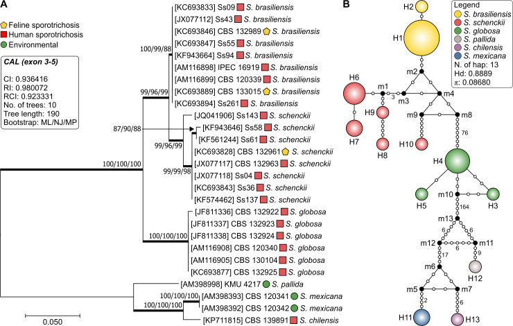
We selected 27 well-characterized Sporothrix isolates and used calmodulin to explore genetic diversity and intraspecific variability in our dataset. The aligned CAL sequences were 623 bp long, including 437 invariable characters, 167 variable parsimony-informative sites (26.8%), and 19 singletons. The clade of pathogenic Sporothrix species was well supported with high bootstrap values (100), including S. brasiliensis (clade I), S. schenckii (clade II), and S. globosa (clade III) (Fig 1A). Comparison of CAL sequences revealed significant polymorphisms among S. schenckii isolates (π = 0.00908), whereas sequences from S. brasiliensis (π = 0.00000) and S. globosa (π = 0.00112) were much less diverse. The haplotype diversity was assessed using the DnaSP software [53], which allowed identifying 13 distinct haplotypes. The overall values of haplotype (Hd = 0.8889) and nucleotide diversities (π = 0.08680) were high for Sporothrix. However, when considering only S. brasiliensis or S. globosa just two (Hd = 0.2222) and three (Hd = 0.6000) haplotypes were found, respectively, suggesting that CAL works as a suitable barcoding marker for diagnosis but may cover cryptic diversity in these species (Fig 1B). The Simpson index of diversity [75] was calculated as 0.2279.
Fig 1.
Phylogenetic tree (A), inferred using the maximum likelihood method and Kimura 2-parameter model of the calmodulin sequences of 27 strains of Sporothrix. Numbers close to the branches represent ML/NJ/MP respectively. Bootstraps higher than 95 based on 1000 replications are represented in bold branches. Haplotype network of Sporothrix (B) was done using the median-joining method. The circumference size is proportional to the frequency of haplotype. The median vectors are displayed by black dots and represent hypothetical unsampled or extinct haplotypes in the population.
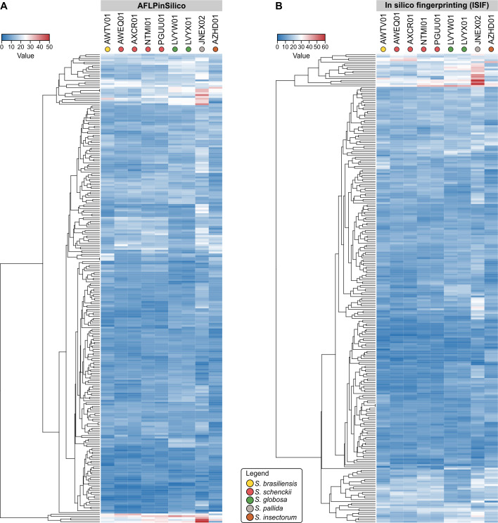
To test the hypothesis of cryptic diversity in S. brasiliensis and S. globosa, we developed AFLP markers to explore genetic variation in these emerging agents. The first step in our strategy involved the in silico characterization of Sporothrix genomes deposited with NCBI, comprising medically relevant members as well as environmental species. AFLPinSilico and ISIF were used to scan restriction sites for EcoRI and MseI. Afterward, a subset of modified genomic fragments was created by adding EcoRI and MseI adaptors, and an enriched population of modified genetic fragments was chosen based on two selective bases for EcoRI+2 and MseI+2 primers. Therefore, 256 possible combinations were investigated for nine genomes, producing a matrix of 2,304 virtual AFLP profiles, which are presented as a heatmap in Fig 2. A remarkable diversity of fragments was generated, which ranged from 0–56 and 0–62 in 256 combinations tested in AFLPinSilico and ISIF, respectively (Fig 3, S1 Table). Sporothrix pallida presented the largest genome core (~37.8 Mb) and it was represented in our in silico scan by the largest number of AFLP markers in 62.5–70.7% combinations (Pearson correlation = 0.861, r2 = 0.7421, P = 0.00283) (Fig 3).
Fig 2.
Heatmap of fragments generated by in silico analyses with 256 combinations of selective primer pairs in two programs–AFLPinSilico (A) and ISIF (B)–for nine genome sequences of Sporothrix retrieved from the GenBank. The highest numbers of fragments are represented by red shading and the lower by blue shading.
Fig 3.
A total of 256 combinations of selective EcoRI+2 and MseI+2 primer pairs were employed to generate 2,304 virtual AFLP profiles AFLPinSilico (A) and ISIF (B). The dots located on the left X-axis represent the number of fragments generated for each combination. The bold bar represents the average of fragments generated for all combinations. The white dots located on the right X-axis represent the genome size, estimated by whole genome sequencing.
Among all Sporothrix species evaluated, S. brasiliensis and S. globosa had little genetic diversity (Fig 1B), so the diversity of fragments generated for these emerging species were fundamental for the selection of putative combinations (Fig 3A). Therefore, we highlighted six combinations (#1–6) to be tested in vitro, which showed the highest number of polymorphic markers (i.e., number and size) with the potential to be used to speciate Sporothrix and explore intraspecific variation (S2 Table).
A total of 685 loci were amplified using the selective primers EcoRI+2 and MseI+2, among them 135, 137, 106, 111, 99 and 96 polymorphic fragments, for combinations #1 to #6, respectively. The averages of fragments varied per isolate for the six combinations between 19.5–32.8 for S. brasiliensis, 19.3–40.6 for S. schenckii, 15.6–31.5 for S. globosa, 14.5–41.5 for S. mexicana, 23–44 for S. pallida and 16–45 for S. chilensis. The details of marker attributes for different AFLP primer combinations are given in Table 3.
Table 3. Summary of polymorphism statistics calculated for different pairs of selective primers (EcoRI+2 and MseI+2) of Sporothrix species.
| #1 EcoRI-GA/MseI-AA | |||||||||
| Species | Scored bands | H | PIC | E | Havp | MI | D | Rp | Error % |
| S. brasiliensis | 62 | 0.4991 | 0.3745 | 29.6667 | 0.0009 | 0.0265 | 0.7715 | 21.3333 | 1.12 |
| S. schenckii | 84 | 0.4995 | 0.3747 | 40.6250 | 0.0007 | 0.0302 | 0.7665 | 38.2500 | 1.23 |
| S. globosa | 43 | 0.3918 | 0.3151 | 31.5000 | 0.0015 | 0.0478 | 0.4641 | 9.6667 | 1.05 |
| S. mexicana | 45 | 0.1435 | 0.1332 | 41.5000 | 0.0016 | 0.0661 | 0.1503 | 7.0000 | 0.00 |
| Overall | 135 | 0.3857 | 0.3113 | 35.2222 | 0.0001 | 0.0037 | 0.9320 | 63.1852 | 1.04 |
| #2 EcoRI-AA/MseI-AA | |||||||||
| Species | Scored bands | H | PIC | E | Havp | MI | D | Rp | Error % |
| S. brasiliensis | 56 | 0.4997 | 0.3749 | 28.6667 | 0.0010 | 0.0284 | 0.7385 | 16.8889 | 0.00 |
| S. schenckii | 63 | 0.4854 | 0.3676 | 26.1250 | 0.0010 | 0.0252 | 0.8285 | 26.7500 | 0.47 |
| S. globosa | 34 | 0.4192 | 0.3313 | 23.8333 | 0.0021 | 0.0490 | 0.5097 | 10.3333 | 0.64 |
| S. mexicana | 37 | 0.0267 | 0.0263 | 36.5000 | 0.0004 | 0.0132 | 0.0270 | 1.0000 | 0.00 |
| Overall | 137 | 0.3287 | 0.2747 | 28.4074 | 0.0001 | 0.0025 | 0.9570 | 52.0741 | 0.28 |
| #3 EcoRI-GA/MseI-TT | |||||||||
| Species | Scored bands | H | PIC | E | Havp | MI | D | Rp | Error % |
| S. brasiliensis | 52 | 0.4974 | 0.3737 | 27.8889 | 0.0011 | 0.0296 | 0.7129 | 19.7778 | 0.00 |
| S. schenckii | 52 | 0.4833 | 0.3665 | 21.2500 | 0.0012 | 0.0247 | 0.8336 | 17.5000 | 0.58 |
| S. globosa | 28 | 0.3501 | 0.2888 | 21.6667 | 0.0021 | 0.0451 | 0.4023 | 4.0000 | 0.00 |
| S. mexicana | 21 | 0.0465 | 0.0454 | 20.5000 | 0.0011 | 0.0227 | 0.0476 | 1.0000 | 0.00 |
| Overall | 106 | 0.3449 | 0.2854 | 23.4815 | 0.0001 | 0.0028 | 0.9510 | 40.8889 | 0.16 |
| #4 EcoRI-AA/MseI-TT | |||||||||
| Species | Scored bands | H | PIC | E | Havp | MI | D | Rp | Error % |
| S. brasiliensis | 44 | 0.4999 | 0.3750 | 21.7778 | 0.0013 | 0.0275 | 0.7557 | 16.4444 | 0.51 |
| S. schenckii | 52 | 0.4967 | 0.3733 | 23.8750 | 0.0012 | 0.0285 | 0.7898 | 19.7500 | 0.52 |
| S. globosa | 38 | 0.4038 | 0.3223 | 27.3333 | 0.0018 | 0.0484 | 0.4835 | 8.6667 | 0.00 |
| S. mexicana | 28 | 0.0000 | 0.0000 | 28.0000 | 0.0000 | 0.0000 | 0.0000 | 0.0000 | 0.00 |
| Overall | 111 | 0.3483 | 0.2876 | 24.9259 | 0.0001 | 0.0029 | 0.9496 | 45.6296 | 0.32 |
| #5 EcoRI-GA/MseI-AG | |||||||||
| Species | Scored bands | H | PIC | E | Havp | MI | D | Rp | Error % |
| S. brasiliensis | 55 | 0.4993 | 0.3746 | 28.5556 | 0.0010 | 0.0288 | 0.7309 | 18.6667 | 0.78 |
| S. schenckii | 52 | 0.4958 | 0.3729 | 28.3750 | 0.0012 | 0.0338 | 0.7028 | 14.2500 | 0.00 |
| S. globosa | 29 | 0.4096 | 0.3257 | 20.6667 | 0.0024 | 0.0486 | 0.4933 | 10.0000 | 0.00 |
| S. mexicana | 28 | 0.0000 | 0.0000 | 28.0000 | 0.0000 | 0.0000 | 0.0000 | 0.0000 | 0.00 |
| Overall | 99 | 0.3908 | 0.3145 | 26.3704 | 0.0001 | 0.0039 | 0.9291 | 40.9630 | 0.29 |
| #6 EcoRI-TA/MseI-AA | |||||||||
| Species | Scored bands | H | PIC | E | Havp | MI | D | Rp | Error % |
| S. brasiliensis | 49 | 0.4787 | 0.3641 | 19.4444 | 0.0011 | 0.0211 | 0.8431 | 21.7778 | 1.14 |
| S. schenckii | 53 | 0.4639 | 0.3563 | 19.3750 | 0.0011 | 0.0212 | 0.8669 | 22.2500 | 0.00 |
| S. globosa | 36 | 0.4928 | 0.3714 | 15.8333 | 0.0023 | 0.0361 | 0.8077 | 18.3333 | 0.00 |
| S. mexicana | 15 | 0.0644 | 0.0624 | 14.5000 | 0.0021 | 0.0311 | 0.0667 | 1.0000 | 0.00 |
| Overall | 97 | 0.3003 | 0.2552 | 17.8519 | 0.0001 | 0.0020 | 0.9662 | 33.6296 | 0.44 |
D = discriminating power; E = effective multiplex ratio; H = expected heterozygosity; Havp = mean heterozygosity; MI = marker index; PIC = polymorphism information content; Rp = resolving power.
The PIC determined for each primer pair was both comparable between species and between markers. Overall, PIC values ranged from 0.2552 to 0.3145, and all markers presented high discrimination power. The highest PIC value was observed for primer combination 5 and the lowest was recorded for primer combination 6, indicating good diversity among the studied Sporothrix. Interestingly, the highest PIC value for S. globosa was obtained in combination 6 (0.3714), demonstrating the potential use of this primer pair to explore diversity in S. globosa. In general, S. brasiliensis and S. schenckii showed slightly higher values than S. globosa (Table 3).
Marker index (MI) as a feature of marker diversity representing the product of the effective multiplex ratio (E) and the arithmetic mean heterozygosity (Havp) was also calculated for all the primer combinations. The MI values ranged from 0.002 to 0.0039. The highest value (0.0039) was obtained with primer pair 5 and the lowest values (0.002) for primer pair 6. A positive correlation was observed between MI and PIC values (Pearson correlation = 0.9950171, r2 = 0.9901, P = 0.00001). The resolving power, Rp, is a feature that indicates the discriminatory potential of the marker to distinguish between large numbers of genotypes. Rp ranged from 33.6296 to 63.1852. The highest value (63.1852) was scored with primer combination 1 and the lowest (33.6296) for primer combination 6. The Rp values were not positively correlated with MI (Pearson correlation = 0.4797601, r2 = 0.2302, P = 0.335). Nevertheless, combination 6 provided the highest values of Rp (18.3333) and MI (0.0361) for S. globosa.
We also determined expected heterozygosity (H), which is defined as the probability that an individual is heterozygous for the locus in the population. It is equivalent to Nei’s unbiased gene diversity (HS), as adapted for dominant markers under the assumptions of Hardy-Weinberg equilibrium and the Lynch-Milligan model [76]. The overall average expected heterozygosity for Sporothrix species ranged between 0.3003–0.3908 (Table 4). The high combined expected heterozygosity for S. brasiliensis (H = 0.4787–0.4999) and S. globosa (H = 0.3501–0.4928) is surprising given previous reports [14, 20, 42, 44, 77–79], which showed little or no genetic variation within these pathogens based on DNA sequencing data. Indeed, the indices reported here are comparable to that of S. schenckii (0.4639–0.4995), a species described as more diverse than S. brasiliensis and S. globosa. Based on these criteria, the high number of loci generated for S. brasiliensis, S. schenckii, and S. globosa is sufficient to reveal the true fine-scale population genetic structure, and we recommend the use of combinations #5 (EcoRI-FAM-GA/MseI-AG), #6 (EcoRI-FAM-TA/MseI-AA) and #3 (EcoRI-FAM-GA/MseI-TT) to explore genetic diversity in Sporothrix species.
Table 4. Comparison among the clustering methods used for phylogenetic analysis and AFLP-derived dendrograms for combinations #1 to #6 generated by congruence index values (Icong).
| Tree comparison | Leaves | MASTa | Icong | P-value | Congruent |
|---|---|---|---|---|---|
| AFLP 1 vs AFLP 2 | 27 | 13 | 1.714 | 2.19e-05 | Yes |
| AFLP 1 vs AFLP 3 | 26 | 11 | 1.479 | 0.0009 | Yes |
| AFLP 1 vs AFLP 4 | 27 | 12 | 1.583 | 0.0001 | Yes |
| AFLP 1 vs AFLP 5 | 26 | 13 | 1.748 | 1.46e-05 | Yes |
| AFLP 1 vs AFLP 6 | 26 | 13 | 1.748 | 1.46e-05 | Yes |
| AFLP 2 vs AFLP 3 | 27 | 13 | 1.714 | 2.19e-05 | Yes |
| AFLP 2 vs AFLP 4 | 27 | 14 | 1.846 | 2.79e-05 | Yes |
| AFLP 2 vs AFLP 5 | 27 | 14 | 1.846 | 2.79e-05 | Yes |
| AFLP 2 vs AFLP 6 | 27 | 13 | 1.714 | 2.19e-05 | Yes |
| AFLP 3 vs AFLP 4 | 27 | 16 | 2.110 | 4.54e-08 | Yes |
| AFLP 3 vs AFLP 5 | 26 | 12 | 1.613 | 0.0001 | Yes |
| AFLP 3 vs AFLP 6 | 26 | 11 | 1.479 | 0.0009 | Yes |
| AFLP 4 vs AFLP 5 | 27 | 15 | 1.978 | 3.56e-07 | Yes |
| AFLP 4 vs AFLP 6 | 27 | 11 | 1.451 | 0.0013 | Yes |
| AFLP 5 vs AFLP 6 | 26 | 14 | 1.882 | 1.82e-06 | Yes |
| AFLP 1 vs Cal | 26 | 11 | 1.479 | 0.0009 | Yes |
| AFLP 2 vs Cal | 27 | 12 | 1.583 | 0.0001 | Yes |
| AFLP 3 vs Cal | 26 | 10 | 1.344 | 0.0075 | Yes |
| AFLP 4 vs Cal | 27 | 23 | 3.034 | 2.46e-14 | Yes |
| AFLP 5 vs Cal | 26 | 12 | 1.613 | 0.0001 | Yes |
| AFLP 6 vs Cal | 26 | 10 | 1.344 | 0.0075 | Yes |
a MAST, Maximum Agreement Subtree.
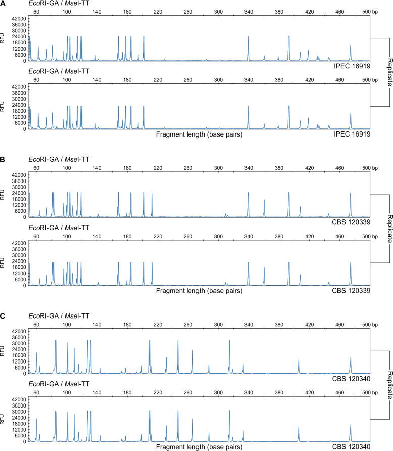
We evaluated the quality of standard fluorescent AFLP genotyping by determining the error rate and reproducibility of our datasets. The suggested and generally acceptable error rate for AFLP data ranges between 2–5% [29, 80]. AFLP electropherograms (50–500 bp) of S. brasiliensis (Fig 4A and 4B) and S. globosa (Fig 4C) show the high reproducibility of the fingerprints obtained using fluorescent capillary electrophoresis, which was crucial to reliably score AFLP markers. A fragment was considered unreliable if it showed any variability within the duplicate tested. For duplicated genotyped Sporothrix isolates, we observed the lowest average error rate across all markers for S. globosa (0.00–1.05%), S. brasiliensis (0.00–1.14%), and S. schenckii (0.00–1.23%). All the fragments scored were reproducible. Error rates never exceeded 1.23%, indicating that our protocol is highly reproducible across Sporothrix species, which are the main agents of human and animal sporotrichosis [80].
Fig 4.
Electropherograms from an ABI3100 depicting the range of fragments between 50 and 500 bp for S. brasiliensis IPEC 16919 (A), S. brasiliensis CBS 120339 (B) and S. globosa CBS 120340 (C), with EcoRI-GA and MseI-TT selective primers labeled with blue (6-FAM). Error rates were never greater than 1.23%, indicating that our protocol is highly reproducible across Sporothrix species.
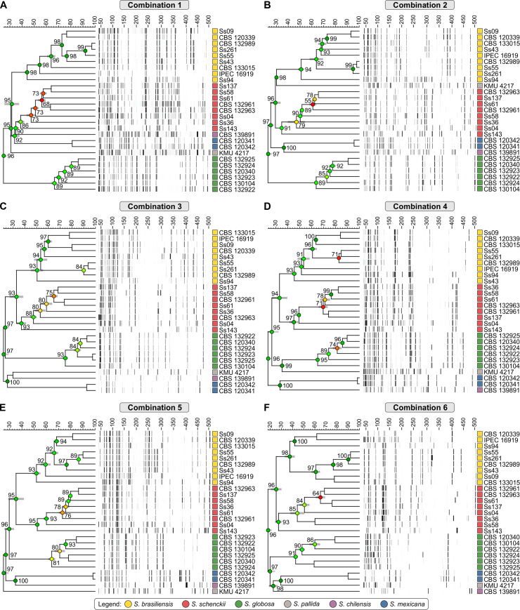
Typical AFLP dendrograms based on Jaccard’s similarity coefficient are depicted in Fig 5. These show six well-supported clades with a similarity level ranging between 40 and 98%, with high cophenetic values (>90) in the majority of branches (S2 Table). This is in accordance with the generally applied calmodulin-based classification of Marimon et al. [47]. The first clade comprises S. brasiliensis isolates recovered from human and animal cases of sporotrichosis. It was possible to reveal cryptic diversity for S. brasiliensis in all datasets, with similarity levels ranging from 37.53% ± 4.06% to 51.96% ± 2.83% (S2 Table). AFLP markers could separate the S. brasiliensis isolates belonging to Rio Grande do Sul, which remained together in all combinations. The polymorphic fragments ranged from 38–50 fragments in the six combinations for all S. brasiliensis isolates, in a total of 44–62 fragments. The second clade comprised S. schenckii, which showed greater diversity, ranging from 27.44% ± 2.55% to 52.56% ± 1.43% (S2 Table), compared to other isolates of clinical relevance, corroborating the classic findings of DNA sequencing. Polymorphic fragments ranged from 37–77 per combination in a total of 52–84 fragments generated. The third clade comprised S. globosa isolates, which showed a similar pattern for the six isolates tested with cryptic diversity (42.79% ± 1.61% to 74.38% ± 1.82%), but lower than S. brasiliensis and S. schenckii isolates. The total of fragments generated for S. globosa varied from 28–43, with a range of 10 to 31 polymorphic fragments per combination. The remaining isolates represent members of the S. pallida complex, including S. chilensis, S. mexicana, and S. pallida s. str. In clade 4, AFLP markers varied from 14–44 fragments, and S. pallida isolate FMR 8803 grouped apart from the other members of the S. pallida complex (i.e. S. chilensis and S. mexicana) in combinations #1, #2 and #4. The two isolates of S. mexicana presented high genetic similarity (above 90% in 5 out of 6 combinations), generating 15–45 fragments. Polymorphic bands were absent in combinations #3 and #4, whereas in combination #1 we detected seven polymorphic bands. Likewise, S. chilensis produced a range of 15–45 fragments, following the same clustering pattern as combinations #2 to #6.
Fig 5.
Dendrograms of combinations 1 to 6 (A to F, respectively). The dendrograms show the clustering profile of the 27 samples of Sporothrix. The dendrograms were constructed by the Jaccard similarity coefficient and UPGMA clustering in the software BioNumerics v.7.6.
To assess the existence of topological congruence between any two dendrograms or between dendrograms and the calmodulin phylogenetic tree, we used the congruence index (Icong) [65]. Multiple comparisons revealed a similar and consistent clustering pattern, as evidenced by Icong values and their significant associated P-values (Table 4). Therefore, the AFLP dendrogram and calmodulin trees are more congruent than expected by chance, supporting the use of new AFLP markers to speciate Sporothrix with the same confidence of DNA-sequencing methods.
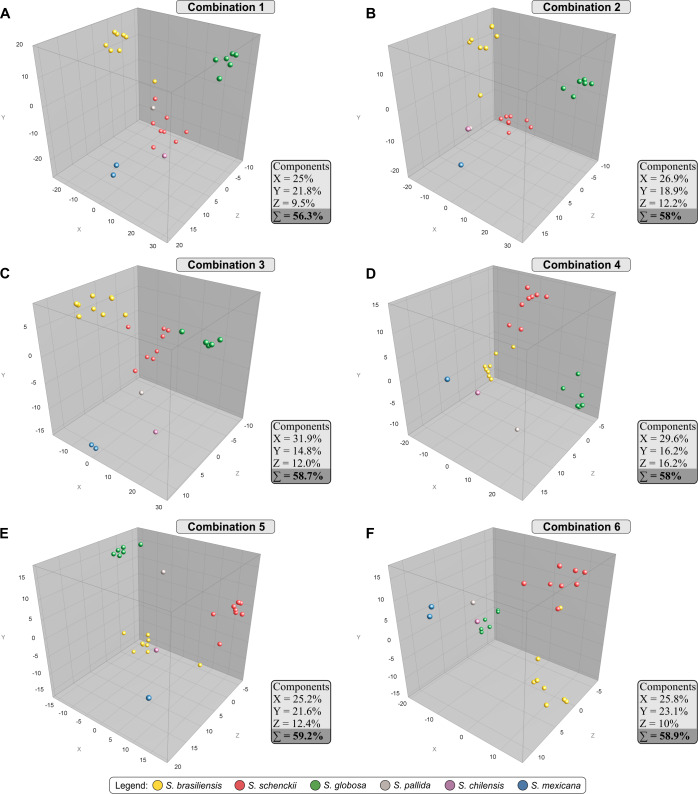
The AFLP markers were used to generate pairwise genetic distance matrices based on Jaccard’s similarity coefficient, which were subjected to a principal component analysis (PCA) with BioNumerics v7.6 to allow 3D graphic visualization of the relationships among each Sporothrix sample with those representing the same taxon. Fig 6 shows the PCA plots for combinations #1–6, and the distribution of 27 Sporothrix isolates among the three co-ordinates agreed with the UPGMA tree. Combinations #3, #5 and #6 revealed the highest cumulative percentage explained, with 58.7–59.2% of the variation explained by the first three components together (coordinates X, Y, and Z). The dimensioning analysis showed clearly both the high degree of intraspecific clustering and the relatively large genetic separation between any individual taxon (interspecific variation). This tight clustering supports cryptic diversity in S. brasiliensis and S. globosa. In all combinations evaluated, S. schenckii continued being a more diverse species than its siblings S. brasiliensis and S. globosa, in line with the higher level of intraspecific variability demonstrated in calmodulin sequencing.
Fig 6.
PCA of combinations 1 to 6 (A to F, respectively). PCAs were constructed in the software BioNumerics v.7.6. The PCAs were used to demonstrate the correlations among the 27 samples of Sporothrix using the six primer pair combinations. The isolates are represented by colored circles.
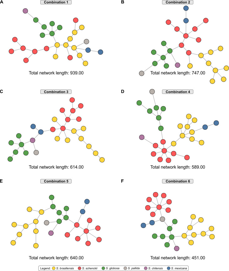
The AFLP-derived MSTs in Fig 7 essentially confirm the diversity structure of S. brasiliensis, S. schenckii, and S. globosa with the majority of isolates having a unique genotype, in contrast with those found for calmodulin sequencing (Fig 1B). In general, AFLP-derived MSTs were similar in their identification of totally different types of genetic variants. This suggests that despite the enhanced inherent variability of AFLP markers, clusters of isolates remain traceable.
Fig 7.
AFLP-derived minimum spanning trees (MSTs) of Sporothrix isolates using combinations 1 to 6 (A to F, respectively). CAL tree and AFLP MSTs were divergent in their identification of different types of genetic variants, since the finer resolution was obtained using AFLP markers.
Discussion
In this study, we develop a new AFLP technique aimed at studying the genetic epidemiology of medically relevant Sporothrix species. The most promising result is the discovery of cryptic diversity in species previously thought to be prevalent clonal types, such as S. brasiliensis, which is responsible for the cat-transmitted sporotrichosis in Brazil, and S. globosa, responsible for large sapronosis taking place in Asia. Our technique, therefore, will enable finer-scale epidemiological patterns to be described than was previously possible.
The first clues that S. brasiliensis might have cryptic genetic diversity emerged from host-pathogen interaction data, where genetically identical isolates (based on CAL sequences) had distinct virulence profiles when inoculated in BALB/c mice for variables such as animal weight loss, death, and capacity to disseminate to organs [81]. For S. schenckii the higher levels of intraspecific genetic variability are associated with more variable virulence profiles in BALB/c [82] suggesting that genotype is an important factor in determining clinically-relevant phenotypes. Considering all molecular markers previously used to explore genetic diversity in Sporothrix, including chitin synthase [83], elongation factor 1α [2], β-tubulin [2, 83], ITS1/2+5.8s [20, 78, 79] and calmodulin [2, 47, 83], the last perform best in estimating diversity by showing up to 31.69% variable parsimony-informative sites and comprising for the largest number of genotypes. However, our study shows that calmodulin (and other markers) underestimated the genotypic diversity of S. brasiliensis. Therefore, we clearly need new, higher-power, markers to study the recent expansion (2015–2019) of epizooties of feline sporotrichosis due to S. brasiliensis towards Northeastern Brazil [1].
Classic studies in the literature describe the use of AFLP markers as one of the most informative and cost-effective DNA fingerprinting methods for genetic characterization of pathogenic Sporothrix. The technique usually shows a strong geographical population structure and is useful to speciate Sporothrix isolates [20, 39, 40]. Through our markers, we were able to identify Sporothrix down to species level, with results similar to the gold standard DNA sequencing of the calmodulin encoding gene [47], CAL-RFLP [41], or species-specific PCR [46]. Moreover, we were able to differentiate Sporothrix down to strain level, surpassing the resolution provided by the gold-standard method (calmodulin) used to investigate diversity in Sporothrix. Our single protocol was easily transferred between distantly related taxa, including members of the clinical and environmental clades.
The main advantages of using AFLP analysis include the use of a standard protocol in combination with different restriction endonucleases and the choice of adding one or more selective nucleotides in the selective EcoRI and MseI primers to achieve optimal fingerprints without prior knowledge of the organism’s genome sequence. This is useful for non-model organisms such as Sporothrix. Disadvantages include the dominance of alleles, and the possible non-homology of comigrating fragments belonging to different loci, leading to suboptimal reproducibility, particularly across different platforms. However, we took advantage of the growing number of full genome sequences available for Sporothrix [58–63] to generate 2,304 virtual fingerprints of Sporothrix DNA, reducing time and costs related to extensive trials, minimizing possible reproducibility errors.
Therefore, the first step in our standardization included an in silico approach using the programs AFLPinSilico [55] and ISIF [56] to optimize the best combination of selective primers (EcoRI+2 and MseI+2) to produce the largest number of polymorphic AFLP markers and address questions related to speciation, genetic diversity, and population structure. The results revealed that, while the two bioinformatic programs had minor divergences between them, in vitro results were consistent with in silico prediction. The main criterion used was to choose combinations that would reveal cryptic diversity in S. brasiliensis and S. globosa, two species that were previously characterized as having low genetic diversity or clonal population structure using DNA-sequencing methods [14, 20, 42, 44, 77–79].
The size and complexity of the Sporothrix genome can have a tremendous influence on AFLP patterns, due to the amount of DNA that is necessary for the initial restriction/ligation step, as large genomes usually require larger amounts of DNA [84]. Here, 200 ng was enough to produce clear, intense AFLP fragments [85]. Another observation from our Sporothrix genome scan follow-up is that the number of selective bases greatly influenced the number and diversity of fragments. Generating too many fragments is not ideal, since it makes it difficult to unambiguously score AFLP electrophoresis profiles (e.g., owing to comigrating non-homologous fragments). If, however, the primer combination generates a low number of AFLP markers, it will reduce the probability of polymorphism detection. We found that two selective bases (EcoRI+2 and MseI+2) produced the ideal number of AFLP markers (i.e., 96–137 polymorphic fragments) to explore diversity in Sporothrix. This holds true also when dealing with microorganisms having a larger genome size [29]. We found a higher number of AFLP bands in silico in at least 62.5–70.7% of combinations for S. pallida (genome size of ~37.8 Mb) compared to S. brasiliensis, S. schenckii or S. globosa (32.5–33.4 Mb) [85] suggesting that, as expected, AFLP patterns scale in complexity with genome size.
Understanding the spread of Sporothrix species by exploring inter- and intraspecific genetic diversities is fundamental to tackle the increasing number of cases in recent years [86]. Many molecular techniques have been employed for Sporothrix to address these questions [20, 39–41, 46, 85, 87, 88], and have been successful in providing quality data. However, limitations include the low number of polymorphic and reproducible characters evaluated. AFLP has been considered to be highly reproducible and discriminatory, mainly for epidemiological studies [89], and can be employed for any organisms without the need of sequence information [90]. However, only Zhang et al. employed the technique on S. brasiliensis, but they were not able to describe high levels of diversity [20]. Our in vitro AFLP succeeded, demonstrating it is a relevant method for epidemiological studies of Sporothrix, which is in line with previous studies that used the technique [20, 39, 40]. Comparing haplotype and AFLP dendrograms or AFLP-derived MSTs generated here, the DNA fingerprint methods demonstrated meaningful variations in Sporothrix spp.
DNA-sequencing data demonstrated there are at least two different S. brasiliensis populations circulating in Brazil, one belonging to the Rio Grande do Sul cluster and the other related to the long-lasting epidemic taking place in Rio de Janeiro, which is spreading to neighboring states such as São Paulo, Minas Gerais and Espírito Santo [44, 86]. Zhang et al. [20] demonstrated that S. brasiliensis clustered into three different clades in an AFLP study, and one of the clades contained isolates from Southern Brazil, agreeing with the phylogeography proposed by Rodrigues et al. [44]. The same pattern was evidenced in our study, and strains from Rio Grande do Sul (e.g., Ss55, Ss261, and CBS 132989) presented an identical clustering pattern, showing high levels of similarity among the isolates, which differed from those originated from other Brazilian regions. The three clusters of S. brasiliensis found by Zhang et al. [20] were supported by high bootstrap values in phylogenetic analysis (i.e., partial CAL, TEF1, and TEF3), but the AFLP fingerprints showed little variation [20]. Our fingerprints revealed meaningful variations in S. brasiliensis, with a large number of polymorphic bands, showing that the strategy used here based on extensive in silico screening of selective bases was fundamental to guide the choice of the best primer combination.
Among the pathogenic species, S. schenckii is considered to have the highest genetic diversity [20, 47, 83], with evidence of genetic recombination [42]. Diversity in S. schenckii was also found by other researchers, who separated this species into five groups using AFLP [20]. Our AFLP setup was able to detect diversity within S. schenckii.
AFLP markers revealed that S. globosa also presented subtle diversity, but it is important to point out that these isolates were less diverse than S. schenckii and S. brasiliensis, confirming previous findings of Zhang et al., who considered that the DNA fingerprints were identical in S. globosa. Sporothrix globosa is widely distributed in temperate and warm regions [47, 83, 91], and rapid dispersal by unknown vectors could explain the similar genotypes for isolates collected from distant geographical regions [20, 92]. In addition, Zhao et al. [40] also detected cryptic diversity in S. globosa evaluating a higher number of strains, similar to the variation found using our new AFLP markers.
Comparison of the DNA-sequence tree with AFLP dendrogram using the congruence index (Icong) revealed a strong topological congruence between any two AFLP dendrograms or between AFLP dendrograms and a calmodulin tree, supporting the use of all combinations to distinguish Sporothrix species. The congruence observed is in agreement with previous studies, which found that S. brasiliensis, S. schenckii and S. globosa are related in a pathogenic/clinical clade and S. mexicana, S. pallida, and S. chilensis are nested in an environmental clade, supported by high bootstrap values or cophenetic values [2, 42–44, 79]. On one hand, several studies have demonstrated that moderate numbers of AFLP fragments are necessary to recover the correct topology of a DNA-sequence tree with high bootstrap support values (i.e. >70%) [93–95]. On the other hand, a higher number of taxa (i.e., covering all 53 Sporothrix species described so far) may increase the number of possible trees and reduce internode distances for a given tree length, making it less likely to recover the correct tree [95, 96]. However, this size effect is more likely to occur in inferences considering ancient radiations [95, 96], which does not seem to be the case of medically relevant Sporothrix species, especially S. brasiliensis, which likely originated from a recent radiation event (about 3.8–4.9 MYA) based on its geographical distribution and phylogenetic inferences [42, 58, 97].
Using our new AFLP markers, we can now better track routes of disease transmission during epizooties and zoonosis in Brazil, allowing the possibility of linking specific genotypes to antifungal susceptibility profiles as well as addressing links between clinical outcomes in feline and human sporotrichosis. All six primers pairs performed well, but the most precise level of inter-species discrimination and the highest level of intra-species discrimination of the Sporothrix isolates were observed in the AFLP EcoRI-FAM-GA/MseI-TT, EcoRI-FAM-GA/MseI-AG and EcoRI-FAM-TA/MseI-AA sets. These combinations stand out among all combinations for having the best diversity indices and the lowest error rates, and thus may be valuable in human and veterinary diagnostics as well as in epidemiology. Moreover, our DNA fingerprinting assay can be further transferred between laboratories to give insights into the ecology and evolution of pathogenic Sporothrix species and to inform management and mitigation strategies to tackle the advance of sporotrichosis. Although sporotrichosis has a global distribution, pathogenic species are not evenly distributed and individual lineages that vary in pathogenicity still occur in geographically limited ranges, such as S. brasiliensis. Thus, as Sporothrix genotypes continue to expand their range, we need to consider using genome-wide markers to identify, distinguish, and track these emerging pathogens spreading through the mammal hosts.
Supporting information
(PDF)
(PDF)
Data Availability
All relevant data are within the manuscript and its Supporting Information files.
Funding Statement
AMR was supported by grants from the São Paulo Research Foundation (FAPESP 2017/27265-5) http://www.fapesp.br/, the National Council for Scientific and Technological Development (CNPq 433276/2018-5) http://www.cnpq.br/, and Coordination of Improvement of Higher Education Personnel (CAPES 88887.159096/2017-00) https://www.capes.gov.br/. ZPC was supported by grants from FAPESP (2018/21460-3). MCF is a CIFAR fellow in the ‘Fungal Kingdoms’ programme (https://www.cifar.ca/research/program/fungal-kingdom) and the UK Medical Research Council MR/R015600/1 (https://mrc.ukri.org/). The funders had no role in the study design, data collection and analysis, decision to publish, or preparation of the manuscript.
References
- 1.Rodrigues AM, Della Terra PP, Gremiao ID, Pereira SA, Orofino-Costa R, de Camargo ZP. The threat of emerging and re-emerging pathogenic Sporothrix species. Mycopathologia. 2020. 10.1007/s11046-020-00425-0 . [DOI] [PubMed] [Google Scholar]
- 2.Rodrigues AM, Cruz Choappa R, Fernandes GF, De Hoog GS, Camargo ZP. Sporothrix chilensis sp. nov. (Ascomycota: Ophiostomatales), a soil-borne agent of human sporotrichosis with mild-pathogenic potential to mammals. Fungal Biol. 2016;120(2):246–64. 10.1016/j.funbio.2015.05.006 . [DOI] [PubMed] [Google Scholar]
- 3.Rodrigues AM, Orofino-Costa R, de Camargo ZP. Sporothrix spp In: Cordeiro Rde A, editor. Pocket Guide to Mycological Diagnosis. 1st ed Boca Raton: CRC Press; 2019. p. 99–113. [Google Scholar]
- 4.de Beer ZW, Duong TA, Wingfield MJ. The divorce of Sporothrix and Ophiostoma: solution to a problematic relationship. Stud Mycol. 2016;83:165–91. 10.1016/j.simyco.2016.07.001 [DOI] [PMC free article] [PubMed] [Google Scholar]
- 5.Orofino-Costa RC, Macedo PM, Rodrigues AM, Bernardes-Engemann AR. Sporotrichosis: an update on epidemiology, etiopathogenesis, laboratory and clinical therapeutics. An Bras Dermatol. 2017;92(5):606–20. 10.1590/abd1806-4841.2017279 [DOI] [PMC free article] [PubMed] [Google Scholar]
- 6.Rippon JW. Medical Mycology–The pathogenic fungi and the pathogenic actinomycetes 3rd ed Philadelphia, PA: W. B. Saunders Company; 1988. [Google Scholar]
- 7.Rodrigues AM, Fernandes GF, de Camargo ZP. Sporotrichosis In: Bayry J, editor. Emerging and re-emerging infectious diseases of livestock: Cham: Springer International Publishing; 2017. p. 391–421. [Google Scholar]
- 8.de Lima Barros MB, Schubach TM, Galhardo MC, de Oliviera Schubach A, Monteiro PC, Reis RS, et al. Sporotrichosis: an emergent zoonosis in Rio de Janeiro. Mem Inst Oswaldo Cruz. 2001;96(6):777–9. 10.1590/s0074-02762001000600006 . [DOI] [PubMed] [Google Scholar]
- 9.Barros MBdL Schubach AdO, do Valle ACF Galhardo MCG, Conceição-Silva F Schubach TMP, et al. Cat-transmitted sporotrichosis epidemic in Rio de Janeiro, Brazil: Description of a series of cases. Clin Infect Dis. 2004;38(4):529–35. 10.1086/381200 . [DOI] [PubMed] [Google Scholar]
- 10.Schubach TM, Schubach A, Okamoto T, Barros MB, Figueiredo FB, Cuzzi T, et al. Evaluation of an epidemic of sporotrichosis in cats: 347 cases (1998–2001). J Am Vet Med Assoc. 2004;224(10):1623–9. 10.2460/javma.2004.224.1623 . [DOI] [PubMed] [Google Scholar]
- 11.Seyedmousavi S, Bosco SdMG, de Hoog S, Ebel F, Elad D, Gomes RR, et al. Fungal infections in animals: a patchwork of different situations. Med Mycol. 2018;56(Suppl_1):165–87. 10.1093/mmy/myx104 [DOI] [PMC free article] [PubMed] [Google Scholar]
- 12.Schubach TM, Schubach A, Okamoto T, Pellon IV, Fialho-Monteiro PC, Reis RS, et al. Haematogenous spread of Sporothrix schenckii in cats with naturally acquired sporotrichosis. J Small Anim Pract. 2003;44(9):395–8. 10.1111/j.1748-5827.2003.tb00174.x . [DOI] [PubMed] [Google Scholar]
- 13.Rodrigues AM, de Hoog GS, de Camargo ZP. Feline Sporotrichosis In: Seyedmousavi S, de Hoog GS, Guillot J, Verweij PE, editors. Emerging and Epizootic Fungal Infections in Animals. 1 Cham: Springer International Publishing; 2018. p. 199–231. [Google Scholar]
- 14.Rodrigues AM, de Hoog GS, de Camargo ZP. Sporothrix species causing outbreaks in animals and humans driven by animal-animal transmission. PLoS Pathog. 2016;12(7):e1005638 10.1371/journal.ppat.1005638 . [DOI] [PMC free article] [PubMed] [Google Scholar]
- 15.Engle J, Desir J, Bernstein JM. A rose by any other name. Skinmed. 2007;6(3):139–41. 10.1111/j.1540-9740.2007.06683.x . [DOI] [PubMed] [Google Scholar]
- 16.Song Y, Li SS, Zhong SX, Liu YY, Yao L, Huo SS. Report of 457 sporotrichosis cases from Jilin province, northeast China, a serious endemic region. J Eur Acad Dermatol Venereol. 2013;27(3):313–8. 10.1111/j.1468-3083.2011.04389.x [DOI] [PubMed] [Google Scholar]
- 17.Ramírez-Soto M, Aguilar-Ancori E, Tirado-Sánchez A, Bonifaz A. Ecological determinants of sporotrichosis etiological agents. J Fungi. 2018;4(3):95 10.3390/jof4030095 [DOI] [PMC free article] [PubMed] [Google Scholar]
- 18.Bongomin F, Gago S, Oladele R, Denning D. Global and multi-national prevalence of fungal diseases—Estimate precision. J Fungi. 2017;3(4):57 10.3390/jof3040057 [DOI] [PMC free article] [PubMed] [Google Scholar]
- 19.Queiroz-Telles F, Fahal AH, Falci DR, Caceres DH, Chiller T, Pasqualotto AC. Neglected endemic mycoses. Lancet Infect Dis. 2017;17(11):e367–e77. 10.1016/S1473-3099(17)30306-7 [DOI] [PubMed] [Google Scholar]
- 20.Zhang Y, Hagen F, Stielow B, Rodrigues AM, Samerpitak K, Zhou X, et al. Phylogeography and evolutionary patterns in Sporothrix spanning more than 14,000 human and animal case reports. Persoonia. 2015;35:1–20. 10.3767/003158515X687416 [DOI] [PMC free article] [PubMed] [Google Scholar]
- 21.Govender NP, Maphanga TG, Zulu TG, Patel J, Walaza S, Jacobs C, et al. An outbreak of lymphocutaneous sporotrichosis among mine-workers in South Africa. PLoS Negl Trop Dis. 2015;9(9):e0004096 10.1371/journal.pntd.0004096 . [DOI] [PMC free article] [PubMed] [Google Scholar]
- 22.Gremiao ID, Miranda LH, Reis EG, Rodrigues AM, Pereira SA. Zoonotic epidemic of sporotrichosis: Cat to human transmission. PLoS Pathog. 2017;13(1):e1006077 10.1371/journal.ppat.1006077 . [DOI] [PMC free article] [PubMed] [Google Scholar]
- 23.Almeida-Paes R, de Oliveira MM, Freitas DF, do Valle AC, Zancope-Oliveira RM, Gutierrez-Galhardo MC. Sporotrichosis in Rio de Janeiro, Brazil: Sporothrix brasiliensis is associated with atypical clinical presentations. PLoS Negl Trop Dis. 2014;8(9):e3094 10.1371/journal.pntd.0003094 [DOI] [PMC free article] [PubMed] [Google Scholar]
- 24.de Macedo PM, Sztajnbok DC, Camargo ZP, Rodrigues AM, Lopes-Bezerra LM, Bernardes-Engemann AR, et al. Dacryocystitis due to Sporothrix brasiliensis: a case report of a successful clinical and serological outcome with low-dose potassium iodide treatment and oculoplastic surgery. Br J Dermatol. 2015;172(4):1116–9. 10.1111/bjd.13378 . [DOI] [PubMed] [Google Scholar]
- 25.Almeida FP. Micologia Médica: Estudo das micoses humanas e de seus cogumelos. São Paulo: Companhia Melhoramentos de São Paulo; 1939. 710 p. [Google Scholar]
- 26.Almeida F, Sampaio SAP, Lacaz CS, Fernandes JC. [Statistical data on sporotrichosis. Analysis of 344 cases]. An Bras Dermatol. 1955;30(1):9–12. [PubMed] [Google Scholar]
- 27.Macêdo-Sales PA, Souto SRLS, Destefani CA, Lucena RP, Machado RLD, Pinto MR, et al. Domestic feline contribution in the transmission of Sporothrix in Rio de Janeiro State, Brazil: a comparison between infected and non-infected populations. BMC Vet Res. 2018;14(1):19 10.1186/s12917-018-1340-4 [DOI] [PMC free article] [PubMed] [Google Scholar]
- 28.Vuylsteke M, Peleman JD, van Eijk MJT. AFLP technology for DNA fingerprinting. Nat Protoc. 2007;2(6):1387–98. 10.1038/nprot.2007.175 [DOI] [PubMed] [Google Scholar]
- 29.Bonin A, Pompanon F, Taberlet P. Use of amplified fragment length polymorphism (AFLP) markers in surveys of vertebrate diversity. Methods Enzymol. 2005;395:145–61. 10.1016/S0076-6879(05)95010-6 . [DOI] [PubMed] [Google Scholar]
- 30.Boekhout T, Theelen B, Diaz M, Fell JW, Hop WC, Abeln EC, et al. Hybrid genotypes in the pathogenic yeast Cryptococcus neoformans. Microbiology. 2001;147(Pt 4):891–907. Epub 2001/04/03. 10.1099/00221287-147-4-891 . [DOI] [PubMed] [Google Scholar]
- 31.Borst A, Theelen B, Reinders E, Boekhout T, Fluit AC, Savelkoul PH. Use of amplified fragment length polymorphism analysis to identify medically important Candida spp., including C. dubliniensis. J Clin Microbiol. 2003;41(4):1357–62. Epub 2003/04/19. 10.1128/jcm.41.4.1357-1362.2003 [DOI] [PMC free article] [PubMed] [Google Scholar]
- 32.Warris A, Klaassen CH, Meis JF, De Ruiter MT, De Valk HA, Abrahamsen TG, et al. Molecular epidemiology of Aspergillus fumigatus isolates recovered from water, air, and patients shows two clusters of genetically distinct strains. J Clin Microbiol. 2003;41(9):4101–6. Epub 2003/09/06. 10.1128/jcm.41.9.4101-4106.2003 [DOI] [PMC free article] [PubMed] [Google Scholar]
- 33.Bogale M, Wingfield BD, Wingfield MJ, Steenkamp ET. Characterization of Fusarium oxysporum isolates from Ethiopia using AFLP, SSR and DNA sequence analyses. Fungal Divers. 2006;23(6):51–66. [Google Scholar]
- 34.Duran A, Gryzenhout M, Drenth A, Slippers B, Ahumada R, Wingfield BD, et al. AFLP analysis reveals a clonal population of Phytophthora pinifolia in Chile. Fungal Biol. 2010;114(9):746–52. Epub 2010/10/15. 10.1016/j.funbio.2010.06.008 . [DOI] [PubMed] [Google Scholar]
- 35.Gril T, Celar F, Javornik B, Jakse J. Fluorescent AFLP fingerprinting of Monilinia fructicola. J Plant Dis Prot. 2010;117(4):168–72. 10.1007/bf03356355 [DOI] [Google Scholar]
- 36.Najafzadeh MJ, Sun J, Vicente V, Xi L, van den Ende AH, de Hoog GS. Fonsecaea nubica sp. nov, a new agent of human chromoblastomycosis revealed using molecular data. Med Mycol. 2010;48(6):800–6. 10.3109/13693780903503081 . [DOI] [PubMed] [Google Scholar]
- 37.Herkert PF, Hagen F, de Oliveira Salvador GL, Gomes RR, Ferreira MS, Vicente VA, et al. Molecular characterisation and antifungal susceptibility of clinical Cryptococcus deuterogattii (AFLP6/VGII) isolates from Southern Brazil. Eur J Clin Microbiol Infect Dis. 2016;35(11):1803–10. 10.1007/s10096-016-2731-8 [DOI] [PubMed] [Google Scholar]
- 38.Rodrigues AM, Beale MA, Hagen F, Fisher MC, Della Terra PP, de Hoog S, et al. The global epidemiology of emerging Histoplasma species in recent years. Stud Mycol. 2020. 10.1016/j.simyco.2020.02.001 [DOI] [PMC free article] [PubMed] [Google Scholar]
- 39.Neyra E, Fonteyne P-A, Swinne D, Fauche F, Bustamante B, Nolard N. Epidemiology of human sporotrichosis investigated by Amplified Fragment Length Polymorphism. J Clin Microbiol. 2005;43(3):1348–52. 10.1128/JCM.43.3.1348-1352.2005 [DOI] [PMC free article] [PubMed] [Google Scholar]
- 40.Zhao L, Cui Y, Zhen Y, Yao L, Shi Y, Song Y, et al. Genetic variation of Sporothrix globosa isolates from diverse geographic and clinical origins in China. Emerg Microbes Infect. 2017;6:e88 10.1038/emi.2017.75 [DOI] [PMC free article] [PubMed] [Google Scholar]
- 41.Rodrigues AM, de Hoog GS, Camargo ZP. Genotyping species of the Sporothrix schenckii complex by PCR-RFLP of calmodulin. Diagn Microbiol Infect Dis. 2014;78(4):383–7. 10.1016/j.diagmicrobio.2014.01.004 . [DOI] [PubMed] [Google Scholar]
- 42.Rodrigues AM, de Hoog GS, Zhang Y, Camargo ZP. Emerging sporotrichosis is driven by clonal and recombinant Sporothrix species. Emerg Microbes Infect. 2014;3(5):e32 10.1038/emi.2014.33 [DOI] [PMC free article] [PubMed] [Google Scholar]
- 43.Rodrigues AM, de Hoog S, de Camargo ZP. Emergence of pathogenicity in the Sporothrix schenckii complex. Med Mycol. 2013;51(4):405–12. 10.3109/13693786.2012.719648 . [DOI] [PubMed] [Google Scholar]
- 44.Rodrigues AM, de Melo Teixeira M, de Hoog GS, Schubach TMP, Pereira SA, Fernandes GF, et al. Phylogenetic analysis reveals a high prevalence of Sporothrix brasiliensis in feline sporotrichosis outbreaks. PLoS Negl Trop Dis. 2013;7(6):e2281 10.1371/journal.pntd.0002281 [DOI] [PMC free article] [PubMed] [Google Scholar]
- 45.Brilhante RS, Silva NF, Lima RA, Caetano EP, Alencar LP, Castelo-Branco Dde S, et al. Easy storage strategies for Sporothrix spp. strains. Biopreserv Biobank. 2015;13(2):131–4. 10.1089/bio.2014.0071 . [DOI] [PubMed] [Google Scholar]
- 46.Rodrigues AM, de Hoog GS, de Camargo ZP. Molecular diagnosis of pathogenic Sporothrix species. PLoS Negl Trop Dis. 2015;9(12):e0004190 10.1371/journal.pntd.0004190 [DOI] [PMC free article] [PubMed] [Google Scholar]
- 47.Marimon R, Cano J, Gené J, Sutton DA, Kawasaki M, Guarro J. Sporothrix brasiliensis, S. globosa, and S. mexicana, three new Sporothrix species of clinical interest. J Clin Microbiol. 2007;45(10):3198–206. 10.1128/JCM.00808-07 [DOI] [PMC free article] [PubMed] [Google Scholar]
- 48.Marimon R, Gené J, Cano J, Guarro J. Sporothrix luriei: a rare fungus from clinical origin. Med Mycol. 2008;46(6):621–5. 10.1080/13693780801992837 . [DOI] [PubMed] [Google Scholar]
- 49.Kumar S, Stecher G, Tamura K. MEGA7: Molecular evolutionary genetics analysis version 7.0 for bigger datasets. Mol Biol Evol. 2016. 10.1093/molbev/msw054 [DOI] [PMC free article] [PubMed] [Google Scholar]
- 50.Kimura M. A simple method for estimating evolutionary rates of base substitutions through comparative studies of nucleotide sequences. J Mol Evol. 1980;16(2):111–20. 10.1007/BF01731581 . [DOI] [PubMed] [Google Scholar]
- 51.Felsenstein J. Evolution confidence limits on phylogenies: An approach using the bootstrap. Evolution. 1985;39(4):783–91. 10.1111/j.1558-5646.1985.tb00420.x [DOI] [PubMed] [Google Scholar]
- 52.Nei M. Molecular evolutionary genetics. New York: Columbia University Press; 1987. 512 p. [Google Scholar]
- 53.Rozas J, Ferrer-Mata A, Sánchez-DelBarrio JC, Guirao-Rico S, Librado P, Ramos-Onsins SE, et al. DnaSP 6: DNA Sequence Polymorphism Analysis of Large Data Sets. Mol Biol Evol. 2017:msx248–msx. 10.1093/molbev/msx248 [DOI] [PubMed] [Google Scholar]
- 54.Bandelt HJ, Forster P, Röhl A. Median-joining networks for inferring intraspecific phylogenies. Mol Biol Evol. 1999;16(1):37–48. 10.1093/oxfordjournals.molbev.a026036 [DOI] [PubMed] [Google Scholar]
- 55.Rombauts S, Van De Peer Y, Rouze P. AFLPinSilico, simulating AFLP fingerprints. Bioinformatics. 2003;19(6):776–7. 10.1093/bioinformatics/btg090 . [DOI] [PubMed] [Google Scholar]
- 56.Paris M, Bonnes B, Ficetola GF, Poncet BN, Després L. Amplified fragment length homoplasy: in silico analysis for model and non-model species. BMC Genomics. 2010;11(1):287 10.1186/1471-2164-11-287 [DOI] [PMC free article] [PubMed] [Google Scholar]
- 57.Babicki S, Arndt D, Marcu A, Liang Y, Grant JR, Maciejewski A, et al. Heatmapper: web-enabled heat mapping for all. Nucleic Acids Res. 2016;44(W1):W147–53. 10.1093/nar/gkw419 [DOI] [PMC free article] [PubMed] [Google Scholar]
- 58.Teixeira MM, de Almeida LG, Kubitschek-Barreira P, Alves FL, Kioshima ES, Abadio AK, et al. Comparative genomics of the major fungal agents of human and animal Sporotrichosis: Sporothrix schenckii and Sporothrix brasiliensis. BMC Genomics. 2014;15:943 Epub 2014/10/30. 10.1186/1471-2164-15-943 . [DOI] [PMC free article] [PubMed] [Google Scholar]
- 59.Cuomo CA, Rodriguez-Del Valle N, Perez-Sanchez L, Abouelleil A, Goldberg J, Young S, et al. Genome sequence of the pathogenic fungus Sporothrix schenckii (ATCC 58251). Genome Announc. 2014;2(3). 10.1128/genomeA.00446-14 [DOI] [PMC free article] [PubMed] [Google Scholar]
- 60.Gomez OM, Alvarez LC, Muñoz JF, Misas E, Gallo JE, Jimenez MDP, et al. Draft genome sequences of two Sporothrix schenckii clinical isolates associated with human sporotrichosis in Colombia. Genome Announc. 2018;6(24):e00495–18. 10.1128/genomeA.00495-18 . [DOI] [PMC free article] [PubMed] [Google Scholar]
- 61.Huang L, Gao W, Giosa D, Criseo G, Zhang J, He T, et al. Whole-genome sequencing and in silico analysis of two strains of Sporothrix globosa. Genome Biol Evol. 2016;8(11):3292–6. 10.1093/gbe/evw230 . [DOI] [PMC free article] [PubMed] [Google Scholar]
- 62.D'Alessandro E, Giosa D, Huang L, Zhang J, Gao W, Brankovics B, et al. Draft genome sequence of the dimorphic fungus Sporothrix pallida, a nonpathogenic species belonging to Sporothrix, a genus containing agents of human and feline sporotrichosis. Genome Announc. 2016;4(2):e00184–16. 10.1128/genomeA.00184-16 . [DOI] [PMC free article] [PubMed] [Google Scholar]
- 63.Shang Y, Xiao G, Zheng P, Cen K, Zhan S, Wang C. Divergent and convergent evolution of fungal pathogenicity. Genome Biol Evol. 2016;8(5):1374–87. 10.1093/gbe/evw082 [DOI] [PMC free article] [PubMed] [Google Scholar]
- 64.Vos P, Hogers R, Bleeker M, Reijans M, van de Lee T, Hornes M, et al. AFLP: a new technique for DNA fingerprinting. Nucleic Acids Res. 1995;23(21):4407–14. 10.1093/nar/23.21.4407 [DOI] [PMC free article] [PubMed] [Google Scholar]
- 65.de Vienne DM, Giraud T, Martin OC. A congruence index for testing topological similarity between trees. Bioinformatics. 2007;23(23):3119–24. 10.1093/bioinformatics/btm500 [DOI] [PubMed] [Google Scholar]
- 66.Vauterin L, Vauterin P. Integrated databasing and analysis In: Stackebrandt E, editor. Molecular Identification, Systematics, and Population Structure of Prokaryotes. Berlin, Heidelberg: Springer Berlin Heidelberg; 2006. p. 141–217. [Google Scholar]
- 67.Bonin A, Bellemain E, Bronken Eidesen P, Pompanon F, Brochmann C, Taberlet P. How to track and assess genotyping errors in population genetics studies. Mol Ecol. 2004;13(11):3261–73. 10.1111/j.1365-294X.2004.02346.x [DOI] [PubMed] [Google Scholar]
- 68.Pompanon F, Bonin A, Bellemain E, Taberlet P. Genotyping errors: causes, consequences and solutions. Nat Rev Genet. 2005;6(11):847–59. 10.1038/nrg1707 . [DOI] [PubMed] [Google Scholar]
- 69.Botstein D, White RL, Skolnick M, Davis RW. Construction of a genetic linkage map in man using restriction fragment length polymorphisms. Am J Hum Genet. 1980;32(3):314–31. [PMC free article] [PubMed] [Google Scholar]
- 70.Liu BH. Statistical genomics: linkage, mapping, and QTL analysis. 1st Edition ed Boca Raton: CRC press; 1998. [Google Scholar]
- 71.Powell W, Morgante M, Andre C, Hanafey M, Vogel J, Tingey S, et al. The comparison of RFLP, RAPD, AFLP and SSR (microsatellite) markers for germplasm analysis. Mol Breed. 1996;2(3):225–38. 10.1007/BF00564200 [DOI] [Google Scholar]
- 72.Varshney RK, Chabane K, Hendre PS, Aggarwal RK, Graner A. Comparative assessment of EST-SSR, EST-SNP and AFLP markers for evaluation of genetic diversity and conservation of genetic resources using wild, cultivated and elite barleys. Plant Sci. 2007;173(6):638–49. 10.1016/j.plantsci.2007.08.010. [DOI] [Google Scholar]
- 73.Tessier C, David J, This P, Boursiquot JM, Charrier A. Optimization of the choice of molecular markers for varietal identification in Vitis vinifera L. Theor Appl Genet. 1999;98(1):171–7. 10.1007/s001220051054 [DOI] [Google Scholar]
- 74.Prevost A, Wilkinson MJ. A new system of comparing PCR primers applied to ISSR fingerprinting of potato cultivars. Theor Appl Genet. 1999;98(1):107–12. 10.1007/s001220051046 [DOI] [Google Scholar]
- 75.Simpson EH. Measurement of diversity. Nature. 1949;(4148):688–. 10.1038/163688a0 [DOI] [Google Scholar]
- 76.Lynch M, Milligan BG. Analysis of population genetic structure with RAPD markers. Mol Ecol. 1994;3(2):91–9. 10.1111/j.1365-294x.1994.tb00109.x . [DOI] [PubMed] [Google Scholar]
- 77.Montenegro H, Rodrigues AM, Galvão Dias MA, da Silva EA, Bernardi F, Camargo ZP. Feline sporotrichosis due to Sporothrix brasiliensis: an emerging animal infection in São Paulo, Brazil. BMC Vet Res. 2014;10(1):269 10.1186/s12917-014-0269-5 [DOI] [PMC free article] [PubMed] [Google Scholar]
- 78.Rodrigues AM, de Hoog GS, de Cassia Pires D, Brihante RSN, da Costa Sidrim JJ, Gadelha MF, et al. Genetic diversity and antifungal susceptibility profiles in causative agents of sporotrichosis. BMC Infect Dis. 2014;14(1):219 10.1186/1471-2334-14-219 . [DOI] [PMC free article] [PubMed] [Google Scholar]
- 79.Zhou X, Rodrigues AM, Feng P, Hoog GS. Global ITS diversity in the Sporothrix schenckii complex. Fungal Divers. 2014;66(1):153–65. 10.1007/s13225-013-0220-2 000338140300009. [DOI] [Google Scholar]
- 80.Blignaut M, Ellis AG, Le Roux JJ. Towards a transferable and cost-effective plant AFLP protocol. PLoS ONE. 2013;8(4):e61704 10.1371/journal.pone.0061704 [DOI] [PMC free article] [PubMed] [Google Scholar]
- 81.Della Terra PP, Rodrigues AM, Fernandes GF, Nishikaku AS, Burger E, de Camargo ZP. Exploring virulence and immunogenicity in the emerging pathogen Sporothrix brasiliensis. PLoS Negl Trop Dis. 2017;11(8):e0005903 10.1371/journal.pntd.0005903 . [DOI] [PMC free article] [PubMed] [Google Scholar]
- 82.Fernandes GF, dos Santos PO, Rodrigues AM, Sasaki AA, Burger E, de Camargo ZP. Characterization of virulence profile, protein secretion and immunogenicity of different Sporothrix schenckii sensu stricto isolates compared with S. globosa and S. brasiliensis species. Virulence. 2013;4(3):241–9. 10.4161/viru.23112 . [DOI] [PMC free article] [PubMed] [Google Scholar]
- 83.Marimon R, Gené J, Cano J, Trilles L, Dos Santos Lazéra M, Guarro J. Molecular phylogeny of Sporothrix schenckii. J Clin Microbiol. 2006;44(9):3251–6. 10.1128/JCM.00081-06 [DOI] [PMC free article] [PubMed] [Google Scholar]
- 84.Jones CJ, Edwards KJ, Castaglione S, Winfield MO, Sala F, van de Wiel C, et al. Reproducibility testing of RAPD, AFLP and SSR markers in plants by a network of European laboratories. Mol Breed. 1997;3(5):381–90. 10.1023/A:1009612517139 [DOI] [Google Scholar]
- 85.Sasaki AA, Fernandes GF, Rodrigues AM, Lima FM, Marini MM, dos S. Feitosa L, et al. Chromosomal polymorphism in the Sporothrix schenckii complex. PLoS ONE. 2014;9(1):e86819 10.1371/journal.pone.0086819 [DOI] [PMC free article] [PubMed] [Google Scholar]
- 86.Sanchotene KO, Madrid IM, Klafke GB, Bergamashi M, Terra PPD, Rodrigues AM, et al. Sporothrix brasiliensis outbreaks and the rapid emergence of feline sporotrichosis. Mycoses. 2015;58(11):652–8. 10.1111/myc.12414 [DOI] [PubMed] [Google Scholar]
- 87.Rodrigues AM, Najafzadeh MJ, de Hoog GS, de Camargo ZP. Rapid identification of emerging human-pathogenic Sporothrix species with rolling circle amplification. Front Microbiol. 2015;6:1385 10.3389/fmicb.2015.01385 [DOI] [PMC free article] [PubMed] [Google Scholar]
- 88.de Oliveira MME, Sampaio P, Almeida-Paes R, Pais C, Gutierrez-Galhardo MC, Zancope-Oliveira RM. Rapid identification of Sporothrix species by T3B fingerprinting. J Clin Microbiol. 2012;50(6):2159–62. 10.1128/JCM.00450-12 [DOI] [PMC free article] [PubMed] [Google Scholar]
- 89.Savelkoul PH, Aarts HJ, de Haas J, Dijkshoorn L, Duim B, Otsen M, et al. Amplified-fragment length polymorphism analysis: the state of an art. J Clin Microbiol. 1999;37(10):3083–91. Epub 1999/09/17. [DOI] [PMC free article] [PubMed] [Google Scholar]
- 90.Mueller UG, Wolfenbarger LL. AFLP genotyping and fingerprinting. Trends Ecol Evol. 1999;14(10):389–94. 10.1016/s0169-5347(99)01659-6 . [DOI] [PubMed] [Google Scholar]
- 91.Madrid H, Cano J, Gene J, Bonifaz A, Toriello C, Guarro J. Sporothrix globosa, a pathogenic fungus with widespread geographical distribution. Rev Iberoam Micol. 2009;26(3):218–22. 10.1016/j.riam.2009.02.005 . [DOI] [PubMed] [Google Scholar]
- 92.Lopes-Bezerra LM, Mora-Montes HM, Zhang Y, Nino-Vega G, Rodrigues AM, de Camargo ZP, et al. Sporotrichosis between 1898 and 2017: The evolution of knowledge on a changeable disease and on emerging etiological agents. Med Mycol. 2018;56(suppl_1):126–43. 10.1093/mmy/myx103 . [DOI] [PubMed] [Google Scholar]
- 93.Albertson RC, Markert JA, Danley PD, Kocher TD. Phylogeny of a rapidly evolving clade: the cichlid fishes of Lake Malawi, East Africa. Proc Natl Acad Sci U S A. 1999;96(9):5107–10. 10.1073/pnas.96.9.5107 . [DOI] [PMC free article] [PubMed] [Google Scholar]
- 94.Hansen M, Kraft T, Christiansson M, Nilsson N-O. Evaluation of AFLP in Beta. Theor Appl Genet. 1999;98(6–7):845–52. [Google Scholar]
- 95.Garcia-Pereira MJ, Caballero A, Quesada H. The relative contribution of band number to phylogenetic accuracy in AFLP data sets. J Evol Biol. 2011;24(11):2346–56. 10.1111/j.1420-9101.2011.02361.x . [DOI] [PubMed] [Google Scholar]
- 96.Shavit Grievink L, Penny D, Hendy MD, Holland BR. Phylogenetic tree reconstruction accuracy and model fit when proportions of variable sites change across the tree. Syst Biol. 2010;59(3):288–97. 10.1093/sysbio/syq003 . [DOI] [PMC free article] [PubMed] [Google Scholar]
- 97.Teixeira MdM, Rodrigues AM, Tsui CKM, de Almeida LGP, Van Diepeningen AD, Gerrits van den Ende B, et al. Asexual propagation of a virulent clone complex in human and feline outbreak of sporotrichosis. Eukaryot Cell. 2015;14(2):158–69. 10.1128/EC.00153-14 [DOI] [PMC free article] [PubMed] [Google Scholar]


