Abstract
Background
In addition to the clinical parameters, immune-inflammatory markers have emerged as prognostic factors in patients with advanced biliary tract cancer (ABC). The recently proposed A.L.A.N. score combines both in an easily applicable manner. The aim of this study was to perform the first external evaluation of this score.
Methods
All patients from our clinical registry unit who had unresectable ABC underwent first-line chemotherapy from 2006 to 2018 and met the inclusion criteria of the original study were included (n = 74). The A.L.A.N. score comprises the following parameters: actual neutrophil count, lymphocyte-to-monocyte ratio, albumin, and neutrophil-to-lymphocyte ratio (A.L.A.N.). Univariate and multivariate hazard regression analyses were performed to evaluate the score's parameters regarding overall survival (OS). The concordance index (C-index) and integrated Brier score (IBS) were calculated to evaluate the score's predictive performance.
Results
Low, intermediate, and high A.L.A.N. scores corresponded to median OS of 21.9, 11.4, and 4.3 months, respectively, resulting in a significant risk stratification (log-rank p=0.017). In multivariate analysis, a high-risk A.L.A.N. score remained an independent predictor of poor survival (p=0.016). Neutrophil-to-lymphocyte ratio was not a significant factor for poor OS in the analyses in the cohort. The score's ability to predict individual patient survival was only moderate with a C-index of 0.63.
Conclusions
The A.L.A.N. score can be used to identify risk groups with a poor prognosis prior to the start of chemotherapy. However, the ability of the score to predict individual patient outcome was only moderate; thus, it may only serve as a minor component in the complex interdisciplinary discussion.
1. Introduction
Biliary tract cancer consists of a group of heterogeneous cancer entities deriving from the biliary system, including intrahepatic cholangiocarcinoma (iCCA), perihilar cholangiocarcinoma (pCCA), distal cholangiocarcinoma (dCCA), and gallbladder carcinoma (GBC) [1]. The incidence of biliary tract cancer, which accounts for 3% of all gastrointestinal cancer cases, is relatively low in Western countries, with a range of 0.35–2/100,000 annually [2, 3]. A rising trend in iCCA is reported, while incidence rates for extrahepatic CCA remain constant or even show a decrease [4]. However, the increase in iCCA cases is likely influenced by an improvement in diagnosis due to better imaging and diagnostic techniques and by prior misclassification (diagnostic transfer) [5, 6].
Resection remains the only curative option, but it is only available for less than one third of the patients and the majority of patients are diagnosed in advanced stages (advanced biliary tract cancer, ABC) [7, 8]. Moreover, even when biliary tract cancers are suspected early on, imaging evaluation especially of nonmass forming CCAs is challenging [9, 10]. For patients with metastatic or locally advanced disease, chemotherapy is the mainstay of therapy [11]. In the first-line treatment, the regimes are mainly gemcitabine-based, and gemcitabine/cisplatin is the most common combination since publication of the UK-ABC 02 trial results [11]. However, the option of multiple chemotherapy cycles, necessary for an appropriate response, is often limited by high toxicities and requires good reserves of renal and liver function [12, 13]. Considering the poor prognosis of patients with ABC and the potential side effects of aggressive chemotherapy, a risk score providing a priori estimate of survival might have a direct impact on the patient's assessment regarding treatment options.
Several risk factors correlated to overall survival (OS) have emerged in recent studies and led to different stratification approaches, including risk factors like primary tumour location, disease status, metastatic sites, Eastern Cooperative Oncology Group (ECOG) performance status (PS), and biochemical laboratory parameters [14–18]. Moreover, the immune system is increasingly recognized as important [19, 20]: neutrophil, lymphocyte, or monocyte counts and their ratios functioned as predictors for median OS of patients suffering from ABC [14, 21–24]. Recently, Salati et al. proposed their A.L.A.N. (actual neutrophil count, lymphocyte-to-monocyte ratio, albumin, neutrophil-to-lymphocyte ratio) score (for ABC patients receiving first-line chemotherapy [25]. This score combined one function-related biochemical parameter with three immune-inflammatory markers and resulted in a significant stratification regarding OS in their cohorts. All the included immune-inflammatory markers emerged as independent prognostic factors. In particular, this was the first study to show a predictive value of the lymphocyte-to-monocyte ratio for ABC patients.
However, even though the score showed promising results in its original publication, external validation of the A.L.A.N. score is still lacking and is mandatory before the score can be implemented in clinical practice. To the best of our knowledge, no attempt has been made to tackle this issue. Therefore, the aim of this study was to perform the first external validation of the A.L.A.N. score.
2. Materials and Methods
2.1. Patient Selection
The study was approved by the responsible ethics committee for the retrospective analysis of clinical data (permit number: 2018–13618). For primary data collection, all patients with biliary tract cancer treated at our tertiary care centre were identified with the help of our clinical registry unit (CRU). The development of the study was based on the criteria of the TRIPOD statement [26].
To ensure comparability, the inclusion and exclusion criteria were adopted from the original A.L.A.N. publication [25]: all patients with histopathologically confirmed unresectable ABC undergoing first-line chemotherapy were retrospectively analysed. Patients with mixed hepatocellular-cholangiocellular and ampullary carcinoma were excluded. Furthermore, none of the patients received locoregional therapy of the primary tumour or surgery/ablation of the metastatic sites. The baseline parameters before the first chemotherapy cycle, including demographic data, performance status (PS), primary tumour site, disease status, and chemotherapy regimen were derived from the CRU. Missing baseline parameters led to exclusion. All laboratory parameters including haematological and biochemical parameters were gathered from the central laboratory information system.
2.2. Calculation of the A.L.A.N. Score
The A.L.A.N. score is calculated by the summed score of the following variables: actual neutrophil count (ANC) (≤8000/μl, 0 points; >8000/μl, 1 point), lymphocyte-to-monocyte ratio (LMR) (≥2.1, 0 points; <2.1, 1 point), albumin (≥3.5 g/dl, 0 points; <3.5 g/dl, 1 point), and neutrophil-to-lymphocyte ratio (NLR) (≤3.0, 0 point; >3.0, 1 point). For further risk stratification, three risk groups are defined: 0 points, low risk; 1–2 points, intermediate risk; and 3–4 points, high risk.
2.3. Statistical Analysis
Statistical analysis and graphics design were performed in R 3.5.1 (A Language and Environment for Statistical Computing, R Foundation for Statistical Computing, http://www.R-project.org; accessed 2019).
Categorical and binary baseline parameters were reported as absolute numbers and percentages. Continuous data were reported as median plus range. Thresholds for dichotomisation of the laboratory parameters were adapted from the original A.L.A.N. study. The OS as primary end-point was calculated from the start of the first-line chemotherapy to the date of death or last contact in followup. Kaplan–Meier curves were created with the packages “survminer” and “survival” (https://cran.r-project.org/package=survminer, https://CRAN.R-project.org/package=survival, accessed on Oct 31, 2019) and strata compared with the log-rank test. The effect of the risk stratification, as well as an evaluation of the included factors, was performed with multivariate Cox proportional-hazards regression models to assess hazard ratios (HR) and corresponding 95% confidence intervals (CIs). For further validation of the score, Harrell's C concordance index (C-index) was calculated by using the “Hmisc” package (https://cran.r-project.org/package=Hmisc, accessed Oct 31, 2019). The C-index ranges from 0 to 1, where 0.5 indicates no predictive ability and 1 indicates perfect predictive ability [27]. Prediction error curves were based on the Brier score (package “pec,” https://cran.r-project.org/package=pec, accessed on Oct 31, 2019) [28]. The Brier score at time t is the mean squared difference between the observed outcome (1 for event and 0 otherwise) and the predicted outcome probability at time t. The integrated Brier score (IBS) over the interval [0 months, 48 months] was calculated as a summary measure of prediction error. A p value of <0.05 was considered statistically significant for all tests.
3. Results
3.1. Patient Recruitment
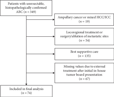
From our CRU, we extracted a total of 349 patients with unresectable ABC, who were treated between January 2006 and June 2018. A total of 275 patients had to be excluded, leaving 74 patients included in the final analysis (Figure 1).
Figure 1.

Strobe flow diagram showing the number of patients included in the final analysis and the reasons for dropout. ABC: advanced biliary tract cancer; HCC/ICC: mixed hepatocellular-cholangiocellular carcinoma.
The median age of our cohort was 65 years (range: 22–86 years). A total of 32 (43%) patients were female. Primary tumour sites were iCCA (n = 49, 66%), pCCA (n = 11, 15%), dCCA (n = 3, 4%), and GBC (n = 11, 15%). Table 1 provides the baseline patient characteristics of our cohort compared with both original A.L.A.N. cohorts.
Table 1.
Baseline characteristics of the patients in this study and in the original A.L.A.N. study∗.
| This study | Original A.L.A.N. exploratory cohort | Original A.L.A.N. validation cohort | |
|---|---|---|---|
| (n = 74) | (n = 123) | (n = 60) | |
| Age, years (median, range) | 65 (22–86) | 67 (29–85) | 64 (54–70) |
| Gender | |||
| Female | 32 (43%) | 65 (53%) | 31 (52%) |
| Male | 42 (57%) | 58 (47%) | 29 (48%) |
|
| |||
| Performance status | |||
| ECOG 0-1 | 69 (93%) | 101 (82%) | 50 (83%) |
| ECOG ≥2 | 5 (7%) | 22 (18%) | 10 (17%) |
|
| |||
| Primary tumour site | |||
| iCCA | 49 (66%) | 61 (50%) | 17 (28%) |
| pCCA | 11 (15%) | 15 (12%) | 18 (30%) |
| dCCA | 3 (4%) | 9 (7%) | 19 (32%) |
| GBC | 11 (15%) | 38 (31%) | 0 |
| Unknown | 0 | 0 | 13 (20%) |
|
| |||
| Cirrhosis among patients with iCCA | |||
| Yes | 7 (14%) | 5 (8%) | — |
| No | 42 (86%) | 56 (92%) | — |
|
| |||
| Disease status | |||
| Locally advanced | 24 (32%) | 15 (12%) | 15 (25%) |
| Metastatic | 50 (68%) | 108 (88%) | 45 (75%) |
|
| |||
| First-line chemotherapy | |||
| Gemcitabine and cisplatin | 30 (40%) | 71 (58%) | 33 (55%) |
| Gemcitabine | 16 (22%) | 8 (6%) | 13 (22%) |
| Others† | 28 (38%) | 44 (36%) | 14 (23%) |
|
| |||
| Combined agents | |||
| Yes | 53 (72%) | 110 (89%) | — |
|
| |||
| No | 21 (28%) | 13 (11%) | — |
| Second-line chemotherapy †† | |||
| Yes | 23 (31%) | 36 (29%) | 24 (40%) |
| No | 51 (69%) | 87 (71%) | 36 (60%) |
|
| |||
| Laboratory test (median, range) | |||
| ANC (cells/μl) | 6012 (1867–15548) | 5504 (1690–36230) | — |
| LMR | 2.19 (0.80–66.83) | — | — |
| Albumin (g/dl) | 3.5 (1.7–4.6) | 3.7 (2.1–4.9) | — |
| NLR | 4.87 (1.11–12.80) | — | — |
ECOG: Eastern Cooperative Oncology Group; iCCA: intrahepatic cholangiocarcinoma; pCCA: perihilar cholangiocarcinoma; dCCA: distal cholangiocarcinoma; GBC: gallbladder cancer; ANC: actual neutrophil count; LMR: lymphocyte-to-monocyte ratio; NLR: neutrophil-to-lymphocyte ratio. ∗Data for the A.L.A.N. cohorts are adapted from the original publication and presented for comparison [25]. †Other chemotherapy regimens for our cohort were: gemcitabine and sorafenib (n = 12); capecitabine and oxaliplatin (n = 5); capecitabine (n = 3); gemcitabine and oxaliplatin (n = 2); fluorouracil and imantinib (n = 2); fluorouracil, folinic acid, and irinotecan (n = 1); cisplatin and fluorouracil (n = 1); irinotecan (n = 1); oxaliplatin (n = 1). ††As further treatment during the course of disease.
The median OS of our patient cohort was 9.0 months (95% CI 6.0–13.2 months) and the 1-year OS was 38%.
3.2. A.L.A.N. Score
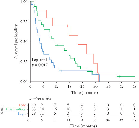
Of the 74 patients, 10 (13.5%) were in the low-risk A.L.A.N. group (score 0), 35 (47.3%) in the intermediate-risk group (score 1-2), and 29 (39.2%) in the high-risk group (score 3-4). The median OS was 21.9 months in the low-risk group, 11.4 months in the intermediate-risk group, and 4.3 months in the high-risk group. The Kaplan–Meier curves are shown in Figure 2.
Figure 2.

Kaplan–Meier curves of OS, beginning with start of first-line chemotherapy for ABC patients and stratified according to the proposed A.L.A.N. score risk groups (low risk: red; intermediate risk: green; high risk: blue).
A comparative presentation of the median survival rates for our study and the original A.L.A.N. study including survival times and confidence intervals is provided in Table 2.
Table 2.
Comparison of median OS among the A.L.A.N. subgroups.
| A.L.A.N. subgroups | Low risk (0 points) | Intermediate risk (1–2 points) | High risk (3-4 points) | p value |
|---|---|---|---|---|
| This study: median OS (95% CI), m | 21.9 (10.3–30.9) | 11.4 (6.4–18.0) | 4.3 (3.0–8.6) | 0.017 |
| Original A.L.A.N. exploratory cohort: median OS (95% CI), m | 22 (14–32) | 12 (8–15) | 5 (2–8) | <0.001 |
| Original A.L.A.N. validation cohort: median OS (95% CI), m | 12.9 (8.7–26.4) | 9.3 (7.4–14.7) | 4.3 (2.6–9.2) | 0.005 |
OS: overall survival; CI: confidence interval; m: months. Significant p values are depicted in bold.
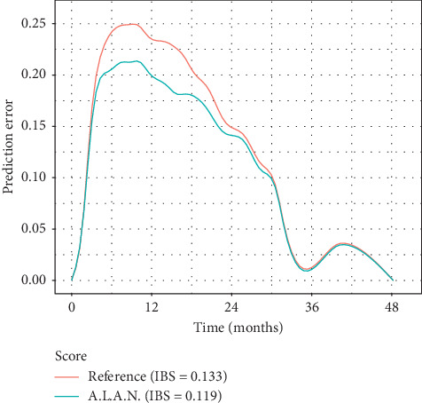
Harrell's C-index was 0.63. Prediction error curves are shown in Figure 3. The IBS for the A.L.A.N. score was 0.119 for the first 48 months, compared to an IBS of 0.133 for the unstratified cohort. As indicated by the prediction errors, the A.L.A.N. score confers a survival discrimination, particularly in the period ranging from 6 to 18 months after initiation of chemotherapy (see Figure 2).
Figure 3.

Prediction error curves and integrated Brier score (IBS) for Kaplan–Meier estimates based on the A.L.A.N. stratification and on the Kaplan–Meier estimates for all patients without any stratification (reference).
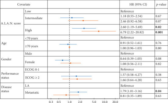
To compare these results with the original study, a Cox regression model including A.L.A.N. score risk groups, age, gender, performance status, and disease status was used. A high-risk A.L.A.N. score was an independent prognostic factor (HR = 2.60, p=0.016). Furthermore, the disease status had a significant prognostic impact (HR = 1.79, p=0.045). However, an intermediate-risk A.L.A.N. score, age, gender, and performance status had no additional predictive value (Figure 4).
Figure 4.

Results of multivariate analysis of the A.L.A.N. score and other risk factors for our (blue) and the original A.L.A.N. validation cohort (red) [25]. ECOG: Eastern Cooperative Oncology Group; LA: locally advanced; HR: hazard ratio; CI: confidence interval. Significant p values depicted in bold.
In univariate analysis, ANC (p=0.003), LMR (p=0.013), and albumin (p=0.045) had a significant influence on median OS (Table 3). However, in a Cox regression including ANC, LMR, albumin, and NLR (A.L.A.N.), only ANC (HR = 2.2, p=0.003) remained an independent prognostic factor.
Table 3.
Univariate and multivariate analysis of the A.L.A.N. score factors.
| Analysis | Univariate | Multivariate | ||||
|---|---|---|---|---|---|---|
| Covariate | HR | 95% CI | p value | HR | 95% CI | p value |
| ANC | ||||||
| >8000 | 2.19 | 1.30–3.70 | 0.003 | 2.23 | 1.31–3.78 | 0.003 |
|
| ||||||
| LMR | ||||||
| <2.1 | 1.87 | 1.14–3.07 | 0.013 | 1.62 | 0.86–3.08 | 0.137 |
|
| ||||||
| Albumin (g/dl) | ||||||
| <3.5 | 1.65 | 1.01–2.69 | 0.045 | 1.27 | 0.67–2.39 | 0.459 |
|
| ||||||
| NLR | ||||||
| >3 | 1.66 | 0.88–3.13 | 0.118 | |||
ANC: actual neutrophil count; LMR: lymphocytes-monocytes ratio; NLR: neutrophil-lymphocytes ratio; HR: hazard ratio; CI: confidence interval. Significant p values depicted in bold.
4. Discussion
To the best of our knowledge, this is the first study to perform an external validation of the recently published A.L.A.N. score for risk stratification of ABC patients receiving first-line chemotherapy. In our cohort, a higher A.L.A.N. score was associated with an increased hazard, and risk stratification yielded median OS of 21.9, 11.4, and 4.3 months for low-risk, intermediate-risk, and high-risk patients, respectively. However, the ability of the score to predict individual patient outcomes was only moderate, as indicated by a Harrell's C-index of 0.63.
To ensure comparability with the original study, we included the same risk factors in a multivariate model [25]. A high A.L.A.N. score and the disease status were independent prognostic factors for median OS. This accords with previously published results: patients with metastatic disease typically have worse prognoses than patients with locally advanced disease [14, 15, 29].
In contrast to prior publications, the performance status was not associated with an increased risk of poor survival in the original study or in our external validation [14, 15, 25, 30]. For our cohort, the main reason for this result might be the low number of patients with ECOG status 2 or higher (n = 5). This may be due to selection bias, as patients with a poor ECOG status are more likely to receive the best supportive care compared with a systemic treatment [31]. However, a larger variety of systemic therapy options and a better understanding of the side effects have led to higher treatment rates among more afflicted patients in recent years [32].
Even though several stratification systems for patients with ABC have been developed [14–16], none has been established in daily clinical practice. Salati et al. used a novel approach and included immune-related markers in their recently proposed risk score.
Of the included immune-inflammatory factors, ANC and LMR showed an influence on OS in univariate analysis. However, ANC remained the only independent risk factor in multivariate analysis. The original A.L.A.N. study was the first to show an independent prognostic effect of the LMR on ABC patients. Our results did not confirm these findings. Contrary to our expectations, a low albumin serum level showed a significant influence on median OS only in univariate analysis and lost its predictive value in multivariate analysis [33, 34]. The univariate analysis of the original A.L.A.N. validation cohort also demonstrated no significance for albumin as a prognostic marker. In contrast to previously reported results, the influence of NLR could not be confirmed in this study [21, 35]. This might be at least partly due to the moderate sample size, and the cut-off used for NLR (NLR > 3.0) might not be optimal for our cohort.
Regarding cut-offs used for stratification, one difficulty new prediction scores face is the risk of “overfitting.” In general, this is described as “a phenomenon occurring when a model maximizes its performance on some set of data, but its predictive performance is not confirmed elsewhere due to random fluctuations of patients' characteristics in different clinical and demographical backgrounds [36].” The low number of patients with ABC, especially, might increase the influence of this effect, as general representation is hard to attain within a single-centre cohort. However, in our cohort, the median OS times according to the three A.L.A.N. risk groups were 21.9, 11.4, and 4.3 months, corresponding extremely well with the median OS of the original A.L.A.N. exploratory cohort (22, 12, and 5 months). Thus, overfitting was not observed as a limiting factor.
However, even though risk stratification according to the A.L.A.N. score resulted in significant divergence of Kaplan–Meier curves in our cohort, concordance index calculation provides the probability that a randomly selected patient who experienced an event (in our case, death) had a higher risk score than a patient who had not experienced the event. Therefore, it can be regarded as a predictive measure of the individual patient outcome. Due to the deaths in all risk groups during the observation period, the discriminative ability of the score to predict which patient would die first when patients were randomly selected was only moderate in our study. Consequently, therapeutic implications from the score on an individual level have to be drawn with caution. This fact has repeatedly been observed in previous evaluations of risk scores [37, 38].
Furthermore, as chemotherapy is the mainstay of treatment for ABC patients and carries a survival advantage compared with the best supportive care, in clinical practice, chemotherapy is started if deemed oncologically reasonable, regardless of certain scoring values [39]. Clear-cut decision-making based only on scores is therefore virtually impossible, and currently no predictive system can replace the decision of an interdisciplinary tumour board.
The different ratio of patients treated with gemcitabine/cisplatin in our cohort compared with the original A.L.A.N. cohort should also be considered. Gemcitabine/cisplatin has been the first-line chemotherapy since 2010, following the results of the UK-ABC 02 trial in that year [11]. Adherence to this regime since then might have improved the patients' outcomes. As our observation period started in 2006, the percentage of patients receiving therapies other than gemcitabine/cisplatin is therefore higher compared with the original A.L.A.N. cohorts (60% vs. 42% in the exploratory cohort and 45% in the validation cohort). However, as the A.L.A.N. score is not created for a distinct regime, we included all suitable patients, independent of their first-line chemotherapy, to better reflect a real-world situation.
Our analysis is limited by several factors. First and most important, the data acquisition was retrospective and based on the patients of a single centre. Second, the sample size was only moderate (n = 74). However, due to the rare incidence and the strict inclusion criteria, our patient cohort was larger than the validation cohort of the original A.L.A.N. study. Moreover, current studies investigating new therapeutic options for ABC patients, for example, the combination of chemotherapy with locoregional treatment, radiation, or targeted and immune therapy, lead to a further diversification of treatment [40–42] and increase interstudy heterogeneity of the patient cohorts. We deliberately did not perform any kind of imputation of missing values and included only patients with the complete data needed for the calculation of the score and comparison with the original A.L.A.N. cohort. This reduced the patients' numbers and statistical power in favour of data completeness.
5. Conclusions
The A.L.A.N. score with its easily applicable parameters was able to differentiate between low-, intermediate-, and high-risk patients, and it can be used to identify risk groups with a poor prognosis prior to the start of chemotherapy. However, the ability of the score to predict individual patient outcome was only moderate; thus, it may only serve as a minor component in the complex interdisciplinary discussion. As chemotherapy is the only therapy option for patients with ABC, no patient for whom chemotherapy is deemed oncologically reasonable should be excluded due to A.L.A.N. score alone.
Acknowledgments
FH was granted protected research time by a University Center for Tumor Diseases/Transmed Fellowship of the Johannes Gutenberg University Mainz Medical Center.
Data Availability
The data that support the findings of this study are included within the article. The primary data are stored in internal clinical registry software specially developed for the clinical characterization of patients with HCC and CCC to ensure participant confidentiality. The datasets used and analysed during the current study are available from the corresponding author upon reasonable request.
Ethical Approval
The study was approved by the responsible ethics committee (Ethics Committee of the Medical Association of Rhineland Palatinate, Mainz, Germany) for the retrospective analysis of clinical data (permit number: 2018-13618). Additional examinations were not performed. Patient records and information were anonymized and deidentified prior to analysis.
Conflicts of Interest
PRG has received grants and personal fees from Bayer and personal fees from Bristol-Myers Squibb, MSD Sharp & Dohme, Lilly, Sillajen, SIRTEX, and AstraZeneca. AW has received speaker fees and travel grants from Bayer. RK has received speaker fees from BTG, Guerbet, and SIRTEX and personal fees from Boston Scientific, Bristol-Myers Squibb, Guerbet, and SIRTEX. None of these companies supported this study, and none of the authors reports conflicts of interest.
Authors' Contributions
Lukas Müller and Aline Mähringer-Kunz contributed equally to this work.
References
- 1.Miyazaki M., Ohtsuka M., Miyakawa S., et al. Classification of biliary tract cancers established by the Japanese society of hepato-biliary-pancreatic surgery: 3rd english edition. Journal of Hepato-Biliary-Pancreatic Sciences. 2015;22(3):181–196. doi: 10.1002/jhbp.211. [DOI] [PubMed] [Google Scholar]
- 2.Rizvi S., Gores G. J. Pathogenesis, diagnosis, and management of cholangiocarcinoma. Gastroenterology. 2013;145(6):1215–1229. doi: 10.1053/j.gastro.2013.10.013. [DOI] [PMC free article] [PubMed] [Google Scholar]
- 3.Bridgewater J. A., Goodman K. A., Kalyan A., Mulcahy M. F. Biliary tract cancer: epidemiology, radiotherapy, and molecular profiling. American Society of Clinical Oncology Educational Book. 2016;36(36):e194–e203. doi: 10.1200/edbk_160831. [DOI] [PubMed] [Google Scholar]
- 4.Khan S. A., Toledano M. B., Taylor-Robinson S. D. Epidemiology, risk factors, and pathogenesis of cholangiocarcinoma. HPB. 2008;10(2):77–82. doi: 10.1080/13651820801992641. [DOI] [PMC free article] [PubMed] [Google Scholar]
- 5.Khan S. A., Taylor-Robinson S. D., Toledano M. B., Beck A., Elliott P., Thomas H. C. Changing international trends in mortality rates for liver, biliary and pancreatic tumours. Journal of Hepatology. 2002;37(6):806–813. doi: 10.1016/S0168-8278(02)00297-0. [DOI] [PubMed] [Google Scholar]
- 6.Welzel T. M., McGlynn K. A., Hsing A. W., O’Brien T. R., Pfeiffer R. M. Impact of classification of hilar cholangiocarcinomas (klatskin tumors) on the incidence of intra- and extrahepatic cholangiocarcinoma in the United States. JNCI: Journal of the National Cancer Institute. 2006;98(12):873–875. doi: 10.1093/jnci/djj234. [DOI] [PubMed] [Google Scholar]
- 7.Khan S. A., Davidson B. R., Goldin R. D., et al. Guidelines for the diagnosis and treatment of cholangiocarcinoma: an update. Gut. 2012;61(12):1657–1669. doi: 10.1136/gutjnl-2011-301748. [DOI] [PubMed] [Google Scholar]
- 8.Blechacz B., Gores G. J. Cholangiocarcinoma: advances in pathogenesis, diagnosis, and treatment. Hepatology. 2008;48(1):308–321. doi: 10.1002/hep.22310. [DOI] [PMC free article] [PubMed] [Google Scholar]
- 9.Principe A., Ercolani G., Bassi F., et al. Diagnostic dilemmas in biliary strictures mimicking cholangiocarcinoma. Hepatogastroenterology. 2003;50(53):1246–1249. [PubMed] [Google Scholar]
- 10.Joo I., Lee J. M., Yoon J. H. Imaging diagnosis of intrahepatic and perihilar cholangiocarcinoma: recent advances and challenges. Radiology. 2018;288(1):7–13. doi: 10.1148/radiol.2018171187. [DOI] [PubMed] [Google Scholar]
- 11.Valle J., Wasan H., Palmer D. H., et al. Cisplatin plus gemcitabine versus gemcitabine for biliary tract cancer. New England Journal of Medicine. 2010;362(14):1273–1281. doi: 10.1056/NEJMoa0908721. [DOI] [PubMed] [Google Scholar]
- 12.Kobayashi S., Ueno M., Ohkawa S., Irie K., Goda Y., Morimoto M. Renal toxicity associated with weekly cisplatin and gemcitabine combination therapy for treatment of advanced biliary tract cancer. Oncology. 2014;87(1):30–39. doi: 10.1159/000362604. [DOI] [PubMed] [Google Scholar]
- 13.Venook A. P., Egorin M. J., Rosner G. L., et al. Phase I and pharmacokinetic trial of gemcitabine in patients with hepatic or renal dysfunction: cancer and leukemia group B 9565. Journal of Clinical Oncology. 2000;18(14):2780–2787. doi: 10.1200/jco.2000.18.14.2780. [DOI] [PubMed] [Google Scholar]
- 14.Bridgewater J., Lopes A., Wasan H., et al. Prognostic factors for progression-free and overall survival in advanced biliary tract cancer. Annals of Oncology. 2016;27(1):134–140. doi: 10.1093/annonc/mdv483. [DOI] [PubMed] [Google Scholar]
- 15.Park I., Lee J.-L., Ryu M.-H., et al. Prognostic factors and predictive model in patients with advanced biliary tract adenocarcinoma receiving first-line palliative chemotherapy. Cancer. 2009;115(18):4148–4155. doi: 10.1002/cncr.24472. [DOI] [PubMed] [Google Scholar]
- 16.Suzuki Y., Kan M., Kimura G., et al. Predictive factors of the treatment outcome in patients with advanced biliary tract cancer receiving gemcitabine plus cisplatin as first-line chemotherapy. Journal of Gastroenterology. 2019;54(3):281–290. doi: 10.1007/s00535-018-1518-3. [DOI] [PMC free article] [PubMed] [Google Scholar]
- 17.Kim B. J., Hyung J., Yoo C., et al. Prognostic factors in patients with advanced biliary tract cancer treated with first-line gemcitabine plus cisplatin: retrospective analysis of 740 patients. Cancer Chemotherapy and Pharmacology. 2017;80(1):209–215. doi: 10.1007/s00280-017-3353-2. [DOI] [PubMed] [Google Scholar]
- 18.Agarwal R., Sendilnathan A., Siddiqi N. I., et al. Advanced biliary tract cancer: clinical outcomes with ABC-02 regimen and analysis of prognostic factors in a tertiary care center in the United States. Journal of Gastrointestinal Oncology. 2016;6(7):996–1003. doi: 10.21037/jgo.2016.09.10. [DOI] [PMC free article] [PubMed] [Google Scholar]
- 19.Leyva-Illades D., McMillin M., Quinn M., DeMorrow S. Cholangiocarcinoma pathogenesis: role of the tumor microenvironment. Translational Gastrointestinal Cancer. 2012;1(1):71–80. [PMC free article] [PubMed] [Google Scholar]
- 20.Ghidini M., Cascione L., Carotenuto P., et al. Characterisation of the immune-related transcriptome in resected biliary tract cancers. European Journal of Cancer. 2017;86:158–165. doi: 10.1016/j.ejca.2017.09.005. [DOI] [PMC free article] [PubMed] [Google Scholar]
- 21.Tang H., Lu W., Li B., Li C., Xu Y., Dong J. Prognostic significance of neutrophil-to-lymphocyte ratio in biliary tract cancers: a systematic review and meta-analysis. Oncotarget. 2017;8(22):36857–36868. doi: 10.18632/oncotarget.16143. [DOI] [PMC free article] [PubMed] [Google Scholar]
- 22.Subimerb C., Pinlaor S., Lulitanond V., et al. Circulating CD14(+) CD16(+) monocyte levels predict tissue invasive character of cholangiocarcinoma. Clinical & Experimental Immunology. 2010;161(3):471–479. doi: 10.1111/j.1365-2249.2010.04200.x. [DOI] [PMC free article] [PubMed] [Google Scholar]
- 23.Atanasov G., Hau H.-M., Dietel C., et al. Prognostic significance of TIE2-expressing monocytes in hilar cholangiocarcinoma. Journal of Surgical Oncology. 2016;114(1):91–98. doi: 10.1002/jso.24249. [DOI] [PubMed] [Google Scholar]
- 24.Grenader T., Nash S., Plotkin Y., et al. Derived neutrophil lymphocyte ratio may predict benefit from cisplatin in the advanced biliary cancer: the ABC-02 and BT-22 studies. Annals of Oncology. 2015;26(9):1910–1916. doi: 10.1093/annonc/mdv253. [DOI] [PubMed] [Google Scholar]
- 25.Salati M., Caputo F., Cunningham D., et al. The A.L.A.N. score identifies prognostic classes in advanced biliary cancer patients receiving first-line chemotherapy. European Journal of Cancer. 1990;117:84–90. doi: 10.1016/j.ejca.2019.05.030. [DOI] [PubMed] [Google Scholar]
- 26.Collins G. S., Reitsma J. B., Altman D. G., Moons K. G. M. Transparent reporting of a multivariable prediction model for individual prognosis or diagnosis (TRIPOD): the TRIPOD statement. Annals of Internal Medicine. 2015;162(1):55–63. doi: 10.7326/m14-0697. [DOI] [PubMed] [Google Scholar]
- 27.Uno H., Cai T., Pencina M. J., D’Agostino R. B., Wei L. J. On the C-statistics for evaluating overall adequacy of risk prediction procedures with censored survival data. Statistics in Medicine. 2011;30(10) doi: 10.1002/sim.4154. [DOI] [PMC free article] [PubMed] [Google Scholar]
- 28.Mogensen U. B., Ishwaran H., Gerds T. A. Evaluating random forests for survival analysis using prediction error curves. Journal of Statistical Software. 2012;50(11):1–23. doi: 10.18637/jss.v050.i11. [DOI] [PMC free article] [PubMed] [Google Scholar]
- 29.Hahn F., Müller L., Stöhr F., et al. The role of sarcopenia in patients with intrahepatic cholangiocarcinoma: prognostic marker or hyped parameter? Liver International. 2019;39(7):1307–1314. doi: 10.1111/liv.14132. [DOI] [PubMed] [Google Scholar]
- 30.Peixoto R. D., Renouf D., Lim H. A population based analysis of prognostic factors in advanced biliary tract cancer. Journal of Gastrointestinal Oncology. 2014;5(6):428–432. doi: 10.3978/j.issn.2078-6891.2014.081. [DOI] [PMC free article] [PubMed] [Google Scholar]
- 31.Furuse J., Takada T., Miyazaki M., et al. Guidelines for chemotherapy of biliary tract and ampullary carcinomas. Journal of Hepato-Biliary-Pancreatic Surgery. 2008;15(1):55–62. doi: 10.1007/s00534-007-1280-z. [DOI] [PMC free article] [PubMed] [Google Scholar]
- 32.Valle J. W., Furuse J., Jitlal M., et al. Cisplatin and gemcitabine for advanced biliary tract cancer: a meta-analysis of two randomised trials. Annals of Oncology. 2014;25(2):391–398. doi: 10.1093/annonc/mdt540. [DOI] [PubMed] [Google Scholar]
- 33.Arkenau H.-T., Barriuso J., Olmos D., et al. Prospective validation of a prognostic score to improve patient selection for oncology phase I trials. Journal of Clinical Oncology. 2009;27(16):2692–2696. doi: 10.1200/jco.2008.19.5081. [DOI] [PubMed] [Google Scholar]
- 34.Wang Y., Pang Q., Jin H., et al. Albumin-bilirubin grade as a novel predictor of survival in advanced extrahepatic cholangiocarcinoma. Gastroenterology Research and Practice. 2018;2018:8. doi: 10.1155/2018/8902146.8902146 [DOI] [PMC free article] [PubMed] [Google Scholar]
- 35.McNamara M. G., Templeton A. J., Maganti M., et al. Neutrophil/lymphocyte ratio as a prognostic factor in biliary tract cancer. European Journal of Cancer. 2014;50(9):1581–1589. doi: 10.1016/j.ejca.2014.02.015. [DOI] [PubMed] [Google Scholar]
- 36.Facciorusso A., Bhoori S., Sposito C., Mazzaferro V. Repeated transarterial chemoembolization: an overfitting effort? Journal of Hepatology. 2015;62(6):1440–1442. doi: 10.1016/j.jhep.2015.01.033. [DOI] [PubMed] [Google Scholar]
- 37.Doussot A., Groot-Koerkamp B., Wiggers J. K., et al. Outcomes after resection of intrahepatic cholangiocarcinoma: external validation and comparison of prognostic models. Journal of the American College of Surgeons. 2015;221(2):452–461. doi: 10.1016/j.jamcollsurg.2015.04.009. [DOI] [PMC free article] [PubMed] [Google Scholar]
- 38.Mähringer-Kunz A., Weinmann A., Schmidtmann I., et al. Validation of the SNACOR clinical scoring system after transarterial chemoembolisation in patients with hepatocellular carcinoma. BMC Cancer. 2018;18(1):p. 489. doi: 10.1186/s12885-018-4407-5. [DOI] [PMC free article] [PubMed] [Google Scholar]
- 39.Razumilava N., Gores G. J. Classification, diagnosis, and management of cholangiocarcinoma. Clinical Gastroenterology and Hepatology. 2013;11(1):13–21.e1. doi: 10.1016/j.cgh.2012.09.009. [DOI] [PMC free article] [PubMed] [Google Scholar]
- 40.Shah U. A., Nandikolla A. G., Rajdev L. Immunotherapeutic approaches to biliary cancer. Current Treatment Options in Oncology. 2017;18(7):p. 44. doi: 10.1007/s11864-017-0486-9. [DOI] [PubMed] [Google Scholar]
- 41.Merla A., Liu K. G., Rajdev L. Targeted therapy in biliary tract cancers. Current Treatment Options in Oncology. 2015;16(10):p. 48. doi: 10.1007/s11864-015-0366-0. [DOI] [PMC free article] [PubMed] [Google Scholar]
- 42.Keane F. K., Zhu A. X., Hong T. S. Radiotherapy for biliary tract cancers. Seminars in Radiation Oncology. 2018;28(4):342–350. doi: 10.1016/j.semradonc.2018.06.003. [DOI] [PubMed] [Google Scholar]
Associated Data
This section collects any data citations, data availability statements, or supplementary materials included in this article.
Data Availability Statement
The data that support the findings of this study are included within the article. The primary data are stored in internal clinical registry software specially developed for the clinical characterization of patients with HCC and CCC to ensure participant confidentiality. The datasets used and analysed during the current study are available from the corresponding author upon reasonable request.


