Upon priming, naive CD4+ helper T (Th) cells differentiate into distinct subsets with specialized functions. The differentiation of Th subsets is driven not only by signals from the T cell receptor (TCR) and costimulatory receptors but is also critically dependent on the specific cytokine milieu. By mimicking such conditions, robust methods have been developed for the in vitro differentiation of type 1 and type 2 Th (Th1 and Th2) cells, and more recently, IL-17-producing Th (Th17) cells and regulatory T (Treg) cells,1 which greatly support the research and applications of these Th subsets. Follicular helper T (Tfh) cells represent another Th subset that specializes in supporting the germinal center (GC) response and regulating the generation of memory B cells and long-lived plasma cells.2 However, current methods for in vitro Tfh differentiation are not optimal. Even in the best practice, only 20% of polarized cells showed the expression of CXCR5, the key Tfh functional marker.3 Here, we report an optimized in vitro differentiation method that generates 50–75% CXCR5+ cells with enhanced B cell helper function. We demonstrate that the priming of antigen-presenting cells (APCs) by lipopolysaccharide (LPS) and the increase of the APC:T cell ratio were key to efficiently generating Tfh cells in vitro.
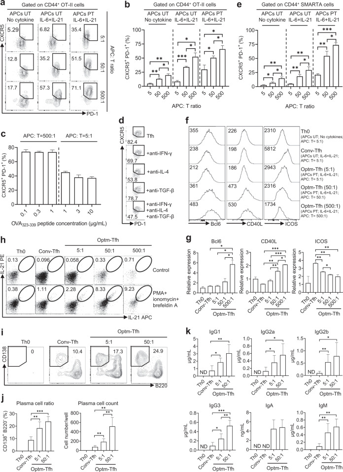
Tfh differentiation is critically dependent on costimulatory signals, including CD28, CD40L, ICOS and OX40,2 which can be upregulated on APCs by LPS treatment.4 A conventional method for in vitro Tfh differentiation is to stimulate TCR transgenic naive CD4+ T cells with MHC II-specific peptides in the presence of IL-6 and IL-21.3,5 We optimized this method by treating splenocytes with 1 µg/mL LPS for 24 h before coculturing with naive OT-II cells with ovalbumin peptide 323–339 (OVA323–339), IL-6 and IL-21. After coculture for 3 days, the pretreatment of splenocytes with APCs and LPS significantly increased CXCR5 expression in OT-II cells from ~5% to more than 30% (Fig. 1a, b).
Fig. 1.
Optimization and characterization of the in vitro differentiation of mouse Tfh cells. a–e APCs were either untreated (UT) or pretreated (PT) with 1 µg/mL LPS for 24 h and cocultured with OT-II or SMARTA cells as indicated for 3 days. Representative FACS plots (a) and statistics (b, e) showing the expression of CXCR5 and PD-1 on CD44+ OT-II or SMARTA cells. The effects on Tfh differentiation of the antigen dose (c) or specific neutralizing antibodies (d) were tested using OT-II cells. The expression of Tfh surface markers (f, g) and signature cytokines (h) in OT-II cells was quantified. i–k After in vitro differentiation for 3 days, total OT-II cells from the indicated conditions were isolated by FACS purification and treated with 50 µg/mL mitomycin-c for 30 min before being cocultured at a 1:1 ratio with purified B cells that had been preactivated with 1 µg/mL LPS for 24 h. Cells were cultured for 6 days in the presence of 1 µg/mL OVA323–339 peptide. Representative FACS plots (i) and statistics (j) showing plasma cell differentiation. The secretion of the indicated antibodies in the culture supernatants was measured by bead-based ELISA (k). The results are representative of at least two independent experiments. Detailed methods are described in the Supplementary information. ND, not detectable. *p < 0.05, **p < 0.01, and ***p < 0.001.
Our second attempt was to increase the APC:T cell ratio since a high dose of antigens has been shown to promote Tfh differentiation.6 By maintaining a consistent number of splenocytes and gradually reducing the number of OT-II cells, APC:T cell ratios were increased from 5:1 (the conventional method) to 50:1 and 500:1. Correspondingly, we observed a more than two-fold increase in CXCR5 expression, which showed a positive correlation with the APC:T cell ratio. When LPS-treated splenocytes and at an APC:T cell ratio of 500:1 were used, over 70% of OT-II cells displayed the CXCR5+ PD-1+ phenotype (Fig. 1a, b). By testing the OVA peptide concentration ranging from 0.1 to 1 µg/mL (high APC:T cell ratio at 500:1) or from 1 to 10 µg/mL (low APC:T cell ratio at 5:1), we found that peptide concentrations had a negligible impact on Tfh cell differentiation (Fig. 1c), suggesting that signaling other than TCR, presumably costimulatory signaling, is the limiting factor for Tfh differentiation in the APC:T cell coculture system.
Despite the utility of anti-IL-4, anti-IFN-γ and, anti-TGF-β antibodies in reported methods for in vitro Tfh differentiation,3,5 we found that the addition of such neutralizing antibodies did not enhance or even inhibit Tfh polarization (Fig. 1d), which could be explained by the plasticity between Tfh and other Th subsets.3,7
Overall, by stimulating APCs with LPS and setting the APC:T cell ratio higher than 50:1, phenotypic CXCR5+PD-1+ Tfh-like cells could be efficiently induced in vitro. To validate the application of this method for other TCR transgenic CD4+ T cells, we examined in vitro Tfh differentiation using naive SMARTA cells with transgenic TCR-specific to lymphocytic choriomeningitis virus (LCMV) glycoprotein residues 61–80 (GP61–80) and obtained comparable results (Fig. 1e).
By comparing Tfh-like cells induced in vitro using the conventional method (Conv-Tfh), we demonstrated that the optimized method (Optm-Tfh) promoted the expression of Bcl6 and CD40L but not ICOS (Fig. 1f, g). Notably, several studies have reported that high expression of ICOS is not always a unique marker for Tfh cells, as shown in culture in vitro3 and in mice with chronic LCMV infection.8 Nevertheless, the Optm-Tfh method significantly enhanced the secretion of IL-21 (Fig. 1h), the signature Tfh cytokine required to support GC reaction and plasma cell differentiation.2
Finally, we assessed the B cell helper function of Tfh cells generated in vitro by the Optm-Tfh method. We tested the Optm-Tfh methods using 5:1 and 50:1 ratios but not the 500:1 condition due to the low number of Tfh cells generated from the low frequency of OT-II seeding of the latter. OT-II CD4+ T cells polarized under the indicated conditions were treated with mitomycin-c to inhibit proliferation and then cocultured for 6 days with isolated B220+ cells that had been primed with LPS. OT-II cells from the Th0 culture condition hardly supported the survival of B cells, while cells from Tfh polarization conditions did so (Fig. 1i). The Optm-Tfh method, especially the APC:T cell ratio at 50:1, induced Tfh cells with superior B cell helper capability, as shown by several fold increases in plasma cell differentiation (Fig. 1j) and antibody secretion (Fig. 1k).
In conclusion, we present an optimized method to induce the in vitro Tfh differentiation of mouse TCR transgenic CD4+ T cells. By stimulating APCs with LPS and setting the APC:T cell ratio higher than 50:1, the Optm-Tfh method efficiently induced Tfh-like cells that expressed higher levels of Tfh markers and possessed superior B cell helper function. We believe that this straightforward and reliable method would greatly support Tfh research and allow test of applications that require a large amount of Tfh cells.
Supplementary information
An Optimised Method to Differentiate Mouse Follicular Helper T Cells in Vitro
Acknowledgements
The study is supported by the National Key Research and Development Program of China (2017YFC0909003), the Australian National Health and Medical Research Council (GNT1147769) and the Bellberry-Viertel Senior Medical Research Fellowship to D.Y.
Author contributions
D.Y. and X.G. conceived and designed the study. X.G. performed the experiments and analyzed the data with the help of W.H., Z.C. and P.Z. X.G. and D.Y. wrote the paper. All authors approved the final paper.
Competing interests
The authors declare no competing interests.
Supplementary information
The online version of this article (10.1038/s41423-019-0329-7) contains supplementary material.
References
- 1.Read KA, Powell MD, Sreekumar BK, Oestreich KJ. In vitro differentiation of effector CD4(+) T helper cell subsets. Methods Mol. Biol. 2019;1960:75–84. doi: 10.1007/978-1-4939-9167-9_6. [DOI] [PubMed] [Google Scholar]
- 2.Vinuesa CG, Linterman MA, Yu D, MacLennan IC. Follicular helper t cells. Annu Rev. Immunol. 2016;34:335–368. doi: 10.1146/annurev-immunol-041015-055605. [DOI] [PubMed] [Google Scholar]
- 3.Lu KT, et al. Functional and epigenetic studies reveal multistep differentiation and plasticity of in vitro-generated and in vivo-derived follicular T helper cells. Immunity. 2011;35:622–632. doi: 10.1016/j.immuni.2011.07.015. [DOI] [PMC free article] [PubMed] [Google Scholar]
- 4.Xu H, et al. The modulatory effects of lipopolysaccharide-stimulated B cells on differential T-cell polarization. Immunology. 2008;125:218–228. doi: 10.1111/j.1365-2567.2008.02832.x. [DOI] [PMC free article] [PubMed] [Google Scholar]
- 5.Nurieva RI, et al. Generation of T follicular helper cells is mediated by interleukin-21 but independent of T helper 1, 2, or 17 cell lineages. Immunity. 2008;29:138–149. doi: 10.1016/j.immuni.2008.05.009. [DOI] [PMC free article] [PubMed] [Google Scholar]
- 6.Baumjohann D, et al. Persistent Antigen and Germinal Center B Cells Sustain T Follicular Helper Cell Responses and Phenotype. Immunity. 2013;38:596–605. doi: 10.1016/j.immuni.2012.11.020. [DOI] [PubMed] [Google Scholar]
- 7.Cannons JL, Lu KT, Schwartzberg PL. T follicular helper cell diversity and plasticity. Trends Immunol. 2013;34:200–207. doi: 10.1016/j.it.2013.01.001. [DOI] [PMC free article] [PubMed] [Google Scholar]
- 8.Crawford A, et al. Molecular and transcriptional basis of CD4(+) T cell dysfunction during chronic infection. Immunity. 2014;40:289–302. doi: 10.1016/j.immuni.2014.01.005. [DOI] [PMC free article] [PubMed] [Google Scholar]
Associated Data
This section collects any data citations, data availability statements, or supplementary materials included in this article.
Supplementary Materials
An Optimised Method to Differentiate Mouse Follicular Helper T Cells in Vitro


