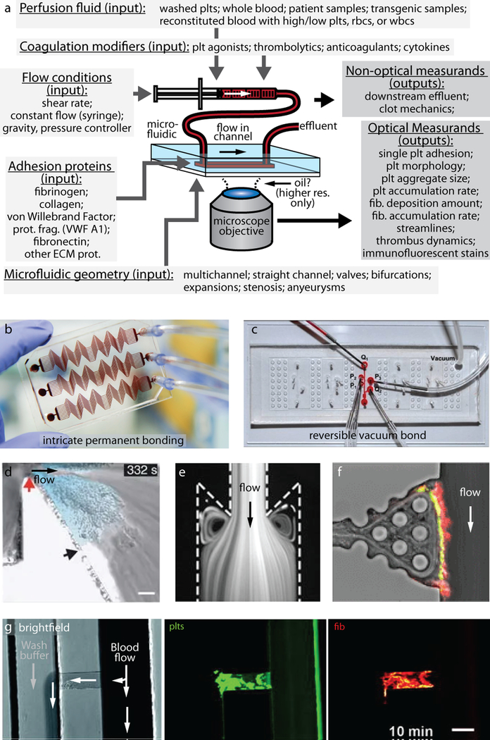
Figure 2: Microfluidic devices represent an extremely versatile tool for studying platelets under flow conditions.
(a) Platelet and fibrin deposition parameters can be measured as a function of perfusion fluid, coagulation modifiers, flow conditions, adhesion proteins, and microfluidic geometry. Permanently bonded microfluidic devices[62] typically achieve more intricate geometries (b) then reversibly bonded microfluidic devices[37] (c), which offer access to the sample for further analysis. A variety of geometries such as stenoses (d) [6], valves (e) [38], or plugs[40] (g) can be created with microfluidic. Clots may be imaged from the side[37] (f) and platelets, fibrin, and coagulation proteins may be visualized utilizing immunofluorescent labels[40] (f, g).

