Abstract
Background:
Intramedullary spinal cord metastases represent 4–8.5% of the central nervous system metastases and affect only 0.1–0.4% of all patients. Those originating from renal cell carcinoma (RCC) are extremely rare. Of the eight patients described in the literature with metastatic RCC and intramedullary cord lesion, only five were found in the cervical spine. Here, the authors add a 6th case involving an RCC intramedullary metastasis at the C1–C2 level.
Case Description:
A 78-year-old male patient presented with intermittent cervicalgia of 5 months duration accompanied by few weeks of a progressive severe right hemiparesis, up to hemiplegia. The magnetic resonance imaging (MRI) examination revealed an intramedullary expansive lesion measuring 10 mm×15 mm at the C1–C2 level; it readily enhanced with contrast. A total body computed tomography (CT) scan documented an 85 mm mass involving the right kidney, extending to the ipsilateral adrenal gland, and posteriorly infiltrating the ipsilateral psoas muscle. The subsequent CT-guided fine-needle biopsy confirmed the diagnosis of an RCC (Stage IV). The patient next underwent total surgical total removal of the C1–C2 intramedullary mass, following which he exhibited a slight motor improvement, with the right hemiparesis (2/5). He died after 14 months due to global RCC tumor progression.
Conclusion:
The present case highlights that a patient without a prior known diagnosis of RCC may present with an intramedullary C1–C2 metastasis. In such cases, global staging is critical to determine whether primary lesion resection versus excision of metastases (e.g., in this case, the C1–C2 intramedullary tumor) are warranted.
Keywords: Craniovertebral junction, Intramedullary, Metastasis, Myelotomy, Renal cell carcinoma

INTRODUCTION
Intramedullary spinal cord metastases (IMSCMs) represent the 4–8.5% of the central nervous system metastases, affecting 0.1–0.4% of all patients.[12] However, those originating from renal cell carcinoma (RCC) are extremely rare. A review of the literature revealed eight cases of RCC IMSCM; five involved the cervical cord.[2] Of interest, the typical mean time interval between the original diagnosis of RCC and the diagnosis of intramedullary spinal metastases is approximately 32.1 months (range 0–180 months).[1,4-7,10,14] Here, a 78-year-old male presented with a C1–C2 cervical intramedullary metastasis that represented the initial manifestation of underlying and previously undiagnosed RCC.
CASE REPORT
Medical history and physical examination
A 78-year-old male patient presented with 5 months of intermittent cervicalgia and several weeks of a progressive right hemiparesis, up to hemiplegia (0/5), brisk upper and lower extremity reflexes, bilateral Hoffmann’s and Babinski signs, left hemisensory dysesthesias, and urinary incontinence.
Diagnostic imaging
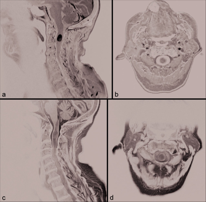
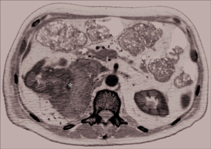
The cervical spinal MRI revealed an intramedullary expansive lesion (10 mm×15 mm) at C1–C2 that markedly enhanced with gadolinium [Figure 1]. As the differential diagnosis included potential metastatic disease, a total body computed tomography (CT) scan was performed that revealed a large mass (about 85 mm in size) involving the upper polar region and the middle third of the right kidney, extending to the adrenal gland, and ipsilateral psoas muscle [Figure 2]. A CT- guided fine-needle ago-biopsy of established the diagnosis of an RCC, also making it most likely that the C1–C2 lesion was an RCC metastasis (Stage IV).
Figure 1:

Sagittal (a) and axial (b) T1-weighted gadolinium- enhanced, sagittal (c), and axial (d) T2-weighted magnetic resonance imaging sequences showing a C1–C2 intramedullary expansive lesion (10 mm×15 mm), T2-hypointense and T1-hyperintense after gadolinium administration.
Figure 2:

Axial abdominal contrast-enhanced computed tomography scan image showing a voluminous mass (about 85 mm) (black asterisk) involving the upper polar region and the middle third of the right kidney, the ipsilateral adrenal gland, and extends posteriorly to infiltrate the ipsilateral psoas muscle. This lesion, which presents an inhomogeneous hypodense aspect with hypervascular foci in this context, is associated with collateral circles in the peri- and pararenal space, with the infiltration of the upper right calyxes. A neoplastic thrombosis of the renal vein and inferior vena cava in the subhepatic tract is also present and may explain hematogenous spread through Batson’s venous plexus.
Surgical treatment
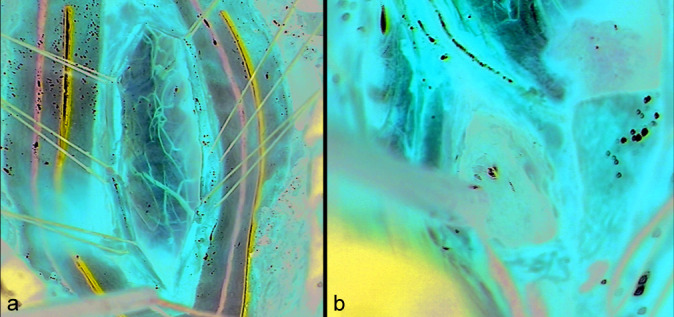
Utilizing intraoperative neurophysiological monitoring, a C1–C2 laminectomy was performed. Through a posterior C2, myelotomy, and the lesion were macroscopically fully resected [Figure 3]. Postoperatively, the patient presented a slight motor improvement, with the right hemiparesis (2/5) and left-sided hemisensory deficit.
Figure 3:

Intraoperative findings during microsurgical removal of the lesion: a good exposure of the posterior surface of the spinal cord at level C1–C2 after opening the dura mater is performed (a). After arachnoid dissection and preservation of the posterior spinal arteries, the posterior median sulcus is identified and the posterior myelotomy is performed, with access to the intramedullary lesion which shows a reddish-gray and highly vascularized appearance (b).
Histology
The histological examination revealed large cells with marked anaplasia. Immunostaining was negative for cytokeratin, GFAP, S-100, and HMB-45 but positive for intermediate vimentin filaments. Together, these studies confirmed the diagnosis of an RCC.
Postoperative course and follow-up
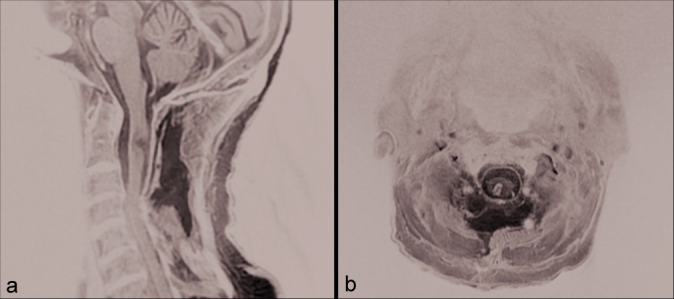
The 1-week postoperative cervical spine MRI showed postoperative changes, but full lesion excision [Figure 4]. The patient was discharged to a neuromotor rehabilitation center and underwent chemotherapy and radiotherapy for RCC. Fourteen months later, the patient died due to metastatic RCC.
Figure 4:

Sagittal (a) and axial (b) T2-weighted magnetic resonance imaging sequences showing a macroscopic total removal of the lesion and a physiological evolution of the operative field with the left median-paramedian malacic area.
DISCUSSION
Patients affected by RCC that develop IMSCMs are usually male (83%). These IMSCMs occur in the cervical spine in 47% of cases. Patients typically present with limb weakness (72%), dysesthesias, and urinary incontinence (50%).[5,8,12] Although chemotherapy is uniformly utilized, surgery is performed in 31% of cases along with adjuvant radiotherapy.[3,9,11,12,13,15] In our patient, as the systemic evaluation revealed a primary RCC, it was most likely that the intramedullary C1–C2 lesion was a metastasis.
CONCLUSION
Patients may present with symptoms/signs of and intramedullary C1–C2 spinal cord metastasis as the first sign of RCC. As performed in this case, global staging should document the origin and extent of the primary lesion and other systemic metastases and help determine whether or not excision of a C1–C2 IMSCM is warranted.
Footnotes
How to cite this article: Ponzo G, Umana GE, Giuffrida M, Furnari M, Nicoletti GF, Scalia G. Intramedullary craniovertebral junction metastasis leading to the diagnosis of underlying renal cell carcinoma. Surg Neurol Int 2020;11:152.
Contributor Information
Giancarlo Ponzo, Email: giancarlo.ponzo@gmail.com.
Giuseppe Emmanuele Umana, Email: umana.nch@gmail.com.
Massimiliano Giuffrida, Email: mass.giuffrida@tiscalinet.it.
Massimo Furnari, Email: massimofurnari@alice.it.
Giovanni Federico Nicoletti, Email: gfnicoletti@alice.it.
Gianluca Scalia, Email: gianluca.scalia@outlook.it.
Declaration of patient consent
The authors certify that they have obtained all appropriate patient consent.
Financial support and sponsorship
Nil.
Conflicts of interest
There are no conflicts of interest.
REFERENCES
- 1.Asadi M, Rokni-Yazdi H, Salehinia F, Allameh FS. Metastatic renal 1 cell carcinoma initially presented with an intramedullary spinal cord lesion: A case report. Cases J. 2009;2:7805. doi: 10.4076/1757-1626-2-7805. [DOI] [PMC free article] [PubMed] [Google Scholar]
- 2.Barrie U, Elguindy M, Pernik M, Adeyemo E, Aoun SG, Hall K, et al. Intramedullary spinal metastatic renal cell carcinoma: Systematic review of disease presentation, treatment, and prognosis with case illustration. World Neurosurg. 2020;134:584–93. doi: 10.1016/j.wneu.2019.11.056. [DOI] [PubMed] [Google Scholar]
- 3.De Meerleer G, Khoo V, Escudier B, Joniau S, Bossi A, Ost P, et al. Radiotherapy for renal-cell carcinoma. Lancet Oncol. 2014;15:e170–7. doi: 10.1016/S1470-2045(13)70569-2. [DOI] [PubMed] [Google Scholar]
- 4.Donovan DJ, Freeman JH. Solitary intramedullary spinal cord tumor presenting as the initial manifestation of metastatic renal cell carcinoma: Case report. Spine (Phila Pa 1976) 2006;31:E460–3. doi: 10.1097/01.brs.0000222022.67502.4e. [DOI] [PubMed] [Google Scholar]
- 5.Fakih M, Schiff D, Erlich R, Logan TF. Intramedullary spinal cord metastasis (ISCM) in renal cell carcinoma: A series of six cases. Ann Oncol. 2001;12:1173–7. doi: 10.1023/a:1011693212682. [DOI] [PubMed] [Google Scholar]
- 6.Gaylor JB, Howie JW. Brown-sequard syndrome: A case of unusual aetiology. J Neurol Psychiatry. 1938;1:301–5. doi: 10.1136/jnnp.1.4.301. [DOI] [PMC free article] [PubMed] [Google Scholar]
- 7.G5.301de la Riva AG, Isla A, Perez-Lopez C, Budke M, Gutierrez M, Frutos R. Intramedullary spinal cord metastasis as the first manifestation of a renal carcinoma. Neurocirugia (Astur) 2005;16:359–64. doi: 10.4321/s1130-14732005000400006. [DOI] [PubMed] [Google Scholar]
- 8.Hrabalek L. Intramedullary spinal cord metastases: Review of the literature. Biomed Pa Med Fac Univ Palacky Olomouc Czech Repub. 2010;154:117–22. doi: 10.5507/bp.2010.018. [DOI] [PubMed] [Google Scholar]
- 9.Kalayci M, Cagavi F, Gul S, Yenidunya S, Acikgoz B. Intramedullary spinal cord31 metastases: Diagnosis and treatment an illustrated review. Acta Neurochir (Wien) 2004;146:1347–54. doi: 10.1007/s00701-004-0386-1. discussion 1354. [DOI] [PubMed] [Google Scholar]
- 10.Kawakami Y, Mair WG. Haematomyelia associated with anticoagulant therapy, an intramedullary ependymoma and Schwann cells. Acta Neuropathol. 1973;26:253–8. doi: 10.1007/BF00684435. [DOI] [PubMed] [Google Scholar]
- 11.Park J, Chung SW, Kim KT, Cho DC, Hwang JH, Sung JK, et al. Intramedullary spinal cord metastasis in renal cell 37 carcinoma: A case report of the surgical experience. J Korean Med Sci. 2013;28:1253–6. doi: 10.3346/jkms.2013.28.8.1253. [DOI] [PMC free article] [PubMed] [Google Scholar]
- 12.Payer S, Mende KC, Westphal M, Eicker SO. Intramedullary spinal cord metastases: An increasingly common diagnosis. Neurosurg Focus. 2015;39:E15. doi: 10.3171/2015.5.FOCUS15149. [DOI] [PubMed] [Google Scholar]
- 13.Saeed H, Patel R, Thakkar J, Hamoodi L, Chen L, Villano JL. Multimodality therapy17 improves survival in intramedullary spinal cord metastasis of lung primary. Hematol Oncol Stem18 Cell Ther. 2017;10:143–50. doi: 10.1016/j.hemonc.2017.07.003. [DOI] [PubMed] [Google Scholar]
- 14.Schijns OE, Kurt E, Wessels P, Luijckx GJ, Beuls EA. Intramedullary spinal cord metastasis as a first manifestation of a renal cell carcinoma: Report of a case and review of the literature. Clin Neurol Neurosurg. 2000;102:249–54. doi: 10.1016/s0303-8467(00)00106-2. [DOI] [PubMed] [Google Scholar]
- 15.Tsai TH, Lin IC, Lin PC, Wu CH, Lin CL, Su YF. Intramedullary spinal cord metastasis 26 from colon cancer: Analysis of 19 reported cases. Spinal Cord Ser Cases. 2016;2:15026. doi: 10.1038/scsandc.2015.26. [DOI] [PMC free article] [PubMed] [Google Scholar]


