Abstract
Background:
The aim of this study was to compare the results of two different titanium cranioplasties for reconstructing skull defects: standard precurved mesh versus custom-made prostheses.
Methods:
Retrospective analysis of 23 patients submitted to titanium cranioplasty between January 2014 and January 2019. Ten patients underwent delayed cranioplasty using custom-made prostheses; and 13 patients were treated using precurved titanium mesh (ten delayed cranioplasties, and three single-stage resection- reconstructions). Demographic, clinical, and radiological data were recorded. Results and complications of the two methods were compared, including duration of surgery, cosmetic results (visual analog scale for cosmesis [VASC]), and costs of the implants.
Results:
Complications: one epidural hematoma in the custom-made group, one delayed failure in precurved group due to wound dehiscence with mesh exposure. There were no infections in either group. All custom-made prostheses perfectly fitted on the defect; eight of 13 precurved mesh prostheses incompletely covered the defect. Custom-made cranioplasty obtained better cosmetic results (average VASC 94 vs. 68), shorter surgical time (141min vs. 186min), and -fewer screws was needed to fix the prostheses in place (6 vs. 15). However, satisfactory results were obtained using precurved mesh in cases of small defects and in single-stage reconstruction. Precurved mesh was found to be cheaper (€1,500 vs. €5,500).
Conclusion:
Custom-made cranioplasty obtained better results and we would suggest that this should be a first choice, particularly for young patients with a large cranial defect. Precurved mesh was cheaper and useful for single-stage resection-reconstruction. Depending on the individual conditions, both prostheses have their place in cranioplasty therapies.
Keywords: Cranioplasty, Custom-made cranioplasty, Decompressive craniectomy, Precurved mesh, Titanium mesh

INTRODUCTION
The management of patients with cranial defects (craniolacunias) has become a very common problem for neurosurgeons over the years. The use of decompressive craniectomy for the emergency treatment of several pathologic conditions such as severe head injuries, ischemic, and hemorrhagic strokes, cerebral venous thrombosis, severe brain infections, and subarachnoid hemorrhages,[5,6,11,16] has led to an increase in patients with cranial defect. After the acute pathological process has ended and the cerebral swelling regressed, patients require neurosurgical treatment to reconstruct the cranial defect (delayed cranioplasty). Patients undergoing craniectomy for neoplasms, infections, or traumas with involvement of the bone (including infectious complications of the previous neurosurgical operations) should be added to this list. These patients, where possible based on location and size of the head defect, can be subjected to either immediate reconstruction (single-stage resection-reconstruction) or delayed reconstruction.
Clinical experience and literature data show that cranioplasty, despite seeming a technically simple procedure, has an average complication rate of 10–20% with a risk of failure (through infection or graft resorption) of about 10%.[3,5,6,26,27] The percentages of complications obviously vary according to the series considered and the type of technique and material used for cranial reconstruction. There was no unanimous consensus on the ideal timing for cranioplasty and the type of material to be used at the time of this study.
Reconstruction of the head defect has two main purposes: to guarantee protection for the brain and to restore adequate cosmetics; the results must be long lasting. The main complications of cranioplasty are represented, in addition to the investigated aesthetic result, by risk of infection, postoperative hematoma, problems with wound healing and even long-term failure by graft resorption or infections with consequent need for removal of the prostheses.
Theoretically, the ideal material for cranial reconstruction is autologous bone flap since it does not present biocompatibility issues, its shape is perfect for restoring normal cosmetics, and it ensures immediate and adequate protection for intracranial structures. Regardless of the difficulties in adequately storing the bone flap, however, it has been found (in several literature reports) that even autologous bone has rather high failure rates related to infection or resorption.[4,11,17,22,26] In both cases, the patient would have to undergo surgery to remove the (infected or resorbed and therefore mobile) bone flap with the need for a second cranioplasty procedure (with the consequent risks of a second procedure beyond the health costs). Over the years, many biomaterials have been used for cranial reconstruction. The most commonly used are hydroxyapatite (HA), polymethyl methacrylate (PMMA), and titanium.[5,11,20,25-27] Each of these materials has its own advantages and disadvantages in terms of results and costs, and the choice of which material to use would depend on the experience and preference of the surgeon, as well as the financial resources of health-care companies. Literature analysis shows that many authors suggest the use of titanium as it has many features that make it suitable for this type of procedure: low rate of infection, high, and immediate biomechanical resistance to direct trauma (and therefore good protection for the brain), and suitability for postoperative imaging techniques.[3,11,13,27]
Titanium cranioplasty can be performed in two ways, using precurved meshes (which are then adapted to the individual patient) or using custom-made prostheses that are specifically reconstructed for the individual patient using computer-aided design/computer-aided manufacturing techniques. The two prostheses differ in terms of costs and results. Within the literature, there are numerous reports about titanium cranioplasty with custom-made prostheses, although relatively few articles discuss the use of precurved meshes. We have, therefore, compared these two types of prostheses.
The aim of this study was to compare the results of two different titanium cranioplasties in reconstructing skull defects following craniectomy: standard precurved mesh versus custom-made prosthesis; the final aim was to assess whether one of the two prostheses should be excluded, or whether both can be useful in different situations.
MATERIALS AND METHODS
Study design
All patients enrolled in the study provided their written consent for anonymous data collection and inclusion in the study. We carried out a retrospective analysis of the patients who underwent cranioplasty surgery at our center in the period between January 2014 and January 2019. We used four types of cranioplasty during this period: cryostored autologous bone flap, manually shaped PMMA cranioplasty, custom-made HA cranioplasty, custom-made titanium cranioplasty, and precurved titanium mesh.
For this study, we selected and included patients undergoing titanium cranioplasty; we reviewed the medical records, radiological data (pre- and postoperative), and operating reports; we noted the demographic data, age at the time of surgery, cause of head defect, time elapsed between defect acquisition and reconstruction, any risk factors for cranioplasty failure, and duration of cranioplasty surgery and any complications (early and late); we finally requested that patients (or family members for patients with persistent neurocognitive deficits) completed a questionnaire covering subjectively-experienced satisfaction with the cosmetic result based on 100-mm-long visual analog scale (VASs) (VAS for cosmesis [VASC]).[3]
Statistical analysis was performed using the t-test. P < 0.05 was considered statistically significant.
Two types of prostheses were used: precurved and custom made. During the study period, the two prostheses were not always available in our department, and the choice of which prostheses to use was partly dependent on the hospital company being able to purchase one or the other.
Description of the two prostheses
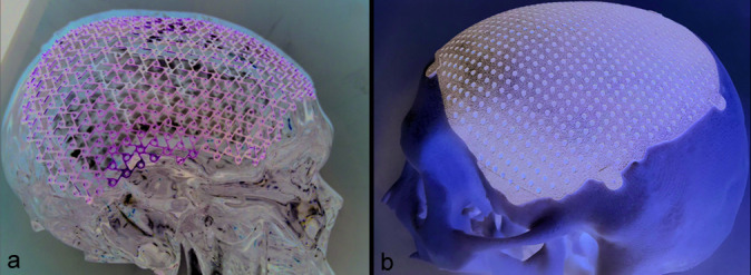
CranioCurve Preformed Mesh (Zimmer Biomet) [example in Figure 1, Panel A] is composed of anatomically preformed titanium nets and is useful for the reconstruction of head defects; the surface is perfectly smooth, with high mechanical strength. The thickness of the titanium mesh is 0.6 mm; it is made in a standard shape to be adapted for cranial defects affecting the frontotemporal and occipital-parietal region. It is available in three models: frontotemporo-parietal (large) right and left, frontotemporal (small) right and left, and occipital (suitable for defects in the occipital and/or parietal region). It can be cut out and partly shaped to best suit the defect to be covered; it is fixed with self-piercing and self- threading 1.5 mm long titanium screws. It is also computed tomography (CT) and magnetic resonance imaging (MRI) compatible. Sold in nonsterile packaging and then sterilized in an autoclave at 134° directly in the hospital, they are equipped with labels for product traceability that can be directly affixed in the operating register. The cost of a single prosthesis is about €1,500. It is secured with 12–15 screws per mesh.
Figure 1:

(a) Panel A: Photograph showing a cranio curve preformed mesh, frontotemporo-parietal (large), right side. (b) Panel B: Photograph showing a custom made titanium prostheses.
Custom-made prostheses (MT Ortho) [example in Figure 1, Panel B]: custom-made prostheses are made using additive 3D technology (Additive Manufacturing) which uses titanium alloy powder (Ti6Al4V - ELI) as raw material. The technology uses the electron beam melting (EBM) process, which is a 3D printing technique in which a high- energy source (composed of a suitably concentrated beam of electrons) hits a bed of titanium powder and causes it to melt. EBM technology offers design freedom in developing and manufacturing a trabecular structure with any 3D shape, customizable for any type and site of the patient’s cranial defect. The three-dimensional architecture and surface characteristics of the prosthetic elements (high porosity and optimal pore size) are able to promote cell migration and vascularization, thus facilitating the transport of oxygen, nutrients, ions, and growth factors that stimulate the process of bone neoformation at the edges. The production process is based on the acquisition of a thin-layer (1 mm) CT scan (DICOM format) and three-dimensional processing by the company’s design office. The latter, working in collaboration with the prescribing neurosurgeon, carries out the final project and consequently starts production on the prosthesis. MT Ortho cranioplasty has the following characteristics: high shape accuracy and adherence to the contours of the defect; guarantees adequate cosmetics (restoring normal and symmetrical shape with the healthy side); no need for intraoperative adaptation; porous and multi-perforated structure (10 holes/cm2); high mechanical resistance; 1 mm thickness, 0.3 mm fixing points; fixed with self-piercing and self-threading 1.5 mm long titanium screws; and compatible with CT and MRI. Sold in nonsterile packaging and then autoclavable at 134° directly in the hospital. The average production time is 20–30 days. Cost is around €5,500 per piece. It is secured with 4–6 screws for each prosthesis.
Surgical procedure
The operation was performed under general anesthesia in the supine position. A first dose of a broad-spectrum antibiotic agent was given intravenously at induction of anesthesia, then one dose every 4 h for the next 24 h. The patient’s hair was completely removed using hair clippers. Great care was taken to avoid skin damage. Skin was thoroughly washed and then disinfected. An iodine- impregnated incision drape was placed over the exposed skin, and care was taken to ensure that all surfaces were covered. In three cases, where cranioplasty was performed at the same time as a tumor removal operation involving the cranial bone (patient number 21, 22, and 23, single- step resection-reconstruction), we used the neuronavigator to plan skin flap and craniectomy and iUS to guide tumor resection (according to our protocol described elsewhere);[23] in all other cases, the previous incision made during the craniectomy operation was reopened; this strategy ensured the best vascularization for the flap (avoiding overlapping or crossed incisions) and allowed adequate exposure of the cranial defect. The wound was then opened using a scalpel and sharp dissection, and the head defect was progressively exposed in the subperiosteal plane. Once the scalp flap was reflected, the temporalis muscle was usually dissected from the dura and reflected laterally. However, if it was densely adherent, it was left attached to the dura, and the cranioplasty was placed on top of both structures. Great care was taken not to open the dura. Once the dissection of the dural plane was completed and all the edges of the craniolacunia exposed, thorough and abundant washing and scrupulous control of hemostasis were carried out. The prostheses were only handled with clean gloves. At this point, the prostheses were positioned. For precurved mesh, it was necessary to adapt it as best as possible by cutting or manipulating it to modify the curvature. The prostheses were secured with titanium screws. Central dural tenting sutures were placed routinely. A wound suction drainage was placed under the skin. A CT scan was obtained the following day and, if there was no postoperative collection, the wound drain was removed.
RESULTS
From January 2014 to January 2019, 23 patients underwent cranioplasty using titanium prostheses. Average age 47.6 years (minimum 16 years, maximum 71 years); 16 males and 7 females. Average follow-up time 813 days (minimum 120 days, maximum 2105 days), [Table 1].
Table 1:
Patients characteristics (sex, age, initial diagnosis, type of cranioplasty) and results (complications, surgical time, cosmetic result, follow-up).
Three patients underwent tumor removal surgery with intradiploic extension (with removal of the involved bone) and immediate reconstruction using precurved titanium mesh (single-stage). The remaining 20 were submitted to delayed cranioplasty after decompressive craniectomy for severe head trauma (14 patients), intraparenchymal hemorrhage (two patients), subarachnoid hemorrhage (two patients), cerebral venous thrombosis (one patient), and subdural empyema (one patient). The mean time interval between craniectomy and cranioplasty was 89 days (minimum 15 days, and maximum 327 days). Twenty patients had cranial defect in the frontotemporo-parietal region, one frontotemporal defect, one frontotemporal- orbital defect, and one biparietal defect at the vertex.
Precurved titanium meshes were used in 13 patients (ten patients undergoing decompressive craniectomy and three undergoing single-step resection-reconstruction); and custom-made titanium cranioplasty was used in ten patients. [Table 2] summarizes the differences in results between the two groups of patients.
Table 2:
Comparison of the results between the two groups.
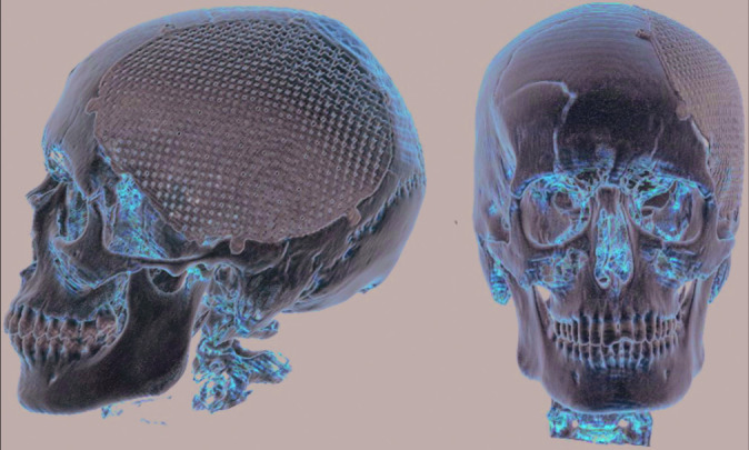
In one patient who underwent decompressive craniectomy for subdural empyema with encephalitis, custom-made mesh was prepared based on a 3D CT scan performed 45 days after surgery. Due to systemic problems related to infectious disease, clinical conditions stabilized very slowly, and cranioplasty was performed after 277 days. Intraoperatively, custom-made prostheses resulted inadequate because of bone defect enlargement (probably due to the progression of infectious disease with osteomyelitis) and therefore the surgery was performed using a precurved mesh. The subsequent postoperative course was uneventful and without complications. This patient was included in the precurved mesh group. In all patient in the custom made group we observed a perfect fit of the mesh on the head defect, this clinical observation was also confirmed by the revision of postoperative imaging [Figure 2].
Figure 2:

Postoperative 3D computed tomography reconstruction of a patient treated using custom-made titanium cranioplasty; the implant completely cover cranial defect and obtain adequate cosmetic result, shape and curvature appear symmetric to contralateral side (patient seven, and visual analog scale for cosmesis 100).
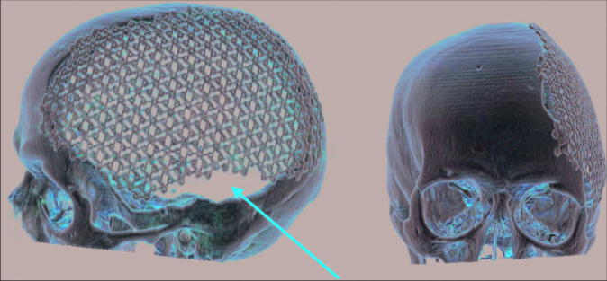
In eight of the 13 patients in whom precurved mesh was used there was no adequate fitting of the mesh on the head defect with persistence of areas not covered by the prostheses [Figure 3]. In all eight cases, the defect was significant on palpation; it was also visible on visual inspection in two cases. In total, three patients complained of muscle pain in the temporal region (two precurved mesh group and one custom-made group).
Figure 3:

Postoperative 3D computed tomography reconstruction of a patient treated using precurved titanium mesh; the cranial defect is not completely covered (red arrow) with persistent small defect in the anterior and inferior portion of the initial defect. Cosmetic result is not satisfactory (patient eight, and visual analog scale for cosmesis 40).
Duration of operation: average surgical time of precurved mesh was 186 min (minmum 110–maximum 240). Average for custom-made prostheses was 141 min (minimum 115– maximum 200); average difference of 45 min with a value of P = 0.04. The three patients who underwent immediate reconstruction after tumor removal were not counted in this evaluation.
Early complications: precurved group: one wound drainage incarceration (removed in the operating room under local anesthesia); and one subcutaneous seroma conservatively treated. Custom-made group: one epidural hematoma surgically evacuated; and one subdural hygroma conservatively treated. Late complications one skin dehiscence, with underlying precurved mesh exposure in patient with multiple cranial surgeries and radiotherapy; dehiscence occurred at a point where the mesh adequately adhered to the cranial defect, no tilt or lump was present; and indication was given for removal of the prostheses but the patient declined. We had no infections. No prostheses were removed. Cosmetic results: precurved VASC average 68 (40– 100), custom-made 94 (80–100); test t P = 0.002.
DISCUSSION
The main objectives of cranioplasty are cosmetics and the protection of the brain. Although there is a great deal of evidence to suggest that cranioplasty can also have a positive effect on neurological outcome (“Syndrome of the Trephined”),[1,5,19,24] in our opinion this aspect was too complex to evaluate as the variables that can influence this result are numerous: type and extent of the initial lesion (trauma, bleeding, and abscess...), clinical conditions of the patient at the time of decompression, postoperative period (any cranial and systemic complications), age of the patient, and neurocognitive conditions at the time of cranioplasty. In our current study, therefore, we have evaluated the results in terms of incidence of complications, surgical times, cosmetics, and failure of the cranioplasty. Failure here means the need to remove the prostheses (prematurely or later), of which the most common causes are infections, exposure of the prostheses to skin lesions, reabsorption of the graft, or of the bone edges on which it is fixed with consequent mobility. Since 2014, the use of titanium cranioplasty has been introduced in our center. The choice of titanium is linked to its features of high biocompatibility, low infection rate, high mechanical resistance to direct trauma, and compatibility with CT and MRI. In our work, we compared the results of two types of titanium cranioplasty (custom-made vs. precurved); the comparison of titanium with autologous bone and other biomaterials (HA, PMMA, and PEEK) is beyond the scope of this article. We therefore analyzed and discussed our results only to evaluate differences in results and indications of the two different titanium cranioplasties used.
Evaluating the major complications, we obtained similar results in the two groups with a major complication for each type of prostheses: a failure (skin dehiscence with prostheses exposure) for the precurved, and an epidural hematoma surgically evacuated for the custom-made group. We had no infections.
Analyzing the single complications, the absence of infections in this series of patients confirmed the literature data showing the low infection rate of titanium compared to other materials.[11,22,27] Cranioplasty infection is one of the most common and most serious complications as it can cause neurological and systemic complications and generally requires the removal of the prostheses (failure). The percentages vary from 0.6% to 25% depending on the series considered.[3,4,5,22,26] Van de Vijfeijken et al. reviewed the literature, including 228 articles, for a total of 10346 cranioplasty procedures; infection was the most common complication (about 6% of the total). In their review, autologous bone and PMMA had the highest rate of infection (6.9% and 7.8%, respectively) compared to HA (3.3%) and titanium (5.4%).[26] Titanium’s low infection rate is confirmed by Matsuno’s work in which titanium prostheses showed significantly lower infection rates than autologous bone, PMMA, and ceramic.[22]
We obtained only one epidural hematoma in a patient with custom-made prostheses who required surgical evacuation. In the whole group, therefore, we had an incidence of 4.3%, which is in line with the data in the literature that report incidences between 2% and 5%,[11,17,21,26,27] although there are also reports with much higher incidence (10%).[12,14,20] Epidural hematoma, in fact, is a rather common complication after cranioplasty (the third in order of frequency after infection and bone resorption).[26] This complication is favored by preoperative depression of the brain at the region of the skull defect. After cranioplasty, the gap between the dura mater and the prostheses facilitates blood retention. In addition, the craniectomy wound contains much scar tissue with rich neovascularization, facilitating intra- and postoperative bleeding. The multi-perforated mesh structure [Figure 1] in both precurved and custom-made meshes reduces the risk of hematoma using subcutaneous blood drainage. This observation is reflected in a randomized clinical trial published by Lindner et al. in 2017, in which the authors compared custom-made cranioplasty in titanium and HA; they found a significantly higher number of epidural hematomas in the HA group compared to none in the titanium group,[20] which could be related to the perforated structure of the prostheses rather than to the biophysical properties of the material used.
We only had one case of failure due to skin dehiscence with exposure of the underlying precurved mesh; although the patient denied to have the prostheses removed due to personal problems, this case must be counted as a failure because the exposed prostheses created a high risk of infection and could lead to the progression of skin dehiscence. The patient had multiple previous surgeries and radiotherapy for skull-base metastases, so we believe that this complication is related to the characteristics of the patient rather than to kind of prostheses. This observation is based on the fact that previous studies have identified several risk factors that contribute toward a poor outcome and failure in cranioplasty, such as previous irradiation, multiple previous operations, communication with the paranasal sinuses, inadequate delay between craniectomy and cranioplasty, and previous infection.[2,27] Our patient had two of these risk factors that may have promoted skin dehiscence, since it also occurred at a point where the mesh had properly adhered to the bone and was not determined by tilts or bumps under the skin.
The main differences between the two prostheses were the duration of the surgery, esthetic result, cost, and time required for production. We did not assessed the postoperative time to discharge because (in our series) it appeared to be mainly dependent on the characteristics of the patient rather than on the type of prostheses used.
The duration of the surgery was on average shorter by around 45 min in the custom-made group [Tables 1 and 2], and this difference reached the threshold of statistical significance evaluated by t-test. This difference is essentially related to the need to adapt the precurved mesh to the defect. The custom-made prostheses perfectly adhered to the defect, precisely reproducing the shape and size. Therefore, once the dissection and exposure of the bone edges was completed, it was enough to position the prostheses and fix it with the appropriate screws. In using the precurved mesh, it had to be adapted partly by cutting off the excess and partly by shaping it to adapt the curvature to the patient. These operations resulted in longer surgical time and greater manipulation of the prostheses. From a theoretical point of view, both data (longer surgical time and prolonged manipulation of the prosthesis) are considered risk factors for infection and failure. Matsuno et al. performed an analysis of the factors influencing bone graft infection after delayed cranioplasty.[22] They reviewed a total of 206 cases, dividing them into two groups: infected (25 cases) and noninfected (181 cases); in their study the mean operation time for the infected group was 146,0 min, whereas that of the noninfected group was 142.2 min. However, there was no statistically significant difference between each group (Student’s t-test), and so the association between increased surgical time and increased infection rate was only suspected but not confirmed.
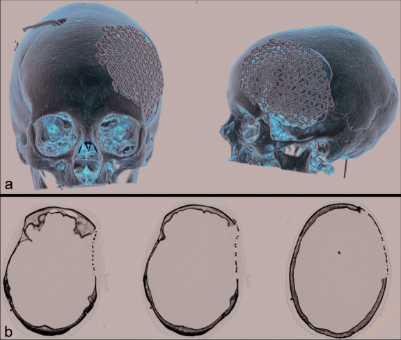
The cosmetic result was assessed using a VASC scale, the score was assigned by the patient (or their family members), but in all cases, the VASC score was consistent with clinicians’ evaluations. Custom-made cranioplasty obtained a higher average score [94 vs. 68, Table 2], with a value of P = 0.002. This result was predictable given the technical difficulties of adequately reproducing the normal shape and cranial conformation; the data were widely discussed in the literature, in which many authors showed the superiority of the cosmetic result of custom-made prostheses, both in titanium[3,13,21] and in other biomaterials such as HA, PMMA, and PEEK.[8,9,14-16,25] An adequate aesthetic result is of great importance both for the psychological perspective of the patient and their relatives and because, in some cases, a prostheses with inadequate curvature can promote pain in the temporal region or skin decubitus which, in turn, can cause the prostheses to fail. It is for these reasons that the cosmetic result is considered one of the main outcomes of cranioplasty.[3,13,21,26,27] Despite the fact that custom-made cranioplasty guarantees optimal esthetic results (regardless of the material used), we must state that both in our experience, and in other literary reports,[7,18,21,28] also manually-shaped cranioplasty can obtain satisfactory results, particularly in cases of small defects or in case of poorly visible defect (posterior location, and long hair) [Figure 4].
Figure 4:

Postoperative computed tomography scan of a patient treated using precurved titanium mesh: (a) 3D reconstruction, (b) three different axial slices of the same exam. The defect is relatively small, although the defect is not completely covered by the mesh, cosmetic result is satisfactory (patient 20, and visual analog scale for cosmesis 90).
The cost of precurved meshes is 1500 € and that of custom- made meshes is about 5500 €; the difference is partly absorbed by the costs of the screws, which are much more numerous in precurved meshes. The high number of screws for this type of prostheses is required to facilitate adhesion with the bone edges and to reduce the risk of tilt under the skin. These costs appear to be in line with what has been reported by other authors. All custom-made cranioplasties have similar costs with differences related to the size of the defect and the material used: PMMA appears to be the least expensive (but with the highest rate of infection), with titanium and PEEK being the most expensive.[3,21,26] Considering this, Gilardino et al. carried out a comparison and cost analysis of cranioplasty techniques comparing autologous bone and custom computer-generated implants. The results of their study demonstrated no significant increase in overall treatment cost associated with the use of the custom-made cranioplasty technique. In addition, the latter was associated with a statistically significant decrease in operative time and need for ICU admission when compared with those patients who underwent autologous bone cranioplasty.[10]
The last difference between the two groups in our current study was the time taken to obtain the prostheses. The precurved meshes, after purchase, were always available in the hospital for implantation even in emergency conditions; the production time for custom-made prostheses (from the execution of the CT to the actual availability in the hospital) was 20–30 days. This time delay is irrelevant in patients who had to undergo delayed cranioplasty after decompressive craniotomy; these patients (who represent the majority in almost all series), required a reasonable period of time before the reconstruction of the cranial defect to promote the stabilization of clinical conditions and the regression of the cerebral swelling. As such, cranioplasty surgery was usually planned in advance, and consequently the 3–4 weeks necessary for obtaining the prostheses caused no problems. There are cases, however, in which it may be useful to reduce the wait for cranioplasty. This would be particularly useful for cases of single stage surgery (resection and reconstruction): neoplasms with bone involvement, head trauma with comminute fracture, and also infectious diseases with bone involvement as proposed by Ehrlich et al. The authors performed immediate titanium cranioplasty for treatment of postcraniotomy infection (manually shaped mesh). In their series (24 patients), only two patients were reoperated (but not due to prostheses infection), and all patients who completed the evaluation questionnaire (20/24) said they were satisfied with the esthetic result.[7] The possibility of immediate reconstruction has many advantages: it would avoid cosmetic deformity caused by the craniotomy defect and the vulnerability of the unprotected brain; furthermore, it would avoid additional stress for the patient that would come from a second surgery with all its medical and surgical risks. In addition, second surgery creates extra costs induced by hospital stay, surgical time, recovery, and unemployability of the patient.[7,18,28]
Study limitations
Our study had limitations, since the study was retrospective and the sample size was rather small. The two groups have similar demographic and etiopathological features, but the small sample number prevented the extrapolation of any definitive conclusions. The results must be integrated with data from literature and on a broader array of cases, particularly for appropriate and complete economic and health observations.
CONCLUSION
In our study, we carried out a retrospective evaluation of two types of titanium cranioplasty: precurved meshes compared with custom-made prostheses. The two groups of patients had similar demographic and clinical characteristics. We evaluated the differences of the two methods, rates of complications, esthetic results, surgical times, and material costs.
In the final comparison, the two prostheses showed overlapping rates of complications in line with the data of the literature; custom-made cranioplasties had a much better aesthetic result, perfect and complete coverage of the head defect, shorter surgical times, and involved an easier operation for the surgeon. Precurved meshes were cheaper, with satisfactory esthetic results in small defects, and were always available even for emergencies or for immediate reconstruction; in case of large defects, the cosmetic result is, on average, lower, with an increased surgical time and incomplete coverage of the craniolacunia in a high percentage of patients. Therefore, despite the greater costs, we believe that a custom-made prostheses should be the first choice, especially in young patients with large cranial defects due to results of decompressive craniotomy; in selected cases, however, precurved meshes may be recommended for small defects in particular, and for single- stage resection reconstruction procedure. In these situations, precurved mesh could be used with good results and with a simultaneous reduction in health-care costs.
Footnotes
How to cite this article: Policicchio D, Casu G, Dipellegrini G, Doda A, Muggianu G, Boccaletti R. Comparison of two different titanium cranioplasty methods: Custom-made titanium prostheses versus precurved titanium mesh. Surg Neurol Int 2020;11:148.
Contributor Information
Domenico Policicchio, Email: domenico.policicchio@aousassari.it.
Gina Casu, Email: casugina@tiscali.it.
Giosuè Dipellegrini, Email: giosuedipellegrini@gmail.com.
Artan Doda, Email: artandoda@gmail.com.
Giampiero Muggianu, Email: giampiero.muggianu@aousassari.it.
Riccardo Boccaletti, Email: riccardo.boccaletti@aousassari.it.
Declaration of patient consent
Patient’s consent not required as patients identity is not disclosed or compromised.
Financial support and sponsorship
Nil.
Conflicts of interest
There are no conflicts of interest.
REFERENCES
- 1.Annan M, De Toffol B, Hommet C, Mondon K. Sinking skin flap syndrome (or syndrome of the trephined): A review. Br J Neurosurg. 2015;29:314–8. doi: 10.3109/02688697.2015.1012047. [DOI] [PubMed] [Google Scholar]
- 2.Baumeister S, Peek A, Friedman A, Levin LS, Marcus JR. Management of postneurosurgical bone flap loss caused by infection. Plast Reconstr Surg. 2008;122:195e–208e. doi: 10.1097/PRS.0b013e3181858eee. [DOI] [PubMed] [Google Scholar]
- 3.Cabraja M, Klein M, Lehmann TN. Long-term results following titanium cranioplasty of large skull defects. Neurosurg Focus. 2009;26:E10. doi: 10.3171/2009.3.FOCUS091. [DOI] [PubMed] [Google Scholar]
- 4.Corliss B, Gooldy T, Vaziri S, Kubilis P, Murad G, Fargen K. Complications after in vivo and ex vivo autologous bone flap storage for cranioplasty: A comparative analysis of the literature. World Neurosurg. 2016;96:510–5. doi: 10.1016/j.wneu.2016.09.025. [DOI] [PubMed] [Google Scholar]
- 5.De Bonis P, Frassanito P, Mangiola A, Nucci CG, Anile C, Pompucci A. Cranial repair: How complicated is filling a hole? J Neurotrauma. 2012;29:1071–6. doi: 10.1089/neu.2011.2116. [DOI] [PubMed] [Google Scholar]
- 6.De Bonis P, Pompucci A, Mangiola A, D’Alessandris QG, Rigante L, Anile C. Decompressive craniectomy for the treatment of traumatic brain injury: Does an age limit exist? J Neurosurg. 2010;112:1150–3. doi: 10.3171/2009.7.JNS09505. [DOI] [PubMed] [Google Scholar]
- 7.Ehrlich G, Kindling S, Wenz H, Hänggi D, Schulte DM, Schmiedek P, et al. Immediate titanium mesh implantation for patients with postcraniotomy neurosurgical site infections: Safe and aesthetic alternative procedure? World Neurosurg. 2017;99:491–9. doi: 10.1016/j.wneu.2016.12.011. [DOI] [PubMed] [Google Scholar]
- 8.Fiaschi P, Pavanello M, Imperato A, Dallolio V, Accogli A, Capra V, et al. Surgical results of cranioplasty with a polymethylmethacrylate customized cranial implant in pediatric patients: A single-center experience. J Neurosurg Pediatr. 2016;17:705–10. doi: 10.3171/2015.10.PEDS15489. [DOI] [PubMed] [Google Scholar]
- 9.Fricia M, Nicolosi F, Ganau M, Cebula H, Todeschi J, Santin MD, et al. Cranioplasty with porous hydroxyapatite custom-made bone flap: Results from a multicenter study enrolling 149 patients over 15 years. World Neurosurg. 2019;121:160–5. doi: 10.1016/j.wneu.2018.09.199. [DOI] [PubMed] [Google Scholar]
- 10.Gilardino MS, Karunanayake M, Al-Humsi T, Izadpanah A, Al-Ajmi H, Marcoux J, et al. A comparison and cost analysis of cranioplasty techniques: Autologous bone versus custom computer-generated implants. J Craniofac Surg. 2015;26:113–7. doi: 10.1097/SCS.0000000000001305. [DOI] [PubMed] [Google Scholar]
- 11.Honeybul S, Morrison DA, Ho KM, Lind CR, Geelhoed E. A randomized controlled trial comparing autologous cranioplasty with custom-made titanium cranioplasty. J Neurosurg. 2017;126:81–90. doi: 10.3171/2015.12.JNS152004. [DOI] [PubMed] [Google Scholar]
- 12.Jaberi J, Gambrell K, Tiwana P, Madden C, Finn R. Long-term clinical outcome analysis of poly-methyl-methacrylate cranioplasty for large skull defects. J Oral Maxillofac Surg. 2013;71:e81–8. doi: 10.1016/j.joms.2012.09.023. [DOI] [PubMed] [Google Scholar]
- 13.Joffe J, Harris M, Kahugu F, Nicoll S, Linney A, Richards R. A prospective study of computer-aided design and manufacture of titanium plate for cranioplasty and its clinical outcome. Br J Neurosurg. 1999;13:576–80. doi: 10.1080/02688699943088. [DOI] [PubMed] [Google Scholar]
- 14.Jonkergouw J, van de Vijfeijken SE, Nout E, Theys T, Van de Casteele E, Folkersma H, et al. Outcome in patient-specific PEEK cranioplasty: A two-center cohort study of 40 implants. J Craniomaxillofac Surg. 2016;44:1266–72. doi: 10.1016/j.jcms.2016.07.005. [DOI] [PubMed] [Google Scholar]
- 15.Kasprzak P, Tomaszewski G, Kotwica Z, Kwinta B, Zwoliński J. Reconstruction of cranial defects with individually formed cranial prostheses made of polypropylene polyester knitwear: An analysis of 48 consecutive patients. J Neurotrauma. 2012;29:1084–9. doi: 10.1089/neu.2011.2247. [DOI] [PubMed] [Google Scholar]
- 16.Kim BJ, Hong KS, Park KJ, Park DH, Chung YG, Kang SH. Customized cranioplasty implants using three-dimensional printers and polymethyl-methacrylate casting. J Korean Neurosurg Soc. 2012;52:541–6. doi: 10.3340/jkns.2012.52.6.541. [DOI] [PMC free article] [PubMed] [Google Scholar]
- 17.Kim JK, Lee SB, Yang SY. Cranioplasty using autologous bone versus porous polyethylene versus custom-made titanium mesh: A retrospective review of 108 patients. J Korean Neurosurg Soc. 2018;61:737–46. doi: 10.3340/jkns.2018.0047. [DOI] [PMC free article] [PubMed] [Google Scholar]
- 18.Kshettry VR, Hardy S, Weil RJ, Angelov L, Barnett GH. Immediate titanium cranioplasty after debridement and craniectomy for postcraniotomy surgical site infection. Neurosurgery. 2012;70(Suppl 1):8–14. doi: 10.1227/NEU.0b013e31822fef2c. [DOI] [PubMed] [Google Scholar]
- 19.Kuo JR, Wang CC, Chio CC, Cheng TJ. Neurological improvement after cranioplasty-analysis by transcranial doppler ultrasonography. J Clin Neurosci. 2004;11:486–9. doi: 10.1016/j.jocn.2003.06.005. [DOI] [PubMed] [Google Scholar]
- 20.Lindner D, Schlothofer-Schumann K, Kern BC, Marx O, Müns A, Meixensberger J. Cranioplasty using custom-made hydroxyapatite versus titanium: A randomized clinical trial. J Neurosurg. 2017;126:175–83. doi: 10.3171/2015.10.JNS151245. [DOI] [PubMed] [Google Scholar]
- 21.Luo J, Liu B, Xie Z, Ding S, Zhuang Z, Lin L, et al. Comparison of manually shaped and computer-shaped titanium mesh for repairing large frontotemporoparietal skull defects after traumatic brain injury. Neurosurg Focus. 2012;33:E13. doi: 10.3171/2012.2.FOCUS129. [DOI] [PubMed] [Google Scholar]
- 22.Matsuno A, Tanaka H, Iwamuro H, Takanashi S, Miyawaki S, Nakashima M, et al. Analyses of the factors influencing bone graft infection after delayed cranioplasty. Acta Neurochir (Wien) 2006;148:535–40. doi: 10.1007/s00701-006-0740-6. [DOI] [PubMed] [Google Scholar]
- 23.Policicchio D, Doda A, Sgaramella E, Ticca S, Santonio FV, Boccaletti R. Ultrasound-guided brain surgery: Echographic visibility of different pathologies and surgical applications in neurosurgical routine. Acta Neurochir (Wien) 2018;160:1175–85. doi: 10.1007/s00701-018-3532-x. [DOI] [PubMed] [Google Scholar]
- 24.Sakamoto S, Eguchi K, Kiura Y, Arita K, Kurisu K. CT perfusion imaging in the syndrome of the sinking skin flap before and after cranioplasty. Clin Neurol Neurosurg. 2006;108:583–5. doi: 10.1016/j.clineuro.2005.03.012. [DOI] [PubMed] [Google Scholar]
- 25.Stefini R, Esposito G, Zanotti B, Iaccarino C, Fontanella MM, Servadei F. Use of custom made porous hydroxyapatite implants for cranioplasty: Postoperative analysis of complications in 1549 patients. Surg Neurol Int. 2013;4:12. doi: 10.4103/2152-7806.106290. [DOI] [PMC free article] [PubMed] [Google Scholar]
- 26.Van de Vijfeijken SE, Münker TJ, Spijker R, Karssemakers LH, Vandertop WP, Becking AG, et al. Autologous bone is inferior to alloplastic cranioplasties: Safety of autograft and allograft materials for cranioplasties, a systematic review. World Neurosurg. 2018;117:443–52.e8. doi: 10.1016/j.wneu.2018.05.193. [DOI] [PubMed] [Google Scholar]
- 27.Williams LR, Fan KF, Bentley RP. Custom-made titanium cranioplasty: Early and late complications of 151 cranioplasties and review of the literature. Int J Oral Maxillofac Surg. 2015;44:599–608. doi: 10.1016/j.ijom.2014.09.006. [DOI] [PubMed] [Google Scholar]
- 28.Wind JJ, Ohaegbulam C, Iwamoto FM, Black PM, Park JK. Immediate titanium mesh cranioplasty for treatment of postcraniotomy infections. World Neurosurg. 2013;79:207.e11–3. doi: 10.1016/j.wneu.2011.02.013. [DOI] [PMC free article] [PubMed] [Google Scholar]


