Abstract
Chromosomal rearrangements (CR) such as translocations, duplications and inversions play a decisive role in the adaptation of microorganisms to specific environments. In enological Saccharomyces cerevisiae strains, CR involving the promoter region of the gene SSU1 lead to a higher sulfite tolerance by enhancing the SO2 efflux. To date, three different SSU1 associated CR events have been described, including translocations XV-t-XVI and VIII-t-XVI and inversion inv-XVI. In the present study, we developed a multiplex PCR method (SSU1 checkup) that allows a rapid characterization of these three chromosomal configurations in a single experiment. Nearly 600 S. cerevisiae strains collected from fermented grape juice were genotyped by microsatellite markers. We demonstrated that alleles of the SSU1 promoter are differently distributed according to the wine environment (cellar versus vineyard) and the nature of the grape juice. Moreover, rearranged SSU1 promoters are significantly enriched among commercial starters. In addition, the analysis of nearly isogenic strains collected in wine related environments demonstrated that the inheritance of these CR shapes the genetic diversity of clonal populations. Finally, the link between the nature of SSU1 promoter and the tolerance to sulfite was statistically validated in natural grape juice containing various SO2 concentrations. The SSU1 checkup is therefore a convenient new tool for addressing population genetics questions and for selecting yeast strains by using molecular markers.
Keywords: sulfite resistance, domestication, translocation, multiplex PCR, marker assisted selection
Introduction
Microorganisms develop various strategies for being better adapted to various environments. Among them, the yeast Saccharomyces cerevisiae is a noteworthy example of a microorganism whose evolution led to specialized genetic groups associated with different human-related environments (Sicard and Legras, 2011; Borneman and Pretorius, 2014; Marsit and Dequin, 2015). In a winemaking context, this species has been exposed to stressful conditions (high alcohol content, high osmotic pressure, low pH, etc.) for millennia, potentially resulting in adaptive differentiation.
In wine production, sulfite addition is widely used since the middle age as a preservative because of its antimicrobial, antioxidant, and antioxydasic activities. Produced by dissolution of sulfur dioxide (SO2), sulfite inhibits key glycolytic enzymes like Tdh and Adh proteins, binds carbonyl compounds such as pyruvate and acetaldehyde (Hinze and Holzer, 1986) affects transporter activity by binding membrane proteins (Divol et al., 2012) and down-regulates the expression of many central metabolism genes (Park and Hwang, 2008). Therefore, sulfite tolerance has been unconsciously selected by wine making practices and constitutes a desired trait in Saccharomyces wine yeast strains. Cellular mechanisms of sulfite tolerance have been extensively reviewed in S. cerevisiae (Divol et al., 2012; García-Ríos and Guillamón, 2019). They include the overproduction of acetaldehyde (Cheraiti et al., 2010) the regulation of sulfite reduction systems and more generally of the sulfur metabolic pathway (Divol et al., 2012). Moreover, sulfite tolerance mostly depends on the pumping of SO2 through the plasma membrane. This sulfite efflux involves the sulfite pump Ssu1p which is encoded by the SSU1 gene. This gene shows a high level of polymorphism (Aa et al., 2006) and deleterious mutations in its coding sequence cause SO2 susceptibility (Avram and Bakalinsky, 1997; Park and Bakalinsky, 2000).
The expression level of SSU1 has a direct consequence on sulfite tolerance and has been widely studied (Pérez-Ortín et al., 2002; Nardi et al., 2010; Engle and Fay, 2012; Zimmer et al., 2014; García-Ríos et al., 2019). Interestingly, the SSU1 promoter sequence is involved in three Chromosomal Rearrangements (CR) (i.e., XV-t-XVI, VIII-t-XVI, and inv-XVI) that increase its expression leading to a more efficient sulfite pumping over (Pérez-Ortín et al., 2002; Zimmer et al., 2014; García-Ríos et al., 2019). These three independent CR events constitute a hallmark on parallel evolutionary routes driven by human selection. In the VIII-t-XVI translocation, the native promoter of SSU1 is replaced by tandem repeated sequences of the ECM34 promoter from chromosome VIII (Pérez-Ortín et al., 2002). In the XV-t-XVI translocation, the upstream region of SSU1 is placed head to tail with the ADH1 promoter from chromosome XV (Zimmer et al., 2014). The inversion of chromosome XVI (inv-XVI) involves the SSU1 and GCR1 regulatory regions, increasing the expression of SSU1 (García-Ríos et al., 2019).
To date, the distribution of translocation (XV-t-XVI and VIII-t-XVI) and inversion (inv-XVI) events of the SSU1 gene have been investigated for a small number of strains (Pérez-Ortín et al., 2002; Zimmer et al., 2014; García-Ríos et al., 2019). Here, we set up a multiplex method (SSU1 checkup) based on labeled primers with different fluorochromes, to identify in a single assay the three types of SSU1 associated CR (VIII-t-XVI, XV-t-XVI, and inv-XVI) as well as the wild type forms of these chromosomes (VIII-wt, XV-wt, and XVI-wt). The SSU1 was applied to nearly 600 yeast strains, including natural isolates and commercial starters, and provides new insights on the allele frequency of rearranged SSU1 promoters. In addition, by using microsatellite genotyping, the genetic relationships between strains of the collection were established allowing the study of CR occurrence in nearly isogenic clones. Finally, for a subset of strains, the phenotypic impact of different CR was evaluated by measuring their parameters of growth in grape juice containing different concentrations of SO2.
Materials and Methods
Origin of Samples
A total of 628 S. cerevisiae isolates were collected from grapes and fermented must (white, red, and sweet) originating from five different countries (France, Lebanon, Argentina, Spain, and Italy), two different Vitis species (mostly V. vinifera and to a lesser extent V. labrusca) and nine different varieties. Two different procedures were used for strain isolation depending on the environment considered: vineyard or cellar. For vineyard isolates, around 2 kg of healthy and mostly undamaged grapes were collected a few days before the harvest in the vineyard, crushed in sterile conditions and macerated for 2 h with 50 mg/L of SO2. The juice was fermented at 21°C in small glass-reactors (500 mL). For cellar isolates, yeast colonies were obtained from spontaneous fermentation vats containing sulfited grape juices according to local enological practices (ranging from 20 to 50 mg/L of sulfur dioxide) except for sweet wines for which no sulfur dioxide was added. For both sampling procedures, fermentations were allowed to proceed until 2/3 of the must sugars were consumed and fermented juices were plated onto YPD plates (yeast extract, 1% w/v; peptone, 1% w/v; glucose, 2% w/v; agar 2% w/v) with 100 μg/mL of chloramphenicol and 150 μg/mL of biphenyl to delay bacterial and mold growth. Around 30 colonies per sample were randomly chosen and after sub-cloning on YPD plates, each yeast colony was stored in 30% (v/v) glycerol at −80°C. Additionally, a collection of 103 industrial S. cerevisiae starters was constituted by streaking on YPD plates a small aliquot of Active Dry Yeast obtained from different commercial suppliers.
Microsatellite Analysis
Strains were genotyped using fifteen polymorphic microsatellite loci (C3, C4, C5, C6, C8, C9, C11, SCAAT1, SCAAT2, SCAAT3, SCAAT5, SCAAT6, SCYOR267C, YKL172W, YPL009C) developed for estimating the genetic relationships among S. cerevisiae strains (Legras et al., 2007). Most of the strains were previously genotyped in our lab (Börlin et al., 2016; Raymond et al., 2018; Peltier et al., 2018a; Borlin, 2015) and the additional 82 strains were genotyped in this work using identical experimental conditions. Briefly, two multiplex PCRs were carried out in a final volume of 12.5 μL containing 6.25 μL of the Qiagen Multiplex PCR master mix (Qiagen, France), 1 μL of DNA template, and 1.94 μL of each mix, using the conditions previously reported (Peltier et al., 2018b). Both reactions were run using an initial denaturation step at 95°C for 5 min, followed by 35 cycles of 95°C for 30 s, 57°C for 2 min, 72°C for 1 min, and a final extension step at 60°C for 30 min. The size of PCR products was determined by the MWG company (Ebersberg, Germany), using 0.2 μL of 600 LIZ GeneScan (Applied Biosystems, France) as a standard marker, and chromatograms were analyzed with the GeneMarker (V2.4.0, Demo) program. Only strains that amplified at least 12 of 15 loci were kept. On the 735 strains collected, 586 met this criterion and were used in this study (listed on Supplementary Table S1). The microsatellite data set was analyzed by means of the poppr R package using the Bruvo’s distance matrix. Strains showing a strong similarity were identified by applying a cut of value of 0.15 to the Bruvo’s genetic distance matrix. In this way, 194 very closely related strains were identified and considered as “clones.” This cut off value was defined in order to restore the normality of the distribution (Supplementary Figure S1). The assignment of clustering methods was achieved by using the find.clusters function (adegenet package). The selection of the optimal groups was computed by the Ward’s clustering method using the Bayesian Information Criterion (BIC) as statistical criterion (Avramova et al., 2018).
The SSU1 Checkup Method
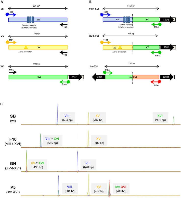
In order to experimentally detect in a single multiplex PCR test all the CR involving the gene SSU1, labeled primers were designed using a specific dye per chromosome position as follows: 6-FAM (Chr8: VIII-14558), ATTO550 (Chr15: XV-160994), HEX (Chr16: XVI-373707), and ATTO565 (Chr16: XVI-412453) (Table 1). All the primers (Table 1 and Supplementary Table S2) were synthesized by Eurofins genomics (Ebersberg, Germany). A multiplex PCR was carried out in a final volume of 20 μL using 100 nM of each primer, 1 μL of template DNA and the Qiagen PCR multiplex PCR kit (Qiagen, France) on a T100TM Thermal cycler (Bio-Rad, France). The following PCR program allows the amplification of all the expected fragments from the rearranged and the wild type VIII, XV and XVI chromosomes: initial denaturation at 95°C for 15 min, followed by 35 cycles of 94°C for 30 s, 55°C for 90 s, 72°C for 90 s, ending with a hold at 60°C for 30 min. DNA templates for PCR were extracted in 96-well microplates using the previously described LiAc-SDS protocol (Chernova et al., 2018). Before analysis, PCR products were diluted 60 times in ddH2O and 1 μL of this solution was mixed with 0.2 μL of the internal size standard GenScanTM 1200 LIZ (Applied Biosystems, France) and 9.8 μL of highly deionized Hi-DiTM formamide (Applied Biosystems, France). Samples were analyzed by Eurofins genomics (Ebersberg, Germany) on an ABI-3710 Genetic Analyzer. Each peak was identified according to the color and size and attributed to the alleles (Figure 1). Each allele was also sequenced by amplifying both strands with non-labeled primers. The sequences were released on GenBank with the following accession numbers: ID MT028493-MT028507.
TABLE 1.
Primers used for the SSU1 checkup method and the strains used as positive controls.
| Chromosome | Strain | Primer F |
Primer R |
Sizea | Positionb | ||||
| Name | Sequence | Dye | Name | Sequence | Dye | ||||
| VIII | SB | 1189 | ATGGCAGCTTCTAAGTTGTGG | FAM | 1190 | GTTTATGTTTGGTTTGGGGG | na | 604 | VIII-14558 |
| GN | 667 | to VIII-15162 | |||||||
| XV | SB | 1191 | AAAGAAGTTGCATGCGCCTA | ATTO550 | 1192 | ACCTGAGTGCATTTGCAACA | na | 702 | XV-160994 |
| F10 | 702 | to XV-161695 | |||||||
| XVI | SB | 1193 | TGTCAAGTTGAGACAAACCGA | na | 1194 | GGGGAAAGCTGTAATTTGTGT | Hex | 991 | XVI-372717 to XVI-373707 |
| VIII-t-XVI | F10 | 1189 | ATGGCAGCTTCTAAGTTGTGG | FAM | 1194 | GGGGAAAGCTGTAATTTGTGT | Hex | 555 | VIII-14558 to XVI-373707 |
| XV-t-XVI | GN | 1191 | AAAGAAGTTGCATGCGCCTA | ATTO550 | 1194 | GGGGAAAGCTGTAATTTGTGT | Hex | 496 | XV-160994 to XVI-373707 |
| inv-XVI | P5 | 1196 | TGCATAAGCAGGCAACTCCT | ATTO565 | 1194 | GGGGAAAGCTGTAATTTGTGT | Hex | 781 | XVI-373707-412453 |
aPCR product lengths in base pairs; bposition in the chromosome for reference strain S288c; na: not applicable.
FIGURE 1.
(A) Position of labeled primers for on the non-rearranged chromosomes VIII, XV, and XVI. The position of the promoter regions ECM34 and ADH1 were represented by a multiple blue bar and a yellow triangle, respectively. The gene SSU1 is represented by a black arrow. The tandem repeats on chromosome VIII (indicated by an *) generates multiple type of amplicons. Labeled primers are represented by a star and the colors blue, yellow, and green represent the specific dye used :FAM, ATO550, and HEX. (B) Rearranged chromosome XVI investigated in this study (VIII-t-XVI, XV-t-XVI, and inv-XVI). The relative position of the gene SSU1 and the modified promoter regions (ECM34, ADH1, and GCR1) are indicated. The hatched line represents the chromosomal break point in rearranged strains F10 (VIII-t-XVI), GN (XV-t-XVI), and P5 (inv-XVI), respectively. The red star represents the primer specific to the inv-XVI chromosome labeled with the fluorochrome ATO 565. (C) Chromatograms of multiplexed PCR reactions for the reference strains. The wt and rearranged XVI chromosomes were detected by co migration of fragment of different size labeled with specific dyes. For Chromosome VIII different sizes were obtained according to the strain due to the differential number of tandem repeats in ECM34 promoter.
SO2 Tolerance Assessment
To assess SO2 tolerance, a subset of 34 strains (Supplementary Table S3) was cultivated in white grape juice (Sauvignon blanc from the Bordeaux area, France). This must had a total SO2 concentration of 14 mg/L and was spiked with 0, 25, 50, and 75 mg/L of total SO2. Cultures were achieved in 96-well plates (U flat well, Greiner, France) filled with 200 μL of grape juice sterilized by a nitrate-cellulose membrane filtration (Millipore, France). Yeasts were pre-cultivated in YPD media (yeast extract, 1% w/v; peptone, 1% w/v; glucose, 2% w/v) for 16 h at 28°C and inoculated into the grape juice to a final concentration of 1 × 10⁶ cells/mL. Growth was monitored by OD600 measurements for 96 h at 28°C using a microplate spectrophotometer (Synergy HT Multi-Mode Reader, BioTek Instruments, Inc., United States). Culture plates were shaken every 25 min for 30 s prior to the OD600 measurements. The well position on the microplate was randomized and six replicates were done for each strain∗media condition. Data from the microplate reader were transformed with the polynomial curve y = −0.0018∗x3+0.1464∗x2+0.7757∗x+0.0386 to correct the non-linearity of the optical recording at higher cell densities as previously reported (Martí-Raga et al., 2016). Growth kinetic data were fitted using the Richards flexible inflection point model implemented by the fit growthmodel function, R package growthrates. This model allows the estimation of the maximal growth rate (μmax). A second parameter, Lag Time, was manually computed from raw data by considering the time necessary to reach twice the OD600 of the inoculum. A linear model was applied for estimating effects of the SO2 concentration and type of chromosome XVI and their possible interactions:
-
(1)
Lm1: Yik = m + ChrXVI i + SO2 k + (ChrXVI:SO2) ik + E ijk
Where Y are the values of the trait (μmax and Lag Time), for j ChrXVI configurations (i = 1 to 6), and k SO2(k=1 to4) concentrations, m was the overall mean and Eijk the residual error. Homoscedasticity of the ANOVA was tested by LeveneTest function (car package) while the normal distribution of models’ residuals was estimated by visual inspection (qq plot).
Results and Discussion
Assessment of the Genetic Diversity of Starters and Natural Isolates Populations of S. cerevisiae
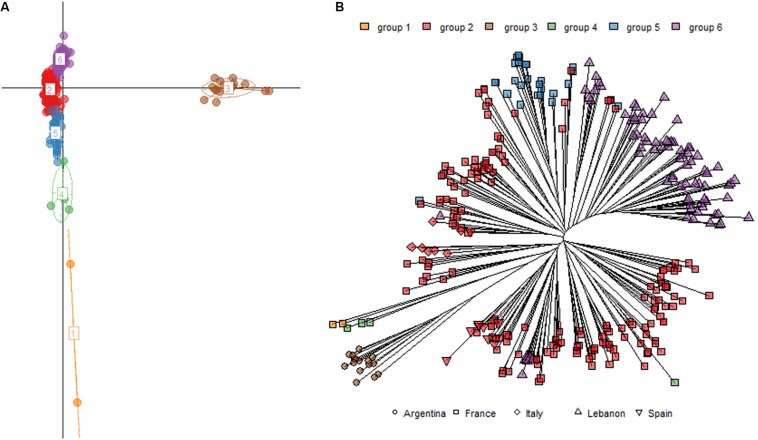
In this study, we analyzed a large dataset of 586 isolates that were genotyped using 15 microsatellite loci. This collection includes 103 industrial starters and 483 indigenous isolates from different origins (sampling mode, red or white grape must, country). Since many natural isolates were sampled in the same juice, some of them could have originated from clonal expansion and be very similar from a genetic point of view. A filtering procedure was applied for keeping only one representative genotype of each clonal population by removing all but one strain having a Bruvo’s genetic distance lower than 0.15 (Supplementary Figure S1). By this procedure, many natural isolates closely related to industrial starters were identified (Supplementary Figure S2) demonstrating the wide dissemination of commercial yeasts in vineyard and winery environments as previously reported (Valero et al., 2005; Borlin, 2015). In addition, 21 isogenic strains were found among commercial starters. This filtering procedure defined three subpopulations: “starters = 82,” “natural isolates = 310,” and “closely related clones = 194” (Supplementary Table S1). The genetic relationships for each strain within the starters and natural isolates subpopulations were then analyzed by a principal component analysis (k = 6). The constitution of genetic groups based on microsatellites inheritance was carried out by using a k-mean based algorithm (see section “Materials and Methods”).
This genetic analysis clustered the 82 commercial strains in three groups with a group C clearly separated from the other two (Supplementary Figure S3) and corresponding to “Champenoise” strains, a particular wine yeast group previously described (Legras et al., 2007; Novo et al., 2009; Borneman et al., 2016). Its detection validated our clustering analysis based on the use of k-mean clustering. The structure of the 310 natural isolates collected was also investigated and six subgroups were defined. Figure 2A shows the first two dimensions of the PCA; axis one clearly identified a group of isolates from Argentina, while axis 2 broadly discriminated the five other groups. The assignment of subgroups on neighbor-joining tree (unrooted) illustrates that isolates are mostly clustered according to their geographical origins (Figure 2B). Some groups are specific to sampling zones such as group 3 (n = 16) and group 6 (n = 84) that only contain strains sampled in Argentina and Lebanon, respectively. In contrast, group 2 (n = 177) encompassed isolates from different geographic origins (Italy, Spain, France, and Lebanon) (Supplementary Table S4). Although not perfectly discriminating, this first analysis filtered the redundancy of our collection and provided a clear overview of the genetic diversity of non-redundant strains.
FIGURE 2.
(A) Principal component analysis of 310 natural isolates discriminated by 15 polymorphic loci. The six groups represented (1 to 6) were inferred by k-mean clustering. The (B) represents the position of the strains according to the inferred groups and the country origin of their sampling.
Development of the SSU1 Checkup, a Simple Method for Genotyping SSU1 Chromosomal Rearrangements in S. cerevisiae
To date, translocation events (i.e., XV-t-XVI and VIII-t-XVI) have been detected by classical PCR experiments narrowing the chromosomal break points identified by two original works (Pérez-Ortín et al., 2002; Zimmer et al., 2014). Recently, an additional chromosomal rearrangement involving the gene SSU1 (inv-XVI) was also described (García-Ríos et al., 2019). Because these classical PCR amplifications are scarcely adapted to screen multiple genotypes in large populations, a multiplexed method (SSU1 checkup) was set up aiming to identify, in a single PCR reaction, these three types of chromosomal rearrangements (VIII-t-XVI, XV-t-XVI, and inv-XVI) as well as the wild type alleles of the corresponding chromosomes (VIII-wt, XV-wt, and XVI-wt). Since primers are labeled with different fluorochromes they allow the identification of the different allelic combinations. Primers used for amplifying the wild type chromosomes (VIII-wt, XV-wt, and XVI-wt) were labeled with a single fluorophore (FAM, ATTO 550, Hex), providing blue, yellow, and green peaks, respectively (Figure 1A). With this set of primers, the amplifications of the rearranged SSU1 promoter regions result to be labeled with two fluorophores, allowing the easy identification of the recombined forms (Figure 1B). The allele sizes amplified range between 388 and 991 bp and were analyzed by following the fluorescence of PCR products with an ABI sequencer (Figure 1C). In preliminary studies, we used reference strains SB (XVI-wt), GN (XV-t-XVI), F10 (VIII-t-XVI), and P5 (inv-XVI), to design and validate primers. Primers position, as well as the length of the DNA fragments amplified for the reference strains, are summarized in Table 1. The sequences of all the alleles identified were submitted to GenBank (ID 2310529). The strain P5 is a commercial starter able to sporulate (data not shown) and would be therefore diploid. The ploidy level of strains SB, GN, and F10 has been previously determined by several genetic analyses (Albertin et al., 2009). These strains are fully homozygous diploids (2n) and were obtained by the sporulation of the commercial starters Actiflore BO213, Zymaflore VL1 and Zymaflore F10, Laffort, France) as previously reported (Marullo et al., 2006, 2009). Their associated chromatograms showed the different types of alleles reported in this study (Figure 1C). In order to simplify the chromatographic patterns, the detection of reciprocal translocation events (XVI-t-XV and XVI-t-VIII) was not included in the SSU1 checkup. However, the strains GN and F10 harbor reciprocal translocations that have been verified by PCR using the primers given in Supplementary Table S2.
The SSU1 checkup was used for tracking the two translocation events (VIII-t-XVI and XV-t-XVI) as well as the chromosomal inversion (inv-XVI) in a large collection of strains (n = 586). In the VIII-t-XVI translocation, the native promoter of SSU1 is replaced by DNA sequences of the ECM34 promoter (located on chromosome VIII) (Pérez-Ortín et al., 2002). Alleles for this CR (i.e., VIII-t-XVI388, VIII-t-XVI478, VIII-t-XVI555, and VIII-t-XVI631 bp) result from the alternative number of units of tandem repeated motifs (76 bp and/or 47 bp) localized in the promoter region of the gene ECM34 (Supplementary Figure S4). In the XV-t-XVI translocation, the upstream region of SSU1 is placed head to tail with the ADH1 promoter (located on chromosome XV) (Zimmer et al., 2014) and a single allele has been recognized (XV-t-XV496). Finally, the chromosomal rearrangement (inv-XVI) has been recently reported by García-Ríos et al. (2019) and consist in a chromosome XVI inversion generating a new SSU1 promoter placed head to tail with the GCR1 promoter. This event was detected in only 19 natural isolates and showed a single allele of 781 bp (inv-XVI781). Surprisingly, twenty strains definitively failed to amplify any fragment even when performing single PCR reactions using alternative primers (Supplementary Table S2). This interesting result suggests that these strains could harbor another still uncharacterized chromosomal rearrangement flanking the SSU1 gene. Such possible new CR should be tracked by chromosome walking PCR starting from SSU1 gene or by de novo assembly of whole genome sequences.
Landscape of the Different SSU1-Promoter Alleles in Natural and Selected Populations
The SSU1 checkup method allows the detection of four types of chromosome XVI structures in a single PCR reaction. However, this method is not quantitative, and it is not possible to know the number of copies of each haplotype. Since all the fragments amplified by the primer 1194 (Hex) are physically linked to the chromosome XVI’s centromere (CEN16), they belong to chromosome XVI during the cell division process. Therefore, native and rearranged chromosome XVI alleles can be merged in order to have an integrated overview of the chromosome XVI inheritance. This allows following the inheritance of the different promoter versions of the SSU1 gene.
The different alleles of chromosome XVI were counted among the 392 non-redundant S. cerevisiae strains analyzed. As a first approximation, we considered that all the strains analyzed are diploids. This assumption is based on the fact that 87% the S. cerevisiae strains are diploid and that polyploids/aneuploid strains are mostly observed in ale beer and sake strains (Peter et al., 2018). In contrast, wine yeast strains are generally euploids and diploids likely due to their homothallic character (Mortimer et al., 1994). In our population, 65% of the population genotyped proved to be heterozygous (and therefore diploid) for at least two microsatellite loci (Supplementary Table S1) which is consistent with previous population genetic observations for wine S. cerevisiae strains (Legras et al., 2007). Assuming this hypothesis, when a strain showed a single chromosome XVI allele we assigned two identical genotypes as done for routine microsatellite analysis (Legras et al., 2007). In this way, strains showing more than two distinct alleles for chromosome XVI were considered to have an extra copy of this chromosome. For example, the strain Zymaflore VL2 inherited the alleles XVI-wt991, VIII-t-XVI555, and XV-t-XVI496 (Supplementary Table S1) and was considered as aneuploid for the chromosome XVI (three CEN16 centromeres instead of two).
The allele frequencies computed are given in Table 2; the occurrence of each allele between natural isolates and starters populations was compared by a Chi2 test. Among the 392 non-redundant S. cerevisiae strains analyzed, the most frequent alleles found were VIII-t-XVI555 (0.41) and XVI-wt991 (0.34). However, their allelic frequencies are not evenly distributed. Indeed, the starters group (n = 82) is significantly enriched in alleles VIII-t-XVI388 and XV-t-XVI496 compared to the natural isolates group (n = 310); in contrast, natural isolates mostly harbor the VIII-t-XVI555 allele.
TABLE 2.
Allele frequency and percentage of homozygosity of different chromosome XVI forms within starters and natural isolates populations.
| Chromosome XVI alleles | Allele frequencies |
% of Homozygous strains |
||||||
| Total samples n = 392 | Natural isolates n = 310 | Starters n = 82 | Chi2 test (p-value) | Total samples n = 392 | Natural isolates n = 310 | Starters n = 82 | Chi2 test (p-value) | |
| VIII-t-XVI388 | 0.093 | 0.021 | 0.369 | <2.2.10–16 | 3.8 | 2.1 | 10.0 | 1.9.10–2 |
| VIII-t-XVI478 | 0.027 | 0.034 | 0.000 | nr | 2.2 | 2.8 | 0.0 | nr |
| VIII-t-XVI555 | 0.411 | 0.464 | 0.206 | 5.5.10–8 | 34.6 | 43.2 | 3.8 | 3.1 10–4 |
| VIII-t-XVI631 | 0.008 | 0.010 | 0.000 | nr | 0.8 | 1.0 | 0.0 | nr |
| VIII-t-XVI (all alleles) | 0.539 | 0.529 | 0.575 | 0.18 | 41.4 | 49.1 | 13.8 | 6.7.10–8 |
| XV-t-XVI496 | 0.041 | 0.026 | 0.100 | 8.4.10–3 | 1.4 | 0.6 | 3.6 | nr |
| inv-XVI781 | 0.042 | 0.053 | 0.000 | nr | 4.4 | 5.6 | 0.0 | nr |
| XVI-wt991 | 0.337 | 0.322 | 0.394 | 0.21 | 21.8 | 25.4 | 8.5 | 3.1 10–4 |
nr: not relevant.
Since chromosomal rearrangements lead to more active SSU1 genes, these alleles are supposed to be mostly dominant (Clowers et al., 2015; Peltier et al., 2018a). Therefore, for having a more accurate understanding of the functional impact of chromosome XVI forms, the percentage of homozygous strains for the different alleles is also given in Table 2. The homozygosity level of VIII-t-XVI alleles is much higher among natural isolates (49.1 vs. 13.8%) than among starters. Interestingly, for these two subgroups of strains, we do not find a significant discrepancy for the overall homozygosity level of the 15 microsatellite markers analyzed (23 vs. 23% for natural isolates and starters, respectively). However, the microsatellite marker C6 localized on the chromosome XVI at less than 100 kb of the SSU1 gene, shows a similar homozygous level discrepancy than the VIII-t-XVI alleles (35 vs. 17%, for natural and industrial strains, respectively). In the same way, although allele frequencies of XVI-wt991 are quite similar between the two populations, homozygous strains are more frequent in the natural isolates group (25.4 vs. 8.8%, corrected Chi2 test, p = 1.10–4). Consequently, from a functional point of view, only seven industrial strains lacked any rearranged SSU1 allele (ECM34-SSU1, ADH1-SSU1, or GCR1-SSU1). Furthermore, an overall difference of heterozygosity was not observed for the 15 microsatellite markers but was significative for the marker C6 (42.1 vs. 11.4% for natural and industrial strains, respectively). Altogether, these observations suggest that the different ratio of homozygosity observed between starters and natural isolates in the region of SSU1 could be due to a local loss of heterozygosity that remains unexplained.
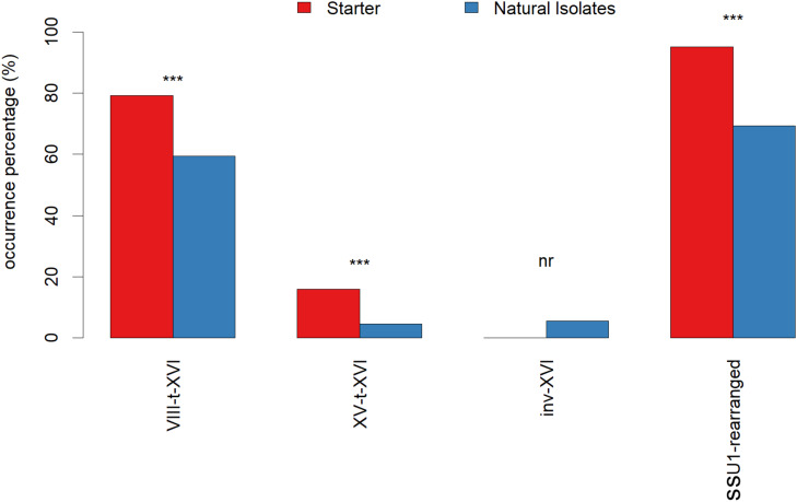
Out of the 21 possible biallelic combinations of the seven chromosome XVI alleles, 18 biallelic combinations were found among the 586 strains typed using the SSU1 checkup. The percentage of strains carrying at least one type of CR is shown in Figure 3. Industrial strains are significantly enriched in translocations VIII-t-XVI and XV-t-XVI compared to natural isolates. In contrast, the inv-XVI allele was rarer and never found in industrial strains (the reference strain P5 was not included here). Interestingly, two industrial starters (3%) carry both translocated chromosomes. In addition, 11 starters (13%) have an extra copy of chromosome XVI (aneuploidy), a fraction much higher than for the natural isolates group (1 out of 310). It has been suggested that an extra-copy number of chromosome XVI would confer a gain of fitness during fermentation (Brion et al., 2013). As shown in Table 2, seven industrial strains are homozygous for the XVI-wt allele (8.5% of the population). These starters are usually recommended for red grape juice winemaking or Cognac distillation, where the SO2 pressure is lower than in white wine production.
FIGURE 3.
Starters are enriched in rearranged chromosome XVI forms respect to natural isolates. The frequency (%) of “Starter” (red) and “Natural” (blue) isolates carrying at least one Chromosomal Rearrangement (CR) is represented. Significant differences are marked with ***.
Linking Chromosomal Rearrangement Events of Chromosome XVI and Yeast Ecology
Wine-related natural isolates allow the study of broad ecological factors influencing the chromosomal configurations of SSU1’s promoter. It has been shown that SSU1 related translocations in wine yeast isolates are advantageous for growth in sulfited grape juice and contribute to a fitness gain respect to oak yeast strains (Clowers et al., 2015). Furthermore, previous studies revealed that variations in the promoter region of SSU1 gene in wine yeasts enhance the SSU1 gene expression during fermentation, and have a remarkable effect on the SO2 resistance levels (Pérez-Ortín et al., 2002; Zimmer et al., 2014; García-Ríos and Guillamón, 2019). The subpopulation of 310 unique strains characterized in this work were split according to the sampling procedure applied: cellar (n = 205) vs. vineyard (n = 105) isolates (Supplementary Table S1). Cellar strains were isolated from spontaneously fermented vats in various wine estates, from sulfited grape musts according to the recommended practices of the area of origin. Vineyard strains were isolated from grapes manually harvested, crushed, and fermented in sterile laboratory conditions (see section “Materials and Methods”).
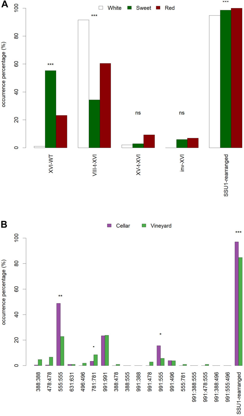
The occurrence percentage of the 18 allelic combinations found in both groups is shown in Figure 4A. The proportion of genotypes (555:555 and 991:555) is significantly higher in the cellar group while vineyard isolates are slightly enriched in 781:781 genotypes (p-value < 0.1, corrected Chi2 test). Moreover, strains having inherited at least one rearranged chromosome XVI are significantly more frequent in the cellar group (Figure 4A). The occurrence percentage observed here could reflect that among wine-related yeast isolates, cellar strains undergo a stronger selective pressure than vineyard strains likely due to winemaking operations.
FIGURE 4.
Grape juice matrixes and sampling methods impact the occurrence of SSU1 alleles. (A) Occurrence frequency (%) of 18 biallelic combinations of the seven chromosome XVI alleles. (B) Occurrence frequency (%) of rearranged chromosome XVI forms and chromosome XVI-wt form based on the type of grape juice matrix (sweet, white and red). *** significance level <0.001; ** significance level <0.05; * significance level <0.1, corrected Chi2 test; ns: not significant.
The impact of the nature of the grape juice from which strains were isolated was also tested. For this study, we focused our investigation only on cellar populations (n = 205) that have been subjected to in situ enological treatments. Indeed, according to the enological practices and doses, grape juices are not equally sulfited. This is consistent with the recommendations of International Organization of the Vine and Wine (OIV) that regulates the limits for total SO2 in wines (150 mg/L for red wines and 200 mg/L for white wines and rosés) (OIV, 2019). Strains were split in three groups depending on the type of grape juice matrix: sweet (n = 67), white (n = 95) and red (n = 43). As shown in Figure 4B, strains isolated from white juice are enriched in the VIII-t-XVI rearrangements and a few of them are homozygous for the XVI-wt991 allele. In contrast, strains isolated from sweet grape juices are strongly enriched in the native chromosome form. For this group, the occurrence percentage of XVI-wt is 0.55, which is twice as much as the overall percentage of cellar population (0.26). These results are consistent with the traditional enological practices used in the Bordeaux area, where the addition of sulfite in the musts is routinely used for dry white wine fermentation, but mostly avoided in the beginning of sweet wine fermentation to limit SO2 binding phenomena. This suggests that the selection of CR is strongly influenced by the winemaking practices used in cellars.
Analysis of SSU1 Allelic Variability in Closely Related Populations
The impact of translocations in the phenotypic adaptation of yeast has been widely investigated by using genetically engineered strains (Tosato and Bruschi, 2015; Fleiss et al., 2019). However, the survey of chromosomal rearrangements in clonal populations is much less described. The SSU1 checkup method provides an indirect opportunity to analyze this CR variability. In this section, the pool of 194 closely related clones was used in order to identify nearly isogenic groups of strains. In order to minimize the genetic distance inside a group, only strains showing less than two VNTR (Variable Number of Tandem Repeat) or LOH (Loss of Heterozygosity) were grouped together. By this way, 16 nearly genetic groups were identified encompassing 125 strains (Supplementary Table S5). Group sizes ranged between 3 and 22 individuals, with a Bruvo’s genetic distance between the strains of each group always lower than 0.106. In most of the cases, strains belonging to the same group were isolated from the same vat/cellar/area samples; however, in groups 6, 11, and 15 strong similarities were found between strains from white and red samples. This is consistent with the fact that isogenic strains can be isolated from different grape juices/cellars as previously demonstrated (Borlin, 2015; Franco-Duarte et al., 2015). The relative distance between each group was illustrated by a Principal Component Analysis (Figure 5A).
FIGURE 5.
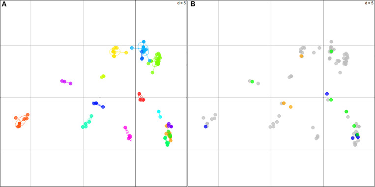
Alleles of chromosome XVI show a rapid evolution in isogenic populations. (A) Principal Component Analysis of 125 closely related clones (nearly isogenic) discriminated by 15 polymorphic loci. The sixteen groups were inferred manually by considering as isogenic the strains having an identical genotype for at least 13 microsatellite loci. The projection of each strain according to the 16 groups represents 14.3% of the total inertia. The Bruvo’s genetic distance within each group is always lower than 0.105. (B) represents the same projection, but strains were colored according to the type of change occurring on chromosome XVI. Gray dots represent the major allelic form found in each subgroup while orange, blue, and green dots represent LOH, VNTR, and CR, respectively.
Considering that each isogenic group is derived from a common clonal population, we analyzed the heterogeneity fraction in each group for three types of loci: microsatellites, chromosome VIII and chromosome XVI. For each group, the most frequent genotype was used as a reference (Supplementary Table S5). For microsatellite loci, few VNTR and LOH variations were detected among individuals. Within the 15 microsatellite markers, the average heterogeneity fraction was 2.4 and 2.1%, for VNTR and LOH, respectively (Table 3). In the same way, the heterogeneity fraction for chromosomes VIII and XVI were computed. Interestingly, a noteworthy variability was found for 10 out the 16 groups. For chromosome VIII, LOH events were observed only in groups 4 and 14 while tandem repeat shifts of the ECM34 promoter (VNTR) impacted in four groups. The overall allelic variability for the chromosome VIII locus was 4.5%, which is slightly higher than the average variability observed for neutral markers (microsatellites) (Table 3).
TABLE 3.
Heterogeneity fraction of VNTR, LOH and CR in isogenic populations.
| Group | Number of strains | Average Bruvo’s distancea | Microsatellites |
Chromosome VIII |
Chromosome XVI |
||||
| VNTRb | LOHb | VNTRb | LOHb | VNTRb | LOHb | CRb | |||
| 1 | 5 | 0.09 | 2.7 | 6.7 | 0 | 0 | 0 | 0 | 0 |
| 2 | 3 | 0.04 | 0 | 0 | 0 | 0 | 0 | 0 | 0 |
| 3 | 12 | 0.03 | 1.7 | 1.7 | 0 | 0 | 0 | 8.3 | 0 |
| 4 | 6 | 0.006 | 2.2 | 0 | 16.7 | 50 | 0 | 0 | 0 |
| 5 | 6 | 0.105 | 7.8 | 0 | 0 | 0 | 16.7 | 0 | 0 |
| 6 | 15 | 0.05 | 3.1 | 2.2 | 6.7 | 0 | 0 | 0 | 6.7 |
| 7 | 4 | 0.07 | 1.7 | 6.7 | 0 | 0 | 0 | 0 | 0 |
| 8 | 3 | 0.05 | 0 | 2.2 | 0 | 0 | 0 | 0 | 0 |
| 9 | 3 | 0.02 | 0 | 2.2 | 0 | 0 | 0 | 0 | 33.3 |
| 10 | 22 | 0.06 | 1.5 | 5.5 | 0 | 0 | 0 | 0 | 0 |
| 11 | 16 | 0.014 | 2.5 | 0 | 0 | 0 | 12.5 | 12.5 | 0 |
| 12 | 10 | 0.037 | 2.7 | 0 | 0 | 0 | 0 | 0 | 0 |
| 13 | 4 | 0.044 | 5 | 0 | 25 | 0 | 0 | 0 | 50 |
| 14 | 3 | 0.023 | 0 | 2.2 | 0 | 33.3 | 33.3 | 0 | 33.3 |
| 15 | 4 | 0.055 | 6.7 | 1.7 | 0 | 0 | 0 | 0 | 0 |
| 16 | 9 | 0.07 | 1.5 | 3 | 11.1 | 0 | 11.1 | 0 | 0 |
| TOTAL | 125 | 0.05 | 2.4 | 2.1 | 3.7 | 5.2 | 2.9 | 1.3 | 7.7 |
aAverage genetic distance computed from the genetic distance matrix between all the strains of the group. bThe average heterogeneity fraction was expressed in percentage of change per locus (15 loci for microsatellites and one locus for chromosomes VIII and XVI). VNTR: Variable Number of Tandem Repeat, LOH: Loss Of Heterozygosity, CR: Chromosomal Rearrangement.
The inheritance of chromosome XVI is more complex to analyze due to the combined influences of VNTR, LOH and the different CR (Figure 5B). These changes were observed in 7 out the 16 groups, being their overall frequency 2.9, 1.3, and 7.7%, respectively. These allelic changes were mostly found within strains isolated at the same place and showing the same microsatellite pattern. For VNTR, four isogenic groups showed allelic variations in the VIII-t-XVI translocation. These variations were due to a different number of tandem repeats on the ECM34 promoter; their frequencies were similar to those observed for chromosome VIII (2.9 vs. 3.7%). LOH variations were observed for two groups (i.e., 3 and 11). For instance, the main genotype observed in the group 11 was 555:555 (12 out 16 strains) but two strains (3bibi6_26 and 3bebi3_13) have the genotype 991:555. This difference suggested that the strains that have inherited the XVI-wt991 form might have resulted from a hybridization event; alternatively the strain group homozygous for the translocation VIII-t-XVI555 could have been the result of meiotic segregation (Mortimer et al., 1994). Interestingly, the C6 microsatellite (localized on chromosome XVI) did not have the same inheritance in the strains 3bibi6_26 and 3bebi3_13. Also, in the same group 11, the strains CLA2016 1 and 2 isolated from Chardonnay (white grape juice) showed a longer VIII-t-XVI allele (617:617) but shared exactly the same microsatellite pattern than the 12 strains isolated from Merlot vats. This supports the idea that alternative numbers of tandem repetitions in the ECM34 promoter can be found in clonal populations. More surprisingly, different CR were also observed within isogenic groups. This is the case of strains 13AQGUICUV1 (555:555) and 14AQGUICUV1 (991:496) isolated from the same area in sweet wines. The first strain is homozygous with a VIII-t-XVI form while the second strain is heterozygous with both XVI-wt and XV-t-XVI forms. These two clones could have resulted from the meiotic segregation of an aneuploid strain carrying the three chromosome XVI alleles (VIII-t-XVI555, XVI-wt991 and XV-t-XVI496). One more time, the microsatellite C6 has not the same inheritance between these two strains supporting this hypothesis. A similar case of heterogeneity was also observed for strains 2duSPO9 (555:555) and 3mabi1_10 (991:496) that belong to group 6. Interestingly, the strain 2duSPO9 (555:555) was isolated from a Sauvignon blanc juice while all the other strains of this group were isolated from red grape juice (Supplementary Table S5). This is consistent with the fact that the translocation VIII-t-XVI555 was more frequently found in samples isolated from white grape juice (Figure 4B). All these findings were verified by simple PCR reactions, after additional DNA extractions. Although the number of events observed is not sufficient for providing robust data, our results illustrate an important heterogeneous fraction among SSU1 alleles in clonal populations that would likely be due to meiotic recombination events. These microevolutionary changes between an industrial strain and its descendants selected after persistence in nature were previously reported using inter-delta markers (Franco-Duarte et al., 2015). In the case of the SSU1 promoter, these allelic changes would play a significant role in the adaptative responses to different environments especially due to the use or not of SO2 in the early stages of vinification.
Impact of Chromosomal Rearrangements on Yeast Fitness Parameters in Sulfited Grape Juice
Finally, we compared groups of unrelated strains harboring six different promoter regions of the gene SSU1. Five representative yeast strains of each group were selected by choosing strains with contrasted microsatellite inheritance and sampling origins. Indeed, strains belonging to each group showed an average Bruvo’s genetic distance higher than 0.50. These genetic distances were similar to those observed for the total population (Supplementary Figure S5). Therefore, the strains selected could be considered as genetically unrelated. To simplify the interpretation of the data, the strains selected are homozygous for the six promoter regions. Growth kinetics of these strains in filtered Sauvignon blanc grape juice spiked with different SO2 concentrations were analyzed by OD600 measurements. Since some strains did not reach an OD600 plateau after 96 h, only μmax (maximal growth rate) and Lag Time (Lag phase time) parameters were analyzed. An overview of the kinetics for the reference strains GN, SB, P5 and Fx10 is given in Supplementary Figure S6. The effect of SO2 concentration and type of chromosome XVI were estimated by a two-way ANOVA (model Lm1, see section “Materials and Methods”). The variance explained by factors is given in Table 4. As expected, SO2 addition to the grape juice significantly impacted the μmax and the Lag Time parameters explaining 14.8 and 18.4% of total variance, respectively. This confirms the selective pressure imposed by increasing SO2 concentrations, which delayed the beginning of exponential growth and reduced the maximum growth rates of S. cerevisiae strains. In addition, the type of chromosome XVI significantly impacted these two parameters (Table 4) contributing in higher proportion to the total variance observed (23.5% for Lag Time and 17.0% for μmax). Finally, a significant interaction was detected between SO2 concentration and the type of chromosome XVI type for the Lag Time parameter (p < 1.10–6).
TABLE 4.
Analysis of variance of SO2 and SSU1 promoter forms for lag time and μmax.
| Trait | SO2 |
Chr XVI |
SO2:Chr XVI interactions |
Residual | Levene test (p-value) | |||
| Effect | p-valuea | Effect | p-valuea | Effect | p-value | |||
| Lag time | 18.4 | <2.2 10–16 | 23.5 | <2.2 10–16 | 7.0 | 1.1 10–5 | 50.7 | 0.06 |
| μmax | 14.8 | 1.8 10–11 | 17.0 | 6.9 10–10 | 2.5 | 0.12 | 65.6 | 0.002 |
aANOVA p-value.
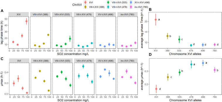
The specific impact of the form of the SSU1 promoter is illustrated in Figure 6. Since five unrelated strains were tested in each group, the impact of the different SSU1 promoters is partially decoupled to the strain effect. As expected, “non-rearranged strains” (i.e., XVI-wt/XVI-wt) present the highest Lag Time and the lowest μmax at high SO2 concentrations. The translocation XV-t-XVI and the inversion (inv-XVI) appear to be very efficient to cope with higher SO2 concentrations. Indeed, strains carrying these alleles were poorly affected by the addition of 75 mg/L of SO2. These findings are consistent with the elevated SSU1 expression levels reported for these two chromosomal rearrangements (Zimmer et al., 2014; García-Ríos and Guillamón, 2019). For the VIII-t-XVI translocations, only three allelic forms (VIII-t-XVI388, VIII-t-XVI478, VIII-t-XVI555) were tested. The fourth allele (VIII-t-XVI631) was found in only three strains, two of them being clearly isogenic. The allele VIII-t-XVI478 was the most efficient in reducing the Lag Time and preserving the μmax. The other two alleles (VIII-t-XVI388 and VIII-t-XVI555) resulted in the least favorable rearrangements for sulfite tolerance (Figure 6).
FIGURE 6.
Phenotypic impact of six SSU1 promoter alleles. (A) Mean of the Lag Times (in hours) of the 5 strains tested at different SO2 concentrations, for each of the chromosome XVI alleles. (B) Mean of the Lag Times (in hours) of the 5 strains tested at all SO2 concentrations, for each of the chromosome XVI alleles. (C) Mean of the μmax of the 5 strains tested at different SO2 concentrations, for each of the chromosome XVI alleles. (D) Mean of the μmax of the 5 strains tested at all SO2 concentrations, for each of the chromosome XVI alleles.
Differences in the length of the VIII-t-XVI translocation alleles mainly result from the variable number of 76 bp tandem repeat units (Goto-Yamamoto et al., 1998). The number of tandem repeats (76 and 47 bp) for the VIII-t-XVI translocation was verified by DNA sequencing and results are summarized in Supplementary Figure S4. Alleles VIII-t-XVI388, VIII-t-XVI478, and VIII-t-XVI555 show two, three, and four 76 bp tandem repeats plus one additional 47 bp tandem repeat, respectively. Since the allele VIII-t-XVI478 out competed the other two forms (Figure 6), we can conclude that the number of tandem repeats and the SO2 tolerance are not fully correlated, as previously reported (Yuasa et al., 2004). Therefore, undetermined natural allelic variations in the SSU1 coding sequence, in flanking regions, or in other genomic loci are possibly also involved in this trait.
By comparing for the first time the effect of six configurations of the SSU1 promoter on yeast fitness we pave the way for screening sulfite tolerance of strains in a simple genetic test. The alleles XV-t-XVI, inv-XVI and VIII-t-XVI478 confer an efficient adaptation for growing in grape juices with high concentrations of SO2. In addition, we illustrate that for the translocation VIII-t-XVI, the sulfite resistance (maximal growth rate and lag phase) is not perfectly related to the number of 76 bp tandem repeats. Indeed, from a technological point of view, the allele VIII-t-XVI478 would be the most efficient form to tolerate high sulfite concentrations in natural grape juice.
Conclusion
In yeast, CR are thought to play a role in adaptation and species evolution and might have physiological consequences (Tosato and Bruschi, 2015). The setup of a SSU1 checkup provides a rapid molecular tool for obtaining a complete overview of three CR involving the promoter region of SSU1. This molecular diagnostic was helpful to address some questions related to the impact of domestication of wine yeast and to progress in the identification of physio-ecological parameters that reshape the genome organization in natural isolates. From an ecological point of view, the SSU1 checkup could be in the future a key molecular tool for addressing different questions in relation to the use of sulfites in wine. Indeed, in the recent years, the consumer-driven push for decreasing the levels of SO2 in the wine industry and the low-sulfite wine market are increasing and could modify the allelic frequency of SSU1 promoter alleles. From a more applied point of view, we established a link between quantitative phenotypes and the inheritance of CR in genetically unrelated groups paving the way for yeast selection programs mediated by molecular markers.
Data Availability Statement
The datasets generated for this study can be found in the GenBank (ID 2310529).
Author Contributions
PM, OC, and IM-P designed the experiment. MR, OC, MBö, NF, MBe, and AR did the experiment. PM, OC, and MR wrote the manuscript. MBö, WA, J-LL, PM, OC, and MR analyzed the data. PM, AR, and IM-P got the grants. All authors contributed to the article and approved the submitted version.
Conflict of Interest
MB and PM are employed by the Biolaffort company. The remaining authors declare that the research was conducted in the absence of any commercial or financial relationships that could be construed as a potential conflict of interest.
Acknowledgments
The authors would like to thank José Manuel Guillamon (Spanish National Research Council) for providing the P5 strain.
Footnotes
Funding. PM recieved funds from the Biolaffort Company and form the Region Aquitaine Grant SESAM for delevopping Marker Assisted Selection. This work was supported by PICT-2014- 3113 from Fondo para la Investigación Científica y Tecnológica (FONCyT) (Argentina) and SUV2015 (Universidad Católica de Córdoba) to AR. MR held a fellowship from the National Research Council of Argentina (CONICET). AR is a Career Research Investigator from CONICET. The funder bodies were not involved in the study design, collection, analysis, interpretation of data, the writing of this article or the decision to submit it for publication.
Supplementary Material
The Supplementary Material for this article can be found online at: https://www.frontiersin.org/articles/10.3389/fmicb.2020.01331/full#supplementary-material
Bruvo’s distance distribution and cut off threshold used for removing very similar strains.
Natural isolates closely related to industrial starters.
(A) Principal Component analysis of 82 commercial starters discriminated by 15 polymorphic loci. The three groups represented (A to C) were inferred by k-mean clustering. The (B) represents the position of the strains according to the inferred groups.
Schematic representation of a VIII allele of S. cerevisiae. The figure shows the location of the 76 bp Tandem Repeats and the 47 bp Tandem Repeats, as well as the translocation point (TP) and the upstream (5′ region; from the end of the forward primer to the beginning of the 76 bp tandem repeats) and downstream (3′region; from the end of the 47 bp tandem repeats to the translocation point) flanking regions. The hybridization position for the primers forward (F; p1189) and reverse (R; either p1190 or p1194), are indicated.
Genetic distance between individuals sharing six SSU1 promoter types, only the strains homozygous for these alleles were considered. The color blue and red indicate the distribution of pair wise Bruvo’s genetic distance for the total set of strains and the subset of five strains selected.
Growth curves of reference strains in different grape juice containing different SO2 concentrations expressed in mg/L. The data presented are the average of two independent replicates for the strain Fx10 (red), GN (green), P5 (cyan), and SB (purple). Standard error was figured out by the shaded area.
List of the 586 strains analyzed in this work.
Additional primers used.
List of the 30 strains phenotyped.
Contingency table of natural isolates origin according to the microsatellite groups.
List of the 16 isogenic subgroups identified encompassing 125 strains.
References
- Aa E., Townsend J. P., Adams R. I., Nielsen K. M., Taylor J. W. (2006). Population structure and gene evolution in Saccharomyces cerevisiae. FEMS Yeast Res. 6 702–715. 10.1111/j.1567-1364.2006.00059.x [DOI] [PubMed] [Google Scholar]
- Albertin W., Marullo P., Aigle M., Bourgais A., Bely M., Dillmann C., et al. (2009). Evidence for autotetraploidy associated with reproductive isolation in Saccharomyces cerevisiae: Towards a new domesticated species. J. Evol. Biol. 22 2157–2170. 10.1111/j.1420-9101.2009.01828.x [DOI] [PubMed] [Google Scholar]
- Avram D., Bakalinsky A. T. (1997). SSU1 encodes a plasma membrane protein with a central role in a network of proteins conferring sulfite tolerance in Saccharomyces cerevisiae. J. Bacteriol. 179 5971–5974. 10.1128/jb.179.18.5971-5974.1997 [DOI] [PMC free article] [PubMed] [Google Scholar]
- Avramova M., Cibrario A., Peltier E., Coton M., Coton E., Schacherer J., et al. (2018). Brettanomyces bruxellensis population survey reveals a diploid-triploid complex structured according to substrate of isolation and geographical distribution. Sci. Rep. 8:4136. 10.1038/s41598-018-22580-7 [DOI] [PMC free article] [PubMed] [Google Scholar]
- Borlin M. (2015). Diversity and Population Structure of Yeast Saccharomyces cerevisiae at the Scale of the Vineyard of Bordeaux?: Impact of Different Factors on Diversity. Available online at: https://tel.archives-ouvertes.fr/tel-01293834 (accessed December 2015). [Google Scholar]
- Börlin M., Venet P., Claisse O., Salin F., Legras J.-L., Masneuf-Pomarede I. (2016). Cellar-associated Saccharomyces cerevisiae population structure revealed high-level diversity and perennial persistence at sauternes wine estates. Appl. Environ. Microbiol. 82 2909–2918. 10.1128/AEM.03627-15 [DOI] [PMC free article] [PubMed] [Google Scholar]
- Borneman A. R., Forgan A. H., Kolouchova R., Fraser J. A., Schmidt S. A. (2016). Whole genome comparison reveals high levels of inbreeding and strain redundancy across the spectrum of commercial wine strains of Saccharomyces cerevisiae. G3 6 957–971. 10.1534/g3.115.025692 [DOI] [PMC free article] [PubMed] [Google Scholar]
- Borneman A. R., Pretorius I. S. (2014). Genomic insights into the Saccharomyces sensu stricto complex. Genetics 199 281–291. 10.1534/genetics.114.173633 [DOI] [PMC free article] [PubMed] [Google Scholar]
- Brion C., Ambroset C., Sanchez I., Legras J.-L., Blondin B. (2013). Differential adaptation to multi-stressed conditions of wine fermentation revealed by variations in yeast regulatory networks. BMC Genomics 14:681. 10.1186/1471-2164-14-681 [DOI] [PMC free article] [PubMed] [Google Scholar]
- Cheraiti N., Guezenec S., Salmon J.-M. (2010). Very early acetaldehyde production by industrial Saccharomyces cerevisiae strains: a new intrinsic character. Appl. Microbiol. Biotechnol. 86 693–700. 10.1007/s00253-009-2337-5 [DOI] [PubMed] [Google Scholar]
- Chernova M., Albertin W., Durrens P., Guichoux E., Sherman D. J., Masneuf-Pomarede I., et al. (2018). Many interspecific chromosomal introgressions are highly prevalent in Holarctic Saccharomyces uvarum strains found in human-related fermentations. Yeast 35 141–156. 10.1002/yea.3248 [DOI] [PubMed] [Google Scholar]
- Clowers K. J., Heilberger J., Piotrowski J. S., Will J. L., Gascha P. (2015). Ecological and genetic barriers differentiate natural populations of Saccharomyces cerevisiae. Mol. Biol. Evol. 32 2317–2327. 10.1093/molbev/msv112 [DOI] [PMC free article] [PubMed] [Google Scholar]
- Divol B., Du Toit M., Duckitt E. (2012). Surviving in the presence of sulphur dioxide: Strategies developed by wine yeasts. Appl. Microbiol. Biotechnol. 95 601–613. 10.1007/s00253-012-4186-x [DOI] [PubMed] [Google Scholar]
- Engle E. K., Fay J. C. (2012). Divergence of the yeast transcription factor FZF1 affects sulfite resistance. PLoS Genet. 8:e2763. 10.1371/journal.pgen.1002763 [DOI] [PMC free article] [PubMed] [Google Scholar]
- Fleiss A., O’Donnell S., Fournier T., Lu W., Agier N., Delmas S., et al. (2019). Reshuffling yeast chromosomes with CRISPR/Cas9. PLoS Genet. 15:e1008332. 10.1371/journal.pgen.1008332 [DOI] [PMC free article] [PubMed] [Google Scholar]
- Franco-Duarte R., Bigey F., Carreto L., Mendes I., Dequin S., Santos M. A., et al. (2015). Intrastrain genomic and phenotypic variability of the commercial Saccharomyces cerevisiae strain Zymaflore VL1 reveals microevolutionary adaptation to vineyard environments. FEMS Yeast Res. 15:fov063. 10.1093/femsyr/fov063 [DOI] [PubMed] [Google Scholar]
- García-Ríos E., Guillamón J. M. (2019). Sulfur dioxide resistance in Saccharomyces cerevisiae: beyond SSU1. Microb. Cell 6 527–530. 10.15698/mic2019.12.699 [DOI] [PMC free article] [PubMed] [Google Scholar]
- García-Ríos E., Nuévalos M., Barrio E., Puig S., Guillamón J. M. (2019). A new chromosomal rearrangement improves the adaptation of wine yeasts to sulfite. Environ. Microbiol. 21 1771–1781. 10.1111/1462-2920.14586 [DOI] [PubMed] [Google Scholar]
- Goto-Yamamoto N., Kitano K., Shiki K., Yoshida Y., Suzuki T., Iwata T., et al. (1998). SSU1-R, a sulfite resistance gene of wine yeast, is an allele of SSU1 with a different upstream sequence. J. Ferment. Bioeng. 86 427–433. 10.1016/S0922-338X(98)80146-3 [DOI] [Google Scholar]
- Hinze H., Holzer H. (1986). Analysis of the energy metabolism after incubation of Saccharomyces cerevisiae with sulfite or nitrite. Arch. Microbiol. 145 27–31. 10.1007/BF00413023 [DOI] [PubMed] [Google Scholar]
- Legras J.-L. L., Merdinoglu D., Cornuet J.-M. M., Karst F. (2007). Bread, beer and wine: Saccharomyces cerevisiae diversity reflects human history. Mol. Ecol. 16 2091–2102. 10.1111/j.1365-294X.2007.03266.x [DOI] [PubMed] [Google Scholar]
- Marsit S., Dequin S. (2015). Diversity and adaptive evolution of Saccharomyces wine yeast: a review. FEMS Yeast Res. 15:fov067. 10.1093/femsyr/fov067 [DOI] [PMC free article] [PubMed] [Google Scholar]
- Martí-Raga M., Marullo P., Beltran G., Mas A., Marti-Raga M., Marullo P., et al. (2016). Nitrogen modulation of yeast fitness and viability during sparkling wine production. Food Microbiol. 54 106–114. 10.1016/j.fm.2015.10.009 [DOI] [Google Scholar]
- Marullo P., Bely M., Masneuf-Pomarède I., Pons M., Aigle M., Dubourdieu D., et al. (2006). Breeding strategies for combining fermentative qualities and reducing off-flavor production in a wine yeast model. FEMS Yeast Res. 6 268–279. 10.1111/j.1567-1364.2006.00034.x [DOI] [PubMed] [Google Scholar]
- Marullo P., Mansour C., Dufour M., Albertin W., Sicard D., Bely M., et al. (2009). Genetic improvement of thermo-tolerance in wine Saccharomyces cerevisiae strains by a backcross approach. FEMS Yeast Res. 9 1148–1160. 10.1111/j.1567-1364.2009.00550.x [DOI] [PubMed] [Google Scholar]
- Mortimer R. K., Romano P., Suzzi G., Polsinellif M. (1994). Genome renewal: a new phenomenon revealed from a genetic study of 43 Strains of Saccharomyces cerevisiae derived from natural fermentation of grape musts. YEAST 10 1543–1552. 10.1002/yea.320101203 [DOI] [PubMed] [Google Scholar]
- Nardi T., Corich V., Giacomini A., Blondin B. (2010). A sulphite-inducible form of the sulphite efflux gene SSU1 in a Saccharomyces cerevisiae wine yeast. Microbiology 156 1686–1696. 10.1099/mic.0.036723-0 [DOI] [PubMed] [Google Scholar]
- Novo M., Bigey F., Beyne E., Galeote V., Gavory F., Mallet S., et al. (2009). Eukaryote-to-eukaryote gene transfer events revealed by the genome sequence of the wine yeast Saccharomyces cerevisiae EC1118. Proc. Natl. Acad. Sci. U.S.A. 106 16333–16338. 10.1073/pnas.0904673106 [DOI] [PMC free article] [PubMed] [Google Scholar]
- OIV (2019). Compendium of International Methods of Analysis of Wine and Musts. Paris: OIV. [Google Scholar]
- Park H., Bakalinsky A. T. (2000). SSU1 mediates sulphite efflux in Saccharomyces cerevisiae. Yeast 16 881–888. [DOI] [PubMed] [Google Scholar]
- Park H., Hwang Y. S. (2008). Genome-wide transcriptional responses to sulfite in Saccharomyces cerevisiae. J. Microbiol. 46 542–548. 10.1007/s12275-008-0053-y [DOI] [PubMed] [Google Scholar]
- Peltier E., Sharma V., Marti Raga M., Roncoroni M., Bernard M., Jiranek V., et al. (2018a). Genetic basis of genetic x environment interaction in an enological context. BMC Genomics, 19:772. [DOI] [PMC free article] [PubMed] [Google Scholar]
- Peltier E., Sharma V., Raga M. M., Roncoroni M., Bernard M., Yves G., et al. (2018b). Dissection of the molecular bases of genotype x environment interactions: a study of phenotypic plasticity of Saccharomyces cerevisiae in grape juices. BMC Genomics 19:772. 10.1186/s12864-018-5145-4 [DOI] [PMC free article] [PubMed] [Google Scholar]
- Pérez-Ortín J. E., Querol A., Puig S., Barrio E. (2002). Molecular characterization of a chromosomal rearrangement involved in the adaptive evolution of yeast strains. Genome Res. 12 1533–1539. 10.1101/gr.436602 [DOI] [PMC free article] [PubMed] [Google Scholar]
- Peter J., De Chiara M., Friedrich A., Yue J.-X., Pflieger D., Bergström A., et al. (2018). Genome evolution across 1,011 Saccharomyces cerevisiae isolates. Nature 556 339–344. 10.1038/s41586-018-0030-5 [DOI] [PMC free article] [PubMed] [Google Scholar]
- Raymond E. M. L., Conti F., Rosa A. L. (2018). Differences between indigenous yeast populations in spontaneously fermenting musts from V. vinifera L. and V. labrusca L. Grapes harvested in the same geographic location. Front. Microbiol. 9:1320. 10.3389/fmicb.2018.01320 [DOI] [PMC free article] [PubMed] [Google Scholar]
- Sicard D., Legras J. L. (2011). Bread, beer and wine: Yeast domestication in the Saccharomyces sensu stricto complex. Comptes Rendus Biol. 334 229–236. 10.1016/j.crvi.2010.12.016 [DOI] [PubMed] [Google Scholar]
- Tosato V., Bruschi C. V. (2015). Per aspera ad astra: When harmful chromosomal translocations become a plus value in genetic evolution. lessons from Saccharomyces cerevisiae. Microb. Cell 2 363–375. 10.15698/mic2015.10.230 [DOI] [PMC free article] [PubMed] [Google Scholar]
- Valero E., Schuller D., Cambon B., Casal M., Dequin S. (2005). Dissemination and survival of commercial wine yeast in the vineyard: A large-scale, three-years study. FEMS Yeast Res. 5 959–969. 10.1016/j.femsyr.2005.04.007 [DOI] [PubMed] [Google Scholar]
- Yuasa N., Nakagawa Y., Hayakawa M., Iimura Y. (2004). Distribution of the sulfite resistance gene SSU1-R and the variation in its promoter region in wine yeasts. J. Biosci. Bioeng. 98 394–397. 10.1263/jbb.98.394 [DOI] [PubMed] [Google Scholar]
- Zimmer A., Durand C., Loira N., Durrens P., Sherman D. J., Marullo P. (2014). QTL dissection of lag phase in wine fermentation reveals a new translocation responsible for Saccharomyces cerevisiae adaptation to sulfite. PLoS One 9:e86298. 10.1371/journal.pone.0086298 [DOI] [PMC free article] [PubMed] [Google Scholar]
Associated Data
This section collects any data citations, data availability statements, or supplementary materials included in this article.
Supplementary Materials
Bruvo’s distance distribution and cut off threshold used for removing very similar strains.
Natural isolates closely related to industrial starters.
(A) Principal Component analysis of 82 commercial starters discriminated by 15 polymorphic loci. The three groups represented (A to C) were inferred by k-mean clustering. The (B) represents the position of the strains according to the inferred groups.
Schematic representation of a VIII allele of S. cerevisiae. The figure shows the location of the 76 bp Tandem Repeats and the 47 bp Tandem Repeats, as well as the translocation point (TP) and the upstream (5′ region; from the end of the forward primer to the beginning of the 76 bp tandem repeats) and downstream (3′region; from the end of the 47 bp tandem repeats to the translocation point) flanking regions. The hybridization position for the primers forward (F; p1189) and reverse (R; either p1190 or p1194), are indicated.
Genetic distance between individuals sharing six SSU1 promoter types, only the strains homozygous for these alleles were considered. The color blue and red indicate the distribution of pair wise Bruvo’s genetic distance for the total set of strains and the subset of five strains selected.
Growth curves of reference strains in different grape juice containing different SO2 concentrations expressed in mg/L. The data presented are the average of two independent replicates for the strain Fx10 (red), GN (green), P5 (cyan), and SB (purple). Standard error was figured out by the shaded area.
List of the 586 strains analyzed in this work.
Additional primers used.
List of the 30 strains phenotyped.
Contingency table of natural isolates origin according to the microsatellite groups.
List of the 16 isogenic subgroups identified encompassing 125 strains.
Data Availability Statement
The datasets generated for this study can be found in the GenBank (ID 2310529).


