. 2020 Jun 5;40(4):1003–1019. doi: 10.1148/rg.2020190180

Figure 4a.

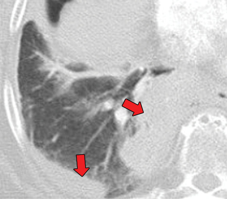
EGPA in a 40-year-old woman. (a) Axial CT image of the chest shows peripheral consolidations in the right lower lobe (arrows), which were the patient’s pulmonary manifestation of EGPA.(b) Maxillofacial CT image shows pansinusitis that includes the left sphenoid sinus (orange arrow) and bilateral ethmoid sinuses (yellow arrows). (c) Axial CT image at 4-year follow-up shows consolidation with cavitation (arrow). Given the new development of cavitation, short-term-interval follow-up or biopsy was recommended, because EGPA consolidations rarely have cavitations. The patient was lost to follow-up and later presented with pelvic pain. CT showed diffuse lytic osseous lesions including a pathologic fracture of the left iliac bone, which was secondary to metastatic non–small cell lung cancer at biopsy.

EGPA in a 40-year-old woman. (a) Axial CT image of the chest shows peripheral consolidations in the right lower lobe (arrows), which were the patient’s pulmonary manifestation of EGPA.(b) Maxillofacial CT image shows pansinusitis that includes the left sphenoid sinus (orange arrow) and bilateral ethmoid sinuses (yellow arrows). (c) Axial CT image at 4-year follow-up shows consolidation with cavitation (arrow). Given the new development of cavitation, short-term-interval follow-up or biopsy was recommended, because EGPA consolidations rarely have cavitations. The patient was lost to follow-up and later presented with pelvic pain. CT showed diffuse lytic osseous lesions including a pathologic fracture of the left iliac bone, which was secondary to metastatic non–small cell lung cancer at biopsy.