. 2020 Jun 5;40(4):1003–1019. doi: 10.1148/rg.2020190180

Figure 6a.

Necrobiotic nodules in a 55-year-old woman with rheumatoid arthritis and a history of smoking. (a, b) Coronal CT images show multiple cavitary nodules (red arrows) that ruptured into the right pleural space (yellow arrows in b) and then into the skin, forming a pleurocutaneous fistula (blue arrow in a). (c) Axial maximum intensity projection reconstruction CT image shows one of the peripheral cavitary necrobiotic nodules (green arrow).

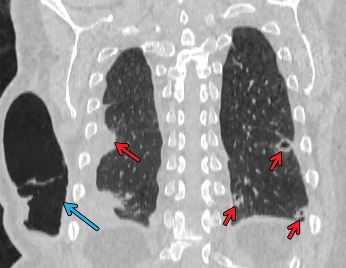
Necrobiotic nodules in a 55-year-old woman with rheumatoid arthritis and a history of smoking. (a, b) Coronal CT images show multiple cavitary nodules (red arrows) that ruptured into the right pleural space (yellow arrows in b) and then into the skin, forming a pleurocutaneous fistula (blue arrow in a). (c) Axial maximum intensity projection reconstruction CT image shows one of the peripheral cavitary necrobiotic nodules (green arrow).