ABSTRACT
The epigenome determines heritable patterns of gene expression in the absence of changes in DNA sequence. The result is programming of different cellular-, tissue- and organ-specific phenotypes from a single organismic genome. Epigenetic marks that comprise the epigenome (e.g. methylation) are placed upon or removed from chromatin (histones and DNA) to direct the activity of effectors that regulate gene expression and chromatin structure. Recently, the cytoskeleton has been identified as a second target for the cell's epigenetic machinery. Several epigenetic ‘readers, writers and erasers’ that remodel chromatin have been discovered to also remodel the cytoskeleton, regulating structure and function of microtubules and actin filaments. This points to an emerging paradigm for dual-function remodelers with ‘chromatocytoskeletal’ activity that can integrate cytoplasmic and nuclear functions. For example, the SET domain-containing 2 methyltransferase (SETD2) has chromatocytoskeletal activity, methylating both histones and microtubules. The SETD2 methyl mark on chromatin is required for efficient DNA repair, and its microtubule methyl mark is required for proper chromosome segregation during mitosis. This unexpected convergence of SETD2 activity on histones and microtubules to maintain genomic stability suggests the intriguing possibility of an expanded role in the cell for chromatocytoskeletal proteins that read, write and erase methyl marks on the cytoskeleton as well as chromatin. Coordinated use of methyl marks to remodel both the epigenome and the (epi)cytoskeleton opens the possibility for integrated regulation (which we refer to as ‘epiregulation’) of other higher-level functions, such as muscle contraction or learning and memory, and could even have evolutionary implications.
KEY WORDS: Actin, Epigenetics, Epiregulation, DMNT, SETD2, Tubulin
Summary: A shared chromatocytoskeletal machinery uses methylation to remodel chromatin and the cytoskeleton; we discuss how this might regulate and integrate cellular functions, and suggest the term ‘epiregulation’ to describe this co-regulation.
Introduction
Epigenetics and the ‘epigenome’
The term ‘epigenetics’ was coined in 1942 by Conrad Waddington to encompass biological concepts and processes that involved genetics and inheritance, but which extended beyond what was then regarded as a shadowy, spectral world of phenotype controlled by unknown external factors (Waddington, 1942). In the modern era of molecular biology, epigenetics is understood as determining heritable patterns of gene expression in the absence of changes in DNA sequence, allowing different cellular-, tissue- and organ-specific phenotypes to be specified by a single organismic genome. Borrowing from the Greek word ‘epi’ (which translates to ‘above’), epigenetics has now become a central pillar upon which rests modern genetics, cell biology, development and even evolution – for an entry into the voluminous literature see: Biswas and Rao (2018); Burggren (2016, 2020); Burggren and Crews (2014); Crews et al. (2014); Gonzalez-Recio et al. (2015); Jablonka (2017); Janssen et al. (2017); Lowdon et al. (2016); Perez and Lehner (2019); Sarkies (2019); Seymour and Becker (2017); Skinner (2015); Vidalis et al. (2016); Wang et al. (2017); Whipple and Holeski (2016); Zhong (2016). (Note: the reader should be advised that numerous variants on the definitions of the terms ‘epigenetics’, and especially the associated ‘epigenetic inheritance’, are to be found in that literature. Fortunately for the authors, sorting out this semantic quagmire is beyond the scope of this Commentary.)
Glossary.
Basal bodies
A mitotic organizing center of the cell at the base of the primary cilium, formed by the centrosome when cells are not in mitosis
Chromatocytoskeletal
Activity of a dual-function epigenetic regulator acting on both chromatin and the cytoskeleton
CpG islands
Regions of DNA ≥200 bp in length with a high frequency of CpG sites
Demethylase
Enzyme that removes methyl groups from target proteins such as DNA or histones
Domain
Modular component of a protein with a specialized function in recognizing or modifying target proteins
Epigenetic machinery
Proteins that add, recognize and/or remove epigenetic modifications of chromatin in a coordinated and circumscribed way
Epigenetic marks
Small chemical modifications to the histones and DNA that affect the function of the genome in a hereditary manner
Epimutations
Heritable changes to the epigenome that can result in abnormal transcriptional repression of active genes and/or abnormal activation of usually repressed genes
Epiregulation
Regulation of structure and function of the genome and cytoskeleton, including but not limited to the reading, writing and erasure of methyl marks, by the epigenetic machinery
Histone code
Combinatorial post-translational modifications that direct gene transcription and/or chromatin conformation
Methyltransferases
Enzymes that add methyl groups to lysine, arginine and histidine (or other) amino acid residues of target proteins
Midbody
The collapsed central spindle microtubules that continue to join daughter cells until cleavage during cytokinesis
Polycomb repressive complex 2 (PRC2)
A multi-component methyltransferase enzyme complex containing the histone methyltransferase EZH2 that writes the H3K27me3 mark on chromatin
Tubulin code
Post-translational modifications that direct tubulin tertiary and quaternary structure and function
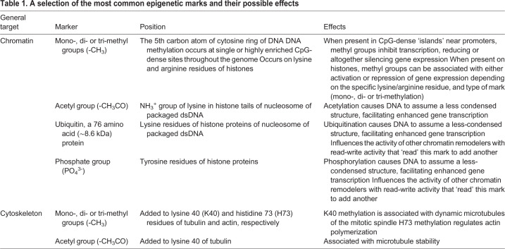
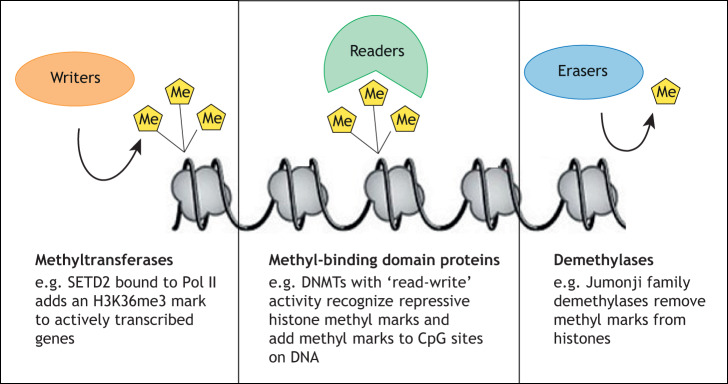
Central to the concept of epigenetics is the ‘epigenome’, defined as the pattern of small chemical modifications made to both the histones and DNA that make up chromatin. Strahl and Allis first proposed the concept that these modifications on histones comprise a ‘histone code’ (see Glossary) that directs gene transcription and creates a ‘language’ that broadly regulates transcription and other aspects of chromatin biology (Strahl and Allis, 2000). We now appreciate the epigenome as the primary mechanism for ‘operationalizing’ the genome. It is of vital importance to within-life development, as well as healthy maturation and aging; if perturbed, it can impact subsequent generations through transgenerational, ‘non-genetic’ inheritance (Booth and Brunet, 2016; Stricker et al., 2017). Table 1 presents a non-exclusive list of the best understood chromatin marks and some of their actions. From a mechanistic perspective, a suite of proteins often termed ‘readers’, ‘writers’ and ‘erasers’ are responsible for programming (and reprogramming) the epigenome (Fig. 1). Recent reviews of these proteins and their actions have been provided by Biswas and Rao (2018), Trevino et al. (2015) and Yang et al. (2016), among others.
Table 1.
A selection of the most common epigenetic marks and their possible effects
Fig. 1.
Writers, readers and erasers of epigenetic markers. As an example, the figure shows information on proteins that can write, read and erase methyl groups (Me). Methyl groups on histones can be sensed (read) by methyl-binding domain proteins (e.g. DNA methyltransferases, DMNTs) acting as readers (green). New methyl groups can be added to chromatin by methyltransferases (e.g. SETD2) acting as writers (orange). Pol II, RNA polymerase II. Alternatively, existing methyl groups can be removed from chromatin by demethylases (e.g. Jumonji demethylases) acting as erasers (blue).
Methylation of the epigenome
Although more than a dozen different post-translational modifications (PTMs) have been identified on chromatin, methylation appears to be the epigenetic mark (see Glossary) that is primarily responsible for the heritable component of the epigenome, i.e. once methyl marks are installed, patterns of methylation on histones and DNA are faithfully copied in daughter cells after each cell division. Methylation on both DNA and chromatin is read, written and erased by effector proteins, methyltransferases and demethylases (see Glossary), respectively, and this methylation is the focus for this Commentary. Most effectors that ‘read’ methyl marks do so using methyl-binding domains (see Glossary) that recognize and bind methyl marks on chromatin (Javaid and Choi, 2017). Epigenetic methyl ‘writers’ are histone and DNA methyltransferases that add methyl groups to histones (e.g. the histone H3 lysine 36 trimethyl mark, H3K36me3) and DNA (e.g. methylation of the 5′ position of cytosine, 5 mC). Methyl erasers are demethylases such as the Jumonji proteins that remove methyl marks from histones, and the Tet enzymes that remove methyl groups from 5 mC.
Highly specific, methyl ‘readers’ can distinguish between different epigenetic marks, and even between the same type of modification located on different residues of DNA or histones, using domains that bind to specific modifications on chromatin. The presence of these domains allows the protein readers to match a surface groove or cavity to a specific epigenetic mark, not unlike the classic ‘lock and key’ mechanism described for enzyme–substrate interaction. For example, even though they are both trimethylated, readers can differentiate between trimethylated lysine 27 and trimethylated lysine 4 on histone H3 (the H3K27me3 and H3K4me3 marks, respectively), with H3K27me3 read as a repressive mark and H3K4me3 read as an active mark for gene expression (Brykczynska et al., 2010). Readers can even differentiate between different modifications on the same residue. For example, histone H3 lysine 27 can be acetylated or trimethylated, with H3K27ac read as an active mark, and H3K27me3 read as a repressive mark for transcription (Pasini et al., 2010). Frequently, reading domains are also utilized by enzymes that function as epigenetic writers or erasers. This allows one epigenetic mark to direct the addition or removal of another mark on chromatin. For example, histone methyl marks can direct the activity of DNA methyltransferases, which read these marks to methylate CpG sites in DNA to silence specific loci (Morselli et al., 2015; Ooi et al., 2007) or mark intergenic regions (Weinberg et al., 2019). Importantly, this close coordination of reading, writing and erasing actions by specialized proteins results in what are termed ‘read-write’ or ‘read-erase’ mechanisms, an important component of what is often referred to as the epigenetic machinery (see Glossary).
As mentioned above, methyl marks on chromatin can provide either positive or negative regulation of gene expression. DNA methylation is particularly important when it occurs on cytosine bases adjacent to guanine bases, which – when clustered – are commonly referred to as CpG islands (see Glossary). Methylation of these islands can effectively silence gene expression, as can repressive histone methyl marks such as H3K27me3 (Vaissiere et al., 2008). Other histone marks can promote gene expression; for example, H3K4me3 on chromatin is associated with active gene promoters (Shilatifard, 2012). Epigenetic methyl marks can also act at a distance; for example, monomethylation of histone H3 at lysine 4 (H3K4me1) is associated with enhancer regions of transcriptionally active domains of chromatin, and the repressive histone H3 lysine 9 trimethyl mark (H3K9me3) causes heterochromatic silencing of long stretches of chromatin associated with X inactivation (Becker et al., 2016).
‘Moonlighting’ by the epigenetic machinery
As a result of several decades of epigenetics research, and more recent initiatives such as the Epigenome Roadmap Project (http://www.roadmapepigenomics.org), we now understand at the molecular and biochemical level how the epigenetic machinery remodels chromatin in the nucleus. We have also learned that these functions are highly conserved from yeast to humans. Consequently, we now have available genome-wide, nucleotide-level detail on epigenomes from many different tissues and cell types. While epigenetic research continues at a robust pace, we are now poised to look outside the nucleus and ask a new question: is the activity of this epigenetic machinery restricted to remodeling chromatin in the nucleus, or might this apparatus also remodel extra-nuclear targets in the cell? Below, we lay out the evidence that the cytoskeleton has emerged as a second, important target for epigenetic readers, writers and erasers. In this Commentary, we consider how methyl marks may function to remodel, regulate and integrate the structure and function of the epigenome and cytoskeleton.
The ever-expanding reach of regulation by methylation
The knowledge of how epigenetic marks regulate gene expression at the molecular and biochemical level is rapidly changing our views on many aspects of biology and medicine. The additional realization that an epigenetic machinery exists with the ability to remodel histones and DNA to regulate chromatin structure and gene expression has provided a major expansion of our understanding of genetics, inheritance, disease pathogenesis (for example, in the fields of cancer and autism) and even evolution (see below). Yet, from this fundamental understanding of the workings of the epigenetic machinery, another key question has emerged: is the coordinated programming of epigenetic marks and the associated remodeling limited to chromatin, or could the epigenetic machinery remodel other structures, and thus regulate other aspects of cell biology, perhaps in coordination with its activity on chromatin? Indeed, as reviewed below, we know that several of the enzymes involved in modifying the epigenome can ‘moonlight’ by catalyzing reactions beyond the epigenome. This raises a second question: which is the ‘main’ and which is the ‘secondary’ function for a machinery known for its activity on chromatin that is also acting on the cytoskeleton?
Tubulin post-translational modifications and the ‘tubulin code’
Following on the heels of the histone code hypothesis, cell biologists have proposed a similar ‘tubulin code’ (see Glossary) for regulation of microtubule structure and function (Barisic and Maiato, 2016; Gadadhar et al., 2017; Janke, 2014; Verhey and Gaertig, 2007). Microtubules are key elements of the cytoskeleton that consist of α- and β-tubulin heterodimers. Polymers of these heterodimers form tubular structures – microtubules – that provide structure and shape to the cell, and are involved in mitosis, motility and intracellular transport (Matamoros and Baas, 2016; Wloga et al., 2017). The tubulin code hypothesis states that tubulin isotype expression and PTMs form a code used by the cell to encrypt the structural, spatial, temporal and functional information that specifies microtubule biology.
One example of a tubulin code modification of microtubules linked to cytoskeletal biology is acetylation, which occurs on lysine 40 of α-tubulin (αTubK40ac) (Sadoul and Khochbin, 2016). αTubK40ac marks microtubules of the mitotic spindle, as well as the extraordinarily stable microtubules of the axoneme of cilia. The αTubK40ac mark provides structural constraints that contribute to microtubule stability (Eshun-Wilson et al., 2019), but whether this mark is ‘read’ by other effectors is not clear. For example, microtubules marked by αTubK40ac are preferred tracks for transport by kinesin-1 motors (Cai et al., 2009; Reed et al., 2006), although kinesin-1 does not directly read this mark (Kaul et al., 2014; Walter et al., 2012). The αTubK40ac mark is written by alpha tubulin acetyltransferase (αTAT) and erased by the deacetylases sirtuin 2 (SIRT2) and histone deacetylase 6 (HDAC6), which – although a member of the histone deacetylase family – does not have significant activity on histones (Howes et al., 2014; Li et al., 2013; Magiera et al., 2018). Thus, although acetylation is a component of both the histone and tubulin codes, there are distinct acetylase/deacetylase enzymes that write and erase this mark on chromatin and microtubules; consequently, until recently, there has been little reason to believe that the histone and tubulin codes were anything other than conceptually linked.
The view that the histone and tubulin codes were distinct was recently revised when one of the key chromatin methyl writers, the histone methyltransferase SETD2, was discovered to also methylate microtubules (Park et al., 2016). This demonstrated for the first time a shared mark (methylation) and a shared writer (SETD2) for that mark on chromatin and the cytoskeleton. SETD2 writes the H3K36me3 mark on chromatin. This writing activity is mediated by an interaction between SETD2 and RNA polymerase II (Pol II), which facilitates SETD2 methylation of histones in conjunction with Pol II transcription of active genes (Hacker et al., 2016; McDaniel and Strahl, 2017). SETD2 also adds a trimethyl mark to lysine 40 of α-tubulin (αTubK40me3) on microtubules in mitotic cells (Park et al., 2016). This newly discovered PTM is located on spindle microtubules, and is required for normal chromosome segregation and cytokinesis (Chiang et al., 2018; Park et al., 2016).
The discovery that SETD2 methylates microtubules as well as chromatin not only adds a new mark to the tubulin code, but now directly links the histone and tubulin codes in an exciting new, functional way. In addition to marking sites of active gene transcription, the H3K36me3 SETD2 mark on chromatin also plays a role in DNA repair, facilitating non-homologous end joining (Fnu et al., 2011), DNA double strand break repair (Carvalho et al., 2014; Li and Wang, 2017), mismatch repair (Li, 2013) and homologous recombination repair (Heyer et al., 2006; Pfister et al., 2014). Thus, the discovery that the αTubK40me3 SETD2 mark on microtubules is required for proper chromosome segregation meant that SETD2 and its methyl mark function on both histones and spindle microtubules to maintain genomic stability (McDaniel and Strahl, 2017; Wagner and Carpenter, 2012), an unexpected convergence of function in two distinct cellular compartments (chromatin and the cytoskeleton).
Methylation of the cytoskeleton: chromatocytoskeletal activity of the epigenetic machinery
The quite unexpected convergence of SETD2 action on both histones and microtubules suggests the intriguing possibility that other components of the epigenetic machinery may act on both chromatin and the cytoskeleton to coordinately regulate, and integrate the functions of, these important elements of the cell. Supporting this concept, many cytoskeletal proteins contain conserved recognition sequences for histone methyltransferases, and several have been shown to be methylated on lysine, arginine or histidine residues (Iwabata et al., 2005). One unbiased proteomic screen for lysine-methylated proteins in the cytoplasm of migrating neural crest cells identified 182 methylated proteins, including cytoskeletal proteins actin, tubulin, myosin, spectrin, filamin and tropomyosin (Vermillion et al., 2014). A more in-depth analysis of one of these, the actin elongation promoting factor EF1a1, identified five sites for lysine methylation that were involved in neural crest migration (Vermillion et al., 2014). Other mass spectrometry screens have identified these and other cytoskeletal proteins as targets for methylation (see Phosphosite.org for an extensive database). Unfortunately, we do not yet know how many of these numerous cytoskeletal methylation events are catalyzed by the same methyltransferases that also modify chromatin structure and function.
In addition to α-tubulin methylation by SETD2, other examples exist of remodelers with chromatocytoskeletal activities (see Glossary). Although methylation is a newly discovered post-translational modification of microtubules, methylation of the actin cytoskeleton (actin and associated proteins) has been previously reported [for reviews, see Song and Brady (2015); Terman and Kashina (2013); Wloga et al. (2017)]. The best-known methyl mark on actin is histidine 73 methylation (actin-H73me), with methylation at this site promoting actin polymerization. Recently, the methyltransferase responsible for this mark was identified as SETD3 (Dai et al., 2019; Guo et al., 2019; Kwiatkowski et al., 2018; Wilkinson et al., 2019). However, whether SETD3 acts as a writer on chromatin is controversial (Eom et al., 2011; Wilkinson et al., 2019).
Another writer acting on the actin cytoskeleton is EZH2, the histone methyltransferase of the polycomb repressive complex 2 (PRC2) (see Glossary). This methyltransferase is responsible for the H3K27me3 mark on chromatin, a repressive mark that inhibits gene expression. EZH2 binds to α-actin (Chen et al., 2017; Su et al., 2005), and localizes to sites of membrane ruffling, where actin cytoskeletal reorganization occurs (Su et al., 2005). EZH2 is associated with increased stability of actin filaments, although the site and mechanism of methylation have not been identified (Chen et al., 2017).
In addition to EZH2, chromatocytoskeletal activity has also been reported for WDR5, a scaffolding protein that participates in read-write functions on chromatin (Guarnaccia and Tansey, 2018). As a methyl reader, WDR5 binds to di-methylated arginine (H3R2me2) residues on histone tails, a mark of genes poised for active transcription (Migliori et al., 2012). As a writer, WDR5 is a component of the COMPASS complex, which writes the histone H3 lysine 4 mono-, di- and tri-methyl marks that are associated with actively transcribed genes (Bochynska et al., 2018). WDR5 has a non-chromatin role on the actin cytoskeleton, binding to basal bodies (see Glossary) and acting as an organizing center to stabilize and maintain filamentous (F)-actin lattice architecture in multi-ciliated cells such as mucociliary epithelial cells (Kulkarni et al., 2018). WDR5 also localizes to microtubules of the mitotic spindle and midbody (see Glossary), where it promotes abscission (cleavage) to maintain genomic stability (Ali et al., 2017; Bailey et al., 2015). Unfortunately, it is unknown whether these non-chromatin functions of WDR5 involve methylation of the actin cytoskeleton.
Cytoskeleton-associated proteins can also be the targets of methylation. The actin-binding protein talin, which plays a role in cell migration, is a target for EZH2 methylation (Gunawan et al., 2015). Methylation disrupts the interaction of talin with actin and promotes the turnover of adhesion structures. Arginine methylation by protein arginine methyltransferase 2 (PRMT2) of the actin nucleator Cobl promotes actin binding and dendritogenesis, a major cytoskeletal process shaping neuronal cells (Hou et al., 2018). PRMT2 can methylate histones (Lakowski and Frankel, 2009) and is responsible for asymmetrical dimethylation of histone H3 at arginine 8 (H3R9me2a) (Dong et al., 2018). Finally, the cytoskeleton-associated protein tau, which plays a causal role in fibril development in Alzheimer's disease, is also lysine methylated (Kontaxi et al., 2017), although the responsible ‘writer’ is unknown. Interestingly, this PTM changes qualitatively with aging and disease pathogenesis (Funk et al., 2014; Huseby et al., 2019), suggesting that methylation may play a role in these processes.
‘Epiregulation’ – from the genome to the cytoskeleton and beyond?
Structure is an important commonality between chromatin and the cytoskeleton. The cytoskeleton provides structural integrity for the cell, and the structure of the cytoskeleton itself determines much of its function (Bodakuntla et al., 2019; Fletcher and Mullins, 2010). This is also the case for chromatin, where epigenetic marks direct chromatin structure (looping, condensation, etc.), which in turn influences function, i.e. transcription. For example, acetylation and active histone methyl marks promote an open chromatin conformation permissive for gene expression, whereas repressive histone methyl marks and other repressive epigenetic marks such as DNA methylation cause a closed chromatin conformation that represses gene expression.
As our appreciation of the importance of the epigenetic machinery beyond its traditional chromatin activity expands, and its remodeling functions are extended to the cytoskeleton, the term ‘epigenetic’ becomes unduly limiting and even inaccurate in conceptualizing and describing how this machinery functions in a broader context. Provocatively, Fletcher and Mullins in their review of a decade ago speculated ‘long-lived cytoskeletal structures can function as a cellular “memory” that integrates past interactions with the mechanical microenvironment and influences future cellular behavior’ and provide ‘epigenetic determinants of cell shape, function and fate’ (Fletcher and Mullins, 2010).
Given the increasingly far reach beyond the genome of marks ‘traditionally’ recognized as only altering chromatin function and structure, we suggest that the terminology needs to keep pace. Consequently, here we employ the new term ‘epiregulation’ (see Glossary) as more accurate than ‘epigenetic regulation’ when discussing the expanding functions of dual-function readers, writers and erasers that determine not only chromatin, but also cytoskeleton structure and function. The identification of their chromatocytoskeletal activity expands what was formerly thought to be a purely epigenetic machinery to cellular epiregulators that act on and coordinate the structure and function of chromatin and the cytoskeleton.
The expanding role of methylation in modifying the structure of cytoskeletal proteins leads to another intriguing question: could epiregulation be acting beyond the cell to coordinate tissue, organ and organismal level functions? Actin, as an expanded target of epiregulation, is a nearly ubiquitous protein that exists as monomeric globular (G) protein or polymerized filamentous (F) structures in the cell. Frequent state changes occur between G- and F-actin (Copeland, 2019; Le Floc'h and Huse, 2015; Li et al., 2018; Yamashiro and Watanabe, 2019). These state changes are involved in functions as diverse as cell motility, cell-to-cell communication and the regulation of cell shape (Dominguez and Holmes, 2011; Pollard, 2016; Rottner et al., 2017). At the tissue level, actin plays an important role in muscle contraction. Together with its partner molecule myosin, actin collectively makes up ∼90% of the proteins within myocytes, forming the thin (actin) and thick (myosin) filaments of the myofibril, the basic unit of muscle contraction. If epiregulation of actin and/or myosin via methyl (or other) marks directs changes in polymerization, tertiary or quaternary structure of the actin cytoskeleton, could epiregulation then actually play a role in modulating muscle contraction?
Expanding on this concept of epiregulation, both microtubules and actin are important components of the neuronal cytoskeleton, where they play key roles in neuron migration, process extension and synaptogenesis. If epiregulators act on microtubules and/or actin of neurons, could epiregulation direct the cytoskeletal remodeling required for these activities? If so, epiregulation could play an as yet undescribed role in brain development and learning and memory. We postulate that epiregulation of methyl (and other) marks on these and other cellular targets might indeed allow chromatocytoskeletal epiregulators to direct somatic tissue-level activity.
Phenotypic plasticity, epiregulation and evolution
Major unanswered questions in epigenetics and evolution include: how has the epigenetic programming itself evolved? How does natural selection act on the epigenome? How do epigenetic alterations (‘epimutations’; see Glossary) become permanently fixed into the epigenome? How is evolution of the epigenome over time coordinated with evolution of its cognate genome? The epigenetic programming (orchestrated changes in DNA and histone methylation) that directs patterns of gene expression to specify tissues (such as epithelia and muscle) and organs (such as liver and kidney) during differentiation is not specified in the genetic sequence of the genome it controls. How, then, does this epigenetic programming evolve?
Traditionally, evolution of the epigenome is understood in two principal ways: (1) via the appearance of epimutations (environmentally induced or spontaneous) and (2) via epigenome-directed mutations in DNA. First appreciated in plants (Furrow, 2014; Rhounim et al., 1992; van der Graaf et al., 2015), but now widely understood to be associated with all epigenomes, epimutations are heritable changes to the epigenome, e.g. DNA or histone methyl marks that can result in changes in gene expression and phenotype upon which natural selection can act (Sarkies, 2019). This model treats epimutations similarly to DNA mutations: epimutations lead to altered gene/protein expression resulting in phenotypic changes that can be acted on by natural selection (Cropley et al., 2012). Satisfying the heredity component of this model requires that epimutations are transmissible to subsequent generations (Burggren, 2016). Similar to DNA mutations, for an epimutation to be inherited by subsequent generations, based on known biology it must be transmitted via the germ cell(s) of the founding generation.
The second way in which epigenetics and evolution interact emerges from the long-appreciated fact that sites for DNA methylation are hypermutable [reviewed in Jones et al. (1992); Storz et al. (2019)]. Methylated cytosines are sensitive to deamination, which results in 5 mC→T mutations. This results, for example, in rates of point mutations of a gene being higher when the CpG islands next to that gene are methylated (Xia et al., 2012), as are meiotic crossover rates in plants (Melamed-Bessudo and Levy, 2012). In this scenario, the epigenome contributes to evolution secondary to facilitating changes in the genome, with DNA methylation acting as an underlying driver for new DNA mutations and phenotypes upon which natural selection could act (Rošić et al., 2018).
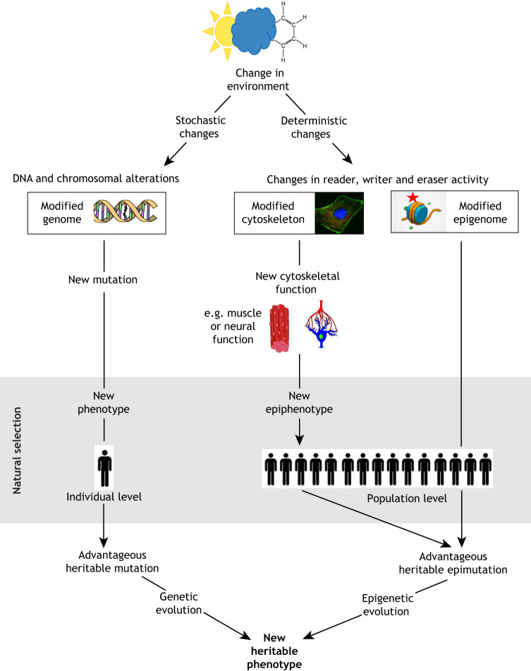
Despite knowing the general outline of these relationships for over a quarter of a century (Holliday and Grigg, 1993; Jackson-Grusby and Jaenisch, 1996), evolutionary biologists and epigeneticists are still struggling to incorporate mechanisms of epigenetic inheritance into the nexus of genetics, phenotypic plasticity, natural selection and evolution. The discovery that the components of the epigenetic machinery discussed here (with the potential for more yet to be discovered) are dual-function proteins with chromatocytoskeletal activity, opens exciting new windows into understanding phenotypic plasticity, evolutionary biology and epigenetics. The potential for epiregulation to coordinate cytoskeletal biology at the cellular and organismal level (such as in muscle contraction and neuronal function) with heritable changes in the epigenome could have profound implications for evolutionary theory. Both mutations and epimutations can be heritable and acted upon by natural selection in fundamentally similar ways. However, coordinated regulation of the cytoskeleton and epigenome by epiregulation may provide heretofore unappreciated ways for natural selection to drive evolution (Fig. 2).
Fig. 2.
A comparison of genetic and epigenetic drivers for evolution. Genetic evolution is driven by stochastic DNA alterations. In contrast, evolution driven by epiregulator activity would be deterministic: environmental perturbation of reading, writing or erasing activity of epiregulators would cause specific and predictable changes in the epigenome and epicytoskeleton. For example, decreased oxygen levels inhibit erasers that remove methyl marks, increasing epigenetic (and, we predict, epicytoskeletal) methylation. Increased cytoskeletal methylation could then produce new phenotypes upon which natural selection could act, and perturbations of the epigenome to drive emergence of hereditary epimutations. Thus, the environment determines the activity of epiregulators, and changes in epiregulator activity determine the nature of the modifications that occur to the epigenome and epicytoskeleton. A second distinction is that whereas stochastic DNA alterations occur at the level of the individual, an environmental perturbation that affects chromatocytoskeletal activity has the potential to affect the epicytoskeleton and epigenome of all individuals within an exposed population. Thirdly, perturbation of chromatocytoskeletal activity would be predicted to have an immediate and direct impact on the epicytoskeleton, potentially giving rise to phenotypes upon which natural selection could immediately act. Finally, in contrast to a new phenotype arising from an irreversible DNA alteration, a new epicytoskeleton-dependent phenotype caused by altered activity of an epiregulator would be reversible until fixed in the population by either a hereditary mutation or epimutation.
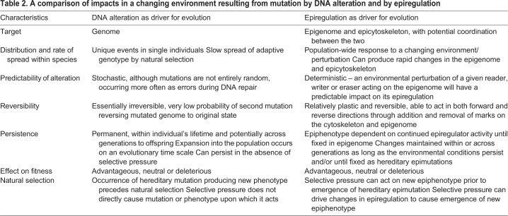
DNA alterations are, of course, the stuff of natural selection and Darwinian evolution, but would differ from epiregulation-driven evolution in several fundamental ways, as shown in Table 2 and Fig. 2. Importantly, an environmental exposure that alters the chromatocytoskeletal activity of an epiregulator to cause a change in phenotype (one mechanism for phenotypic plasticity), could also exert selective pressure upon that epiphenotype, and cause changes in the epigenome that could drive the emergence of hereditary epimutations. Therefore, in contrast to genetic (classical) evolution where DNA alteration necessarily precedes the emergence of a phenotype upon which natural selection can act, changes in epiregulator activity could produce an epiphenotype upon which natural selection could act that precedes the emergence of a hereditary epimutation. In this way, a changing environment could exert directionality by changing chromatocytoskeletal activity to produce an advantageous phenotype while concomitantly changing the epigenome to drive the emergence of new epimutations that encode that phenotype. We would propose the term ‘epigenetic entrainment’ for this process. The literature is replete with examples of environmental exposures that modulate the activity of epiregulators, including availability of dietary nutrients required for 1-carbon metabolites needed as methyl donors, changes in oxygen levels that affect the activity of demethylases, and environmental exposures that change cell signaling pathways and the activity of kinases that phosphorylate epiregulators to increase or decrease their activity (for a review, see Walker, 2016).
Table 2.
A comparison of impacts in a changing environment resulting from mutation by DNA alteration and by epiregulation
What might be the consequences for evolution if environmental changes modulate the activity of chromatocytoskeletal epiregulators? If environmentally induced changes in epiregulator activity result in advantageous changes in cytoskeletal function, for example altered muscular or neural function, then natural selection for that epiphenotype could occur as long as the epiregulator activity responsible persisted. Selective pressure for this altered epiregulator activity could also drive changes in the epigenome, with the potential to become fixed over time as hereditary epimutations. If so, this would predict that over evolutionary time, coordinated functions for marks on the epigenome and epicytoskeleton would arise, such as a role for methyl marks made by SETD2 on both chromatin and the cytoskeleton participating in maintenance of genomic stability.
Equally importantly, regulation of the chromatocytoskeletal activity of epiregulators in response to changes in the environment would be predicted to occur on a population-wide level (Fig. 2). A graphic example of epigenetically driven population-wide modulation of phenotype conversion involves the locust Schistocerca gregaria. This locust converts from its typical solitarious form to a gregarious form – famed for the swarms of locusts that plague Africa and other regions. The morphological, physiological and behavioral transition from solitarious to gregarious form begins within hours of the appropriate environmental cues, and is thought to result from epigenetic changes that simultaneously affect as many as 80 million individuals (Burggren, 2017; Ernst et al., 2015). Fascinatingly, these changes are also accompanied by changes in learning and memory (Simoes et al., 2013). Could these arise by concurrent coupling of epigenetic and epicytoskeletal changes?
Finally, this hypothesis of coordinated regulation between genome and cytoskeleton could be examined experimentally via several approaches, such as altering the activity of a specific writer or eraser and determining whether coordinate changes occur in their cognate methyl marks on both chromatin and the cytoskeleton. Similarly, it would be of interest to determine whether the ‘rules’ governing the histone code also apply to the tubulin (or actin) code. For example, are the same methyltransferase/demethylase pairs that add/remove a specific histone methyl mark on chromatin also involved in addition and removal of that cognate methyl mark on microtubules of the cytoskeleton?
Conclusions and perspectives
Molecular biologists have learned much about the epigenetic machinery, and the epigenome itself is now well defined for numerous cell types, tissues and even disease states. Emerging data are showing that these readers, writers and erasers have chromatocytoskeletal activity, thereby contributing to remodeling functions in the cytoplasm as well as the nucleus. This opens up the exciting possibility that this machinery may not only regulate the structure of chromatin and the cytoskeleton, but also serve to integrate their functions. In light of this possibility, we suggest that the term epiregulation – that is, coordinated regulation of the epigenome and cytoskeleton to integrate key biological processes using a shared machinery – may better capture how this machinery functions in a broader context.
We predict that with the expansion of the concept of epiregulation will come a new perspective to our understanding of both normal and pathophysiological processes. For example, epiregulation may help us understand how cells coordinate epigenetic differentiation programs with acquisition of specialized cytoskeletal structures. Such coordination could affect brain development by coordinating patterns of gene expression with remodeling of the neuronal cytoskeleton. This, in turn, could modify such basic neural functions as learning, memory and behavior in a wide variety of animals, either as adaptations or maladaptations. Similarly, in the study of human diseases such as cancer and autism spectrum disorder (ASD), where mutations in components of the epigenetic machinery (including SETD2) frequently occur, we need to look beyond changes in gene expression caused by these defects to examine their impact on the cytoskeleton. In creating a deeper understanding of cancer this could mean, for example, determining how mutations in chromatocytoskeletal remodelers cause cytoskeletal defects contributing to cell migration and metastasis, and in ASD, how mutations in chromatocytoskeletal remodelers impact neurodevelopment, learning and memory.
We hope and anticipate that physiologists, morphologists, ecologists, evolutionary biologists and others working on molecular and cellular regulation may find useful ways to explore epiregulation as a new paradigm for interpreting their data. Thus, moving both conceptually and pragmatically beyond ‘epigenetic regulation’ to ‘epiregulation’ may lead to experimental paradigms that supplement searches for modified gene expression as a result of addition or removal of epigenetic markers with expanded investigations of methylation beyond the genome itself. Such experiments would establish a new frontier in our investigation of evolution of the epigenome specifically and regulatory mechanisms more broadly, perhaps extending to evolutionary biology itself.
Footnotes
Competing interests
The authors declare no competing or financial interests.
Funding
We acknowledge the financial support of the John Templeton Foundation (61099) and National Cancer Institute (R35 CA2319930) to C.W. and the National Science Foundation to W.B. (1543301). Deposited in PMC for release after 12 months.
References
- Ali A., Veeranki S. N., Chinchole A. and Tyagi S. (2017). MLL/WDR5 complex regulates Kif2A localization to ensure chromosome congression and proper spindle assembly during mitosis. Dev. Cell 41, 605-622.e7. 10.1016/j.devcel.2017.05.023 [DOI] [PubMed] [Google Scholar]
- Bailey J. K., Fields A. T., Cheng K., Lee A., Wagenaar E., Lagrois R., Schmidt B., Xia B. and Ma D. (2015). WD repeat-containing protein 5 (WDR5) localizes to the midbody and regulates abscission. J. Biol. Chem. 290, 8987-9001. 10.1074/jbc.M114.623611 [DOI] [PMC free article] [PubMed] [Google Scholar]
- Barisic M. and Maiato H. (2016). The tubulin code: a navigation system for chromosomes during mitosis. Trends Cell Biol. 26, 766-775. 10.1016/j.tcb.2016.06.001 [DOI] [PMC free article] [PubMed] [Google Scholar]
- Becker J. S., Nicetto D. and Zaret K. S. (2016). H3K9me3-dependent heterochromatin: barrier to cell fate changes. Trends Genet. 32, 29-41. 10.1016/j.tig.2015.11.001 [DOI] [PMC free article] [PubMed] [Google Scholar]
- Biswas S. and Rao C. M. (2018). Epigenetic tools (the writers, the readers and the erasers) and their implications in cancer therapy. Eur. J. Pharmacol. 837, 8-24. 10.1016/j.ejphar.2018.08.021 [DOI] [PubMed] [Google Scholar]
- Bochynska A., Luscher-Firzlaff J. and Luscher B. (2018). Modes of interaction of KMT2 histone H3 lysine 4 methyltransferase/COMPASS complexes with chromatin. Cells 7, E17 10.3390/cells7030017 [DOI] [PMC free article] [PubMed] [Google Scholar]
- Bodakuntla S., Jijumon A. S., Villablanca C., Gonzalez-Billault C. and Janke C. (2019). Microtubule-associated proteins: structuring the cytoskeleton. Trends Cell Biol. 29, 804-819. 10.1016/j.tcb.2019.07.004 [DOI] [PubMed] [Google Scholar]
- Booth L. N. and Brunet A. (2016). The aging epigenome. Mol. Cell 62, 728-744. 10.1016/j.molcel.2016.05.013 [DOI] [PMC free article] [PubMed] [Google Scholar]
- Brykczynska U., Hisano M., Erkek S., Ramos L., Oakeley E. J., Roloff T. C., Beisel C., Schübeler D., Stadler M. B. and Peters A. H. (2010). Repressive and active histone methylation mark distinct promoters in human and mouse spermatozoa. Nat. Struct. Mol. Biol. 17, 679-687. 10.1038/nsmb.1821 [DOI] [PubMed] [Google Scholar]
- Burggren W. W. (2016). Epigenetic inheritance and its role in evolutionary biology: re-evaluation and new perspectives. Biology 5, 24 10.3390/biology5020024 [DOI] [PMC free article] [PubMed] [Google Scholar]
- Burggren W. W. (2017). Epigenetics in insects: mechanisms, phenotypes and ecological and evolutionary implications. In Epigenetics: How the Environment Can Have Impact on Genes and Regulate Phenotypes, Vol. 53 (ed. Verlinden H.), pp. 1-30 Elsevier. [Google Scholar]
- Burggren W. W. (2020). Phenotypic switching resulting from developmental plasticity: fixed or reversible? Front. Physiol. 10 10.3389/fphys.2019.01634 [DOI] [PMC free article] [PubMed] [Google Scholar]
- Burggren W. W. and Crews D. (2014). Epigenetics in comparative biology: why we should pay attention. Integr. Comp. Biol. 54, 7-20. 10.1093/icb/icu013 [DOI] [PMC free article] [PubMed] [Google Scholar]
- Cai D., McEwen D. P., Martens J. R., Meyhofer E. and Verhey K. J. (2009). Single molecule imaging reveals differences in microtubule track selection between Kinesin motors. PLoS Biol. 7, e1000216 10.1371/journal.pbio.1000216 [DOI] [PMC free article] [PubMed] [Google Scholar]
- Carvalho S., Vitor A. C., Sridhara S. C., Martins F. B., Raposo A. C., Desterro J. M., Ferreira J. and de Almeida S. F. (2014). SETD2 is required for DNA double-strand break repair and activation of the p53-mediated checkpoint. Elife 3, e02482 10.7554/eLife.02482 [DOI] [PMC free article] [PubMed] [Google Scholar]
- Chen R., Kong P., Zhang F., Shu Y.-N., Nie X., Dong L.-H., Lin Y.-L., Xie X.-L., Zhao L.-L., Zhang X.-J. et al. (2017). EZH2-mediated alpha-actin methylation needs lncRNA TUG1, and promotes the cortex cytoskeleton formation in VSMCs. Gene 616, 52-57. 10.1016/j.gene.2017.03.028 [DOI] [PubMed] [Google Scholar]
- Chiang Y. C., Park I. Y., Terzo E. A., Tripathi D. N., Mason F. M., Fahey C. C., Karki M., Shuster C. B., Sohn B. H., Chowdhury P. et al. (2018). SETD2 haploinsufficiency for microtubule methylation is an early driver of genomic instability in renal cell carcinoma. Cancer Res. 78, 3135-3146. 10.1158/0008-5472.CAN-17-3460 [DOI] [PMC free article] [PubMed] [Google Scholar]
- Copeland J. (2019). Formins, golgi, and the centriole. Results Probl. Cell Differ. 67, 27-48. 10.1007/978-3-030-23173-6_3 [DOI] [PubMed] [Google Scholar]
- Crews D., Gillette R., Miller-Crews I., Gore A. C. Skinner M. K. (2014). Nature, nurture and epigenetics. Mol. Cell. Endocrinol. 398, 42-52. 10.1016/j.mce.2014.07.013 [DOI] [PMC free article] [PubMed] [Google Scholar]
- Cropley J. E., Dang T. H. Y., Martin D. I. K. and Suter C. M. (2012). The penetrance of an epigenetic trait in mice is progressively yet reversibly increased by selection and environment. Proc. Biol. Sci. 279, 2347-2353. 10.1098/rspb.2011.2646 [DOI] [PMC free article] [PubMed] [Google Scholar]
- Dai S., Horton J. R., Woodcock C. B., Wilkinson A. W., Zhang X., Gozani O. and Cheng X. (2019). Structural basis for the target specificity of actin histidine methyltransferase SETD3. Nat. Commun. 10, 3541 10.1038/s41467-019-11554-6 [DOI] [PMC free article] [PubMed] [Google Scholar]
- Dominguez R. and Holmes K. C. (2011). Actin structure and function. Annu. Rev. Biophys. 40, 169-186. 10.1146/annurev-biophys-042910-155359 [DOI] [PMC free article] [PubMed] [Google Scholar]
- Dong F., Li Q., Yang C., Huo D., Wang X., Ai C., Kong Y., Sun X., Wang W., Zhou Y. et al. (2018). PRMT2 links histone H3R8 asymmetric dimethylation to oncogenic activation and tumorigenesis of glioblastoma. Nat. Commun. 9, 4552 10.1038/s41467-018-06968-7 [DOI] [PMC free article] [PubMed] [Google Scholar]
- Eom G. H., Kim K.-B., Kim J. H., Kim J.-Y., Kim J.-R., Kee H. J., Kim D. W., Choe N., Park H.-J., Son H.-J. et al. (2011). Histone methyltransferase SETD3 regulates muscle differentiation. J. Biol. Chem. 286, 34733-34742. 10.1074/jbc.M110.203307 [DOI] [PMC free article] [PubMed] [Google Scholar]
- Ernst U. R., Van Hiel M. B., Depuydt G., Boerjan B., De Loof A. and Schoofs L. (2015). Epigenetics and locust life phase transitions. J. Exp. Biol. 218, 88-99. 10.1242/jeb.107078 [DOI] [PubMed] [Google Scholar]
- Eshun-Wilson L., Zhang R., Portran D., Nachury M. V., Toso D. B., Löhr T., Vendruscolo M., Bonomi M., Fraser J. S. and Nogales E. (2019). Effects of alpha-tubulin acetylation on microtubule structure and stability. Proc. Natl. Acad. Sci. USA 116, 10366-10371. 10.1073/pnas.1900441116 [DOI] [PMC free article] [PubMed] [Google Scholar]
- Fletcher D. A. and Mullins R. D. (2010). Cell mechanics and the cytoskeleton. Nature 463, 485-492. 10.1038/nature08908 [DOI] [PMC free article] [PubMed] [Google Scholar]
- Fnu S., Williamson E. A., De Haro L. P., Brenneman M., Wray J., Shaheen M., Radhakrishnan K., Lee S.-H., Nickoloff J. A. and Hromas R. (2011). Methylation of histone H3 lysine 36 enhances DNA repair by nonhomologous end-joining. Proc. Natl. Acad. Sci. USA 108, 540-545. 10.1073/pnas.1013571108 [DOI] [PMC free article] [PubMed] [Google Scholar]
- Funk K. E., Thomas S. N., Schafer K. N., Cooper G. L., Liao Z., Clark D. J., Yang A. J. and Kuret J. (2014). Lysine methylation is an endogenous post-translational modification of tau protein in human brain and a modulator of aggregation propensity. Biochem. J. 462, 77-88. 10.1042/BJ20140372 [DOI] [PMC free article] [PubMed] [Google Scholar]
- Furrow R. E. (2014). Epigenetic inheritance, epimutation, and the response to selection. PLoS ONE 9, e101559 10.1371/journal.pone.0101559 [DOI] [PMC free article] [PubMed] [Google Scholar]
- Gadadhar S., Bodakuntla S., Natarajan K. and Janke C. (2017). The tubulin code at a glance. J. Cell Sci. 130, 1347-1353. 10.1242/jcs.199471 [DOI] [PubMed] [Google Scholar]
- Gonzalez-Recio O., Toro M. A. and Bach A. (2015). Past, present, and future of epigenetics applied to livestock breeding. Front. Genet. 6, 305 10.3389/fgene.2015.00305 [DOI] [PMC free article] [PubMed] [Google Scholar]
- Guarnaccia A. D. and Tansey W. P. (2018). Moonlighting with WDR5: a cellular multitasker. J. Clin. Med. 7, E21 10.3390/jcm7020021 [DOI] [PMC free article] [PubMed] [Google Scholar]
- Gunawan M., Venkatesan N., Loh J. T., Wong J. F., Berger H., Neo W. H., Li L. Y., La Win M. K., Yau Y. H., Guo T. et al. (2015). The methyltransferase Ezh2 controls cell adhesion and migration through direct methylation of the extranuclear regulatory protein talin. Nat. Immunol. 16, 505-516. 10.1038/ni.3125 [DOI] [PubMed] [Google Scholar]
- Guo Q., Liao S., Kwiatkowski S., Tomaka W., Yu H., Wu G., Tu X., Min J., Drozak J. and Xu C. (2019). Structural insights into SETD3-mediated histidine methylation on beta-actin. Elife 8, e43676 10.7554/eLife.43676.031 [DOI] [PMC free article] [PubMed] [Google Scholar]
- Hacker K. E., Fahey C. C., Shinsky S. A., Chiang Y. J., DiFiore J. V., Jha D. K., Vo A. H., Shavit J. A., Davis I. J., Strahl B. D. et al. (2016). Structure/function analysis of recurrent mutations in SETD2 protein reveals a critical and conserved role for a SET domain residue in maintaining protein stability and histone H3 Lys-36 trimethylation. J. Biol. Chem. 291, 21283-21295. 10.1074/jbc.M116.739375 [DOI] [PMC free article] [PubMed] [Google Scholar]
- Heyer W. D., Li X., Rolfsmeier M. and Zhang X. P. (2006). Rad54: the Swiss Army knife of homologous recombination? Nucleic Acids Res. 34, 4115-4125. 10.1093/nar/gkl481 [DOI] [PMC free article] [PubMed] [Google Scholar]
- Holliday R. and Grigg G. W. (1993). DNA methylation and mutation. Mutat. Res. 285, 61-67. 10.1016/0027-5107(93)90052-H [DOI] [PubMed] [Google Scholar]
- Hou W., Nemitz S., Schopper S., Nielsen M. L., Kessels M. M. and Qualmann B. (2018). Arginine methylation by PRMT2 controls the functions of the actin nucleator Cobl. Dev. Cell 45, 262-275.e8. 10.1016/j.devcel.2018.03.007 [DOI] [PubMed] [Google Scholar]
- Howes S. C., Alushin G. M., Shida T., Nachury M. V. and Nogales E. (2014). Effects of tubulin acetylation and tubulin acetyltransferase binding on microtubule structure. Mol. Biol. Cell 25, 257-266. 10.1091/mbc.e13-07-0387 [DOI] [PMC free article] [PubMed] [Google Scholar]
- Huseby C. J., Hoffman C. N., Cooper G. L., Cocuron J. C., Alonso A. P., Thomas S. N., Yang A. J. and Kuret J. (2019). Quantification of tau protein lysine methylation in aging and Alzheimer's disease. J. Alzheimer's Dis. 71, 979-991. 10.3233/JAD-190604 [DOI] [PMC free article] [PubMed] [Google Scholar]
- Iwabata H., Yoshida M. and Komatsu Y. (2005). Proteomic analysis of organ-specific post-translational lysine-acetylation and -methylation in mice by use of anti-acetyllysine and -methyllysine mouse monoclonal antibodies. Proteomics 5, 4653-4664. 10.1002/pmic.200500042 [DOI] [PubMed] [Google Scholar]
- Jablonka E. (2017). The evolutionary implications of epigenetic inheritance. Interface Focus 7, 20160135 10.1098/rsfs.2016.0135 [DOI] [PMC free article] [PubMed] [Google Scholar]
- Jackson-Grusby L. and Jaenisch R. (1996). Experimental manipulation of genomic methylation. Semin. Cancer Biol. 7, 261-268. 10.1006/scbi.1996.0034 [DOI] [PubMed] [Google Scholar]
- Janke C. (2014). The tubulin code: molecular components, readout mechanisms, and functions. J. Cell Biol. 206, 461-472. 10.1083/jcb.201406055 [DOI] [PMC free article] [PubMed] [Google Scholar]
- Janssen K. A., Sidoli S. and Garcia B. A. (2017). Recent achievements in characterizing the histone code and approaches to integrating epigenomics and systems biology. Methods Enzymol. 586, 359-378. 10.1016/bs.mie.2016.10.021 [DOI] [PMC free article] [PubMed] [Google Scholar]
- Javaid N. and Choi S. (2017). Acetylation- and methylation-related epigenetic proteins in the context of their targets. Genes (Basel) 8, E196 10.3390/genes8080196 [DOI] [PMC free article] [PubMed] [Google Scholar]
- Jones P. A., Rideout W. M. III, Shen J. C., Spruck C.-H. and Tsai Y. C. (1992). Methylation, mutation and cancer. BioEssays 14, 33-36. 10.1002/bies.950140107 [DOI] [PubMed] [Google Scholar]
- Kaul N., Soppina V. and Verhey K. J. (2014). Effects of α-tubulin K40 acetylation and detyrosination on kinesin-1 motility in a purified system. Biophys. J. 106, 2636-2643. 10.1016/j.bpj.2014.05.008 [DOI] [PMC free article] [PubMed] [Google Scholar]
- Kontaxi C., Piccardo P. and Gill A. C. (2017). Lysine-directed post-translational modifications of tau protein in Alzheimer's disease and related tauopathies. Front. Mol. Biosci. 4, 56 10.3389/fmolb.2017.00056 [DOI] [PMC free article] [PubMed] [Google Scholar]
- Kulkarni S. S., Griffin J. N., Date P. P., Liem K. F. Jr and Khokha M. K. (2018). WDR5 stabilizes actin architecture to promote multiciliated cell formation. Dev. Cell 46, 595-610.e3. 10.1016/j.devcel.2018.08.009 [DOI] [PMC free article] [PubMed] [Google Scholar]
- Kwiatkowski S., Seliga A. K., Vertommen D., Terreri M., Ishikawa T., Grabowska I., Tiebe M., Teleman A. A., Jagielski A. K., Veiga-da-Cunha M. et al. (2018). SETD3 protein is the actin-specific histidine N-methyltransferase. Elife 7, e37921 10.7554/eLife.37921 [DOI] [PMC free article] [PubMed] [Google Scholar]
- Lakowski T. M. and Frankel A. (2009). Kinetic analysis of human protein arginine N-methyltransferase 2: formation of monomethyl- and asymmetric dimethyl-arginine residues on histone H4. Biochem. J. 421, 253-261. 10.1042/BJ20090268 [DOI] [PubMed] [Google Scholar]
- Le Floc'h A. and Huse M. (2015). Molecular mechanisms and functional implications of polarized actin remodeling at the T cell immunological synapse. Cell. Mol. Life Sci. 72, 537-556. 10.1007/s00018-014-1760-7 [DOI] [PMC free article] [PubMed] [Google Scholar]
- Li G. M. (2013). Decoding the histone code: Role of H3K36me3 in mismatch repair and implications for cancer susceptibility and therapy. Cancer Res. 73, 6379-6383. 10.1158/0008-5472.CAN-13-1870 [DOI] [PMC free article] [PubMed] [Google Scholar]
- Li L. and Wang Y. (2017). Cross-talk between the H3K36me3 and H4K16ac histone epigenetic marks in DNA double-strand break repair. J. Biol. Chem. 292, 11951-11959. 10.1074/jbc.M117.788224 [DOI] [PMC free article] [PubMed] [Google Scholar]
- Li Y., Shin D. and Kwon S. H. (2013). Histone deacetylase 6 plays a role as a distinct regulator of diverse cellular processes. FEBS J. 280, 775-793. 10.1111/febs.12079 [DOI] [PubMed] [Google Scholar]
- Li P., Bademosi A. T., Luo J. and Meunier F. A. (2018). Actin remodeling in regulated exocytosis: toward a mesoscopic view. Trends Cell Biol. 28, 685-697. 10.1016/j.tcb.2018.04.004 [DOI] [PubMed] [Google Scholar]
- Lowdon R. F., Jang H. S. and Wang T. (2016). Evolution of epigenetic regulation in vertebrate genomes. Trends Genet. 32, 269-283. 10.1016/j.tig.2016.03.001 [DOI] [PMC free article] [PubMed] [Google Scholar]
- Magiera M. M., Singh P., Gadadhar S. and Janke C. (2018). tubulin posttranslational modifications and emerging links to human disease. Cell 173, 1323-1327. 10.1016/j.cell.2018.05.018 [DOI] [PubMed] [Google Scholar]
- Matamoros A. J. and Baas P. W. (2016). Microtubules in health and degenerative disease of the nervous system. Brain Res. Bull. 126, 217-225. 10.1016/j.brainresbull.2016.06.016 [DOI] [PMC free article] [PubMed] [Google Scholar]
- McDaniel S. L. and Strahl B. D. (2017). Shaping the cellular landscape with Set2/SETD2 methylation. Cell. Mol. Life Sci. 74, 3317-3334. 10.1007/s00018-017-2517-x [DOI] [PMC free article] [PubMed] [Google Scholar]
- Melamed-Bessudo C. and Levy A. A. (2012). Deficiency in DNA methylation increases meiotic crossover rates in euchromatic but not in heterochromatic regions in Arabidopsis. Proc. Natl. Acad. Sci. USA 109, E981-E988. 10.1073/pnas.1120742109 [DOI] [PMC free article] [PubMed] [Google Scholar]
- Migliori V., Muller J., Phalke S., Low D., Bezzi M., Mok W. C., Sahu S. K., Gunaratne J., Capasso P., Bassi C. et al. (2012). Symmetric dimethylation of H3R2 is a newly identified histone mark that supports euchromatin maintenance. Nat. Struct. Mol. Biol. 19, 136-144. 10.1038/nsmb.2209 [DOI] [PubMed] [Google Scholar]
- Morselli M., Pastor W. A., Montanini B., Nee K., Ferrari R., Fu K., Bonora G., Rubbi L., Clark A. T., Ottonello S. et al. (2015). In vivo targeting of de novo DNA methylation by histone modifications in yeast and mouse. Elife 4, e06205 10.7554/eLife.06205 [DOI] [PMC free article] [PubMed] [Google Scholar]
- Ooi S. K., Qiu C., Bernstein E., Li K., Jia D., Yang Z., Erdjument-Bromage H., Tempst P., Lin S. P., Allis C. D. et al. (2007). DNMT3L connects unmethylated lysine 4 of histone H3 to de novo methylation of DNA. Nature 448, 714-717. 10.1038/nature05987 [DOI] [PMC free article] [PubMed] [Google Scholar]
- Park I. Y., Powell R. T., Tripathi D. N., Dere R., Ho T. H., Blasius T. L., Chiang Y. C., Davis I. J., Fahey C. C., Hacker K. E. et al. (2016). Dual chromatin and cytoskeletal remodeling by SETD2. Cell 166, 950-962. 10.1016/j.cell.2016.07.005 [DOI] [PMC free article] [PubMed] [Google Scholar]
- Pasini D., Malatesta M., Jung H. R., Walfridsson J., Willer A., Olsson L., Skotte J., Wutz A., Porse B., Jensen O. N. et al. (2010). Characterization of an antagonistic switch between histone H3 lysine 27 methylation and acetylation in the transcriptional regulation of Polycomb group target genes. Nucleic Acids Res. 38, 4958-4969. 10.1093/nar/gkq244 [DOI] [PMC free article] [PubMed] [Google Scholar]
- Perez M. F. and Lehner B. (2019). Intergenerational and transgenerational epigenetic inheritance in animals. Nat. Cell Biol. 21, 143-151. 10.1038/s41556-018-0242-9 [DOI] [PubMed] [Google Scholar]
- Pfister S. X., Ahrabi S., Zalmas L. P., Sarkar S., Aymard F., Bachrati C. Z., Helleday T., Legube G., La Thangue N. B., Porter A. C. et al. (2014). SETD2-dependent histone H3K36 trimethylation is required for homologous recombination repair and genome stability. Cell Reports 7, 2006-2018. 10.1016/j.celrep.2014.05.026 [DOI] [PMC free article] [PubMed] [Google Scholar]
- Pollard T. D. (2016). Actin and actin-binding proteins. Cold Spring Harbor Perspect. Biol. 8, a018226 10.1101/cshperspect.a018226 [DOI] [PMC free article] [PubMed] [Google Scholar]
- Reed N. A., Cai D., Blasius T. L., Jih G. T., Meyhofer E., Gaertig J. and Verhey K. J. (2006). Microtubule acetylation promotes kinesin-1 binding and transport. Curr. Biol. 16, 2166-2172. 10.1016/j.cub.2006.09.014 [DOI] [PubMed] [Google Scholar]
- Rhounim L., Rossignol J. L. and Faugeron G. (1992). Epimutation of repeated genes in Ascobolus immersus. EMBO J. 11, 4451-4457. 10.1002/j.1460-2075.1992.tb05546.x [DOI] [PMC free article] [PubMed] [Google Scholar]
- Rošić S., Amouroux R., Requena C. E., Gomes A., Emperle M., Beltran T., Rane J. K., Linnett S., Selkirk M. E., Schiffer P. H. et al. (2018). Evolutionary analysis indicates that DNA alkylation damage is a byproduct of cytosine DNA methyltransferase activity. Nat. Genet. 50, 452-459. 10.1038/s41588-018-0061-8 [DOI] [PMC free article] [PubMed] [Google Scholar]
- Rottner K., Faix J., Bogdan S., Linder S. and Kerkhoff E. (2017). Actin assembly mechanisms at a glance. J. Cell Sci. 130, 3427-3435. 10.1242/jcs.206433 [DOI] [PubMed] [Google Scholar]
- Sadoul K. and Khochbin S. (2016). The growing landscape of tubulin acetylation: lysine 40 and many more. Biochem. J. 473, 1859-1868. 10.1042/BCJ20160172 [DOI] [PubMed] [Google Scholar]
- Sarkies P. (2019). Molecular mechanisms of epigenetic inheritance: possible evolutionary implications. Semin. Cell Dev. Biol. 97, 106-115. 10.1016/j.semcdb.2019.06.005 [DOI] [PMC free article] [PubMed] [Google Scholar]
- Seymour D. K. and Becker C. (2017). The causes and consequences of DNA methylome variation in plants. Curr. Opin. Plant Biol. 36, 56-63. 10.1016/j.pbi.2017.01.005 [DOI] [PubMed] [Google Scholar]
- Shilatifard A. (2012). The COMPASS family of histone H3K4 methylases: mechanisms of regulation in development and disease pathogenesis. Annu. Rev. Biochem. 81, 65-95. 10.1146/annurev-biochem-051710-134100 [DOI] [PMC free article] [PubMed] [Google Scholar]
- Simoes P. M., Niven J. E. and Ott S. R. (2013). Phenotypic transformation affects associative learning in the desert locust. Curr. Biol. 23, 2407-2412. 10.1016/j.cub.2013.10.016 [DOI] [PMC free article] [PubMed] [Google Scholar]
- Skinner M. K. (2015). Environmental epigenetics and a unified theory of the molecular aspects of evolution: a neo-Lamarckian concept that facilitates neo-Darwinian evolution. Genome Biol. Evol. 7, 1296-1302. 10.1093/gbe/evv073 [DOI] [PMC free article] [PubMed] [Google Scholar]
- Song Y. and Brady S. T. (2015). Post-translational modifications of tubulin: pathways to functional diversity of microtubules. Trends Cell Biol. 25, 125-136. 10.1016/j.tcb.2014.10.004 [DOI] [PMC free article] [PubMed] [Google Scholar]
- Storz J. F., Natarajan C., Signore A. V., Witt C. C., McCandlish D. M. and Stoltzfus A. (2019). The role of mutation bias in adaptive molecular evolution: insights from convergent changes in protein function. Philos. Trans. R. Soc. Lond. B Biol. Sci. 374, 20180238 10.1098/rstb.2018.0238 [DOI] [PMC free article] [PubMed] [Google Scholar]
- Strahl B. D. and Allis C. D. (2000). The language of covalent histone modifications. Nature 403, 41-45. 10.1038/47412 [DOI] [PubMed] [Google Scholar]
- Stricker S. H., Köferle A. and Beck S. (2017). From profiles to function in epigenomics. Nat. Rev. Genet. 18, 51-66. 10.1038/nrg.2016.138 [DOI] [PubMed] [Google Scholar]
- Su I.-H., Dobenecker M. W., Dickinson E., Oser M., Basavaraj A., Marqueron R., Viale A., Reinberg D., Wulfing C. and Tarakhovsky A. (2005). Polycomb group protein ezh2 controls actin polymerization and cell signaling. Cell 121, 425-436. 10.1016/j.cell.2005.02.029 [DOI] [PubMed] [Google Scholar]
- Terman J. R. and Kashina A. (2013). Post-translational modification and regulation of actin. Curr. Opin. Cell Biol. 25, 30-38. 10.1016/j.ceb.2012.10.009 [DOI] [PMC free article] [PubMed] [Google Scholar]
- Trevino L. S., Wang Q. and Walker C. L. (2015). Phosphorylation of epigenetic ‘readers, writers and erasers’: implications for developmental reprogramming and the epigenetic basis for health and disease. Prog. Biophys. Mol. Biol. 118, 8-13. 10.1016/j.pbiomolbio.2015.02.013 [DOI] [PMC free article] [PubMed] [Google Scholar]
- Vaissiere T., Sawan C. and Herceg Z. (2008). Epigenetic interplay between histone modifications and DNA methylation in gene silencing. Mutat. Res. 659, 40-48. 10.1016/j.mrrev.2008.02.004 [DOI] [PubMed] [Google Scholar]
- van der Graaf A., Wardenaar R., Neumann D. A., Taudt A., Shaw R. G., Jansen R. C., Schmitz R. J., Colome-Tatche M. and Johannes F. (2015). Rate, spectrum, and evolutionary dynamics of spontaneous epimutations. Proc. Natl. Acad. Sci. USA 112, 6676-6681. 10.1073/pnas.1424254112 [DOI] [PMC free article] [PubMed] [Google Scholar]
- Verhey K. J. and Gaertig J. (2007). The tubulin code. Cell Cycle 6, 2152-2160. 10.4161/cc.6.17.4633 [DOI] [PubMed] [Google Scholar]
- Vermillion K. L., Lidberg K. A. and Gammill L. S. (2014). Cytoplasmic protein methylation is essential for neural crest migration. J. Cell Biol. 204, 95-109. 10.1083/jcb.201306071 [DOI] [PMC free article] [PubMed] [Google Scholar]
- Vidalis A., Živković D., Wardenaar R., Roquis D., Tellier A. and Johannes F. (2016). Methylome evolution in plants. Genome Biol. 17, 264 10.1186/s13059-016-1127-5 [DOI] [PMC free article] [PubMed] [Google Scholar]
- Waddington C. H. (1942). The epigenotype. Endeavour 1, 18-20. [Google Scholar]
- Wagner E. J. and Carpenter P. B. (2012). Understanding the language of Lys36 methylation at histone H3. Nat. Rev. Mol. Cell Biol. 13, 115-126. 10.1038/nrm3274 [DOI] [PMC free article] [PubMed] [Google Scholar]
- Walker C. L. (2016). Minireview: epigenomic plasticity and vulnerability to EDC exposures. Mol. Endocrinol. 30, 848-855. 10.1210/me.2016-1086 [DOI] [PMC free article] [PubMed] [Google Scholar]
- Walter W. J., Beranek V., Fischermeier E. and Diez S. (2012). Tubulin acetylation alone does not affect kinesin-1 velocity and run length in vitro. PLoS ONE 7, e42218 10.1371/journal.pone.0042218 [DOI] [PMC free article] [PubMed] [Google Scholar]
- Wang Y., Liu H. and Sun Z. (2017). Lamarck rises from his grave: parental environment-induced epigenetic inheritance in model organisms and humans. Biol. Rev. Camb. Philos. Soc. 92, 2084-2111. 10.1111/brv.12322 [DOI] [PubMed] [Google Scholar]
- Weinberg D. N., Papillon-Cavanagh S., Chen H., Yue Y., Chen X., Rajagopalan K. N., Horth C., McGuire J. T., Xu X., Nikbakht H. et al. (2019). The histone mark H3K36me2 recruits DNMT3A and shapes the intergenic DNA methylation landscape. Nature 573, 281-286. 10.1038/s41586-019-1534-3 [DOI] [PMC free article] [PubMed] [Google Scholar]
- Whipple A. V. and Holeski L. M. (2016). Epigenetic inheritance across the landscape. Front. Genet. 7, 189 10.3389/fgene.2016.00189 [DOI] [PMC free article] [PubMed] [Google Scholar]
- Wilkinson A. W., Diep J., Dai S., Liu S., Ooi Y. S., Song D., Li T. M., Horton J. R., Zhang X., Liu C. et al. (2019). SETD3 is an actin histidine methyltransferase that prevents primary dystocia. Nature 565, 372-376. 10.1038/s41586-018-0821-8 [DOI] [PMC free article] [PubMed] [Google Scholar]
- Wloga D., Joachimiak E. and Fabczak H. (2017). Tubulin post-translational modifications and microtubule dynamics. Int. J. Mol. Sci. 18, E2207 10.3390/ijms18102207 [DOI] [PMC free article] [PubMed] [Google Scholar]
- Xia J., Han L. and Zhao Z. (2012). Investigating the relationship of DNA methylation with mutation rate and allele frequency in the human genome. BMC Genomics 13 Suppl. 8, S7 10.1186/1471-2164-13-S8-S7 [DOI] [PMC free article] [PubMed] [Google Scholar]
- Yamashiro S. and Watanabe N. (2019). Quantitative high-precision imaging of myosin-dependent filamentous actin dynamics. J. Muscle Res. Cell Motil. 41, 163-173. 10.1007/s10974-019-09541-x [DOI] [PubMed] [Google Scholar]
- Yang A. Y., Kim H., Li W. and Kong A. N. (2016). Natural compound-derived epigenetic regulators targeting epigenetic readers, writers and erasers. Curr. Top. Med. Chem. 16, 697-713. 10.2174/1568026615666150826114359 [DOI] [PMC free article] [PubMed] [Google Scholar]
- Zhong X. (2016). Comparative epigenomics: a powerful tool to understand the evolution of DNA methylation. New Phytol. 210, 76-80. 10.1111/nph.13540 [DOI] [PubMed] [Google Scholar]


