Abstract
In this study, four plastomes of Hymenophyllum, distributed in the Korean peninsula, were newly sequenced and phylogenomic analysis was conducted to reveal (1) the evolutionary history of plastomes of early-diverging fern species at the species level, (2) the importance of mobile open reading frames in the genus, and (3) plastome sequence divergence providing support for H. coreanum to be recognized as an independent species distinct from H. polyanthos. In addition, 1C-values of H. polyanthos and H. coreanum were measured to compare the genome size of both species and to confirm the diversification between them. The rrn16-trnV intergenic regions in the genus varied in length caused by Mobile Open Reading Frames in Fern Organelles (MORFFO). We investigated enlarged noncoding regions containing MORFFO throughout the fern plastomes and found that they were strongly associated with tRNA genes or palindromic elements. Sequence identity between plastomes of H. polyanthos and H. coreanum is quite low at 93.35% in the whole sequence and 98.13% even if the variation in trnV-rrn16 intergenic spacer was ignored. In addition, different genome sizes were found for these species based on the 1C-value. Consequently, there is no reason to consider them as a conspecies.
Subject terms: Evolution, Plant sciences
Introduction
Plastomes of vascular plants are varied in size (120–170 kb) and compact with 110–130 genes1–5. In angiosperms, they are normally conserved in sequence, gene contents and organization1 except in heterotrophic plants6,7 or specific lineages8–10. Consequently, single nucleotide polymorphisms (SNPs) and insertions/deletions (indels) among infraspecific taxa or individuals in angiosperms are very low11–14. In contrast to angiosperms, the genetic variation in plastomes between individuals in ferns is almost similar to the interspecific genetic variation in angiosperms (e.g., for Equisetum arvense15). This is likely due to the longer evolutionary history of ferns than angiosperms.
Since the first plastome sequence of Adiantum capillus-veneris L. was reported16, plastomes from 11 orders of extant ferns17 have also been sequenced18–22. These data have given insights into (1) phylogenomics for resolving the deep relationships throughout fern lineages19,23,24, (2) inserted foreign DNA3,25, and (3) lineage-specific structural evolutions3,26,27. However, because a large proportion of plastome sequence data in ferns belongs to the order Polypodiales, which is the most derived order in the extant ferns17, the evolution of plastomes at a low taxonomic level in eusporangiate ferns and basal leptosporangiate ferns is still equivocal.
Hymenophyllaceae, filmy ferns, is a basal family of leptosporangiate ferns17, and it consists of more than 600 species28. Their distinctive feature, single-cell thick laminae, easily distinguishes them from other fern families29. However, infra-familial classification of the filmy ferns has been argued for a long time30–33. Nevertheless, Hymenophyllaceae is traditionally classified into two major clades, “trichomanoid” with obconic or tubular involucre and “hymenophylloid” with bivalvate involucre. This relationship has also been clarified using recent molecular analysis28.
Hymenophyllum s.l. is comprised of more than 300 species with a nearly cosmopolitan distribution34. Pryer et al.28 showed that Serpyllopsis, Cardiomanes, and Microtrichmanes, which were segregated from each other or belonged to Trichomanes by transitional classifications30,32,33,35, were included within Hymenophyllum s.l. Although, they also mentioned that their data set was insufficient in terms of taxon sampling and the number of genes used to evaluate all of the segregates within Hymenophyllum s.l. Hennequin et al.36 reconstructed the phylogenetic tree of Hymenophyllum s.l. using increased sampling and additional genes and divided Hymenophyllum s.l. into eight subgenera. Interestingly, four segregate genera and one section belonging to Trichomanes formed a clade with Hymenophyllum species, in agreement with Pryer et al.28. Recently, Ebihara et al.29 divided Hymenophyllum s.l. into 10 subgenera based on molecular phylogenetic analyses and macroscopic characters.
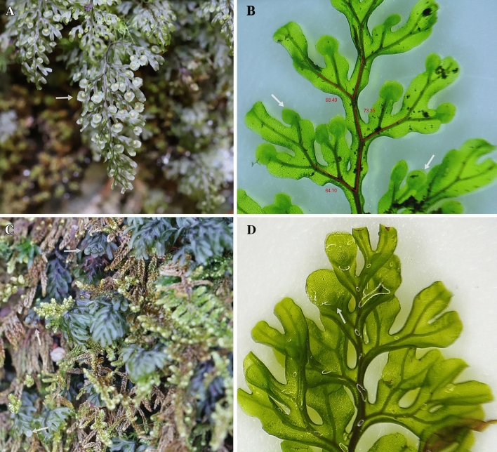
In Korea, three species and one unacceptable taxon of the genus Hymenophyllum have been reported: Hymenophyllum barbatum (Bosch) Baker, H. polyanthos (Sw.) Sw, H. wrightii Bosch and H. coreanum Nakai. Among them, H. coreanum was first recognized by Nakai37 who collected the sample from Kumgangsan in Gangwondo, Korea and described it as a plant with 2–5 mm stipes, 5–15 mm fronds and dense sori in apex fronds. In contrast to H. coreanum, H. polyanthos in Korea is 1.5–3 times longer and has involucres from the apex to the middle of the frond (Fig. 1)38. However, in spite of these different characteristics, H. coreanum has been considered a synonym of H. polyanthos by many researchers. The broadly described species, H. polyanthos has a worldwide distribution but probably includes a number of distant lineages in the subgenus Mecodium that do not have any specialized morphological characters39. The recent phylogeny of Hymenophyllum s.l. using molecular markers revealed that H. polyanthos was highly polyphyletic36,40. Therefore, more clear scientific evidence for considering the correct taxonomical position of H. coreanum and its relationship with H. polyanthos is necessary.
Figure 1.
Phenotypes of Hymenophyllum polyanthos (A and B) and H. coreanum (C and D). (A) Photo of a whole plant of H. polyanthos in the field. (B) Microphotograph of H. polyanthos. (C) H. coreanum in the field. (D) Microphotograph of H. coreanum. White arrow refers to the involucre and red angle refers to angle from rachis to pinna.
In this study, we sequenced four new plastomes of Hymenophyllum species distributed in the Korean Peninsula: (1) to explore the plastome evolution of early-diverging leptosporangiate ferns at the species level, (2) to verify the Mobile Open Reading Frames in Fern Organelles (MORFFO) which were identified by Robison et al.25, and (3) to investigate the possibility that the two morphologically similar species, H. polyanthos and H. coreanum, are distinct species. In addition, 1C-values of both species were measured to compare genome size and to explore the diversification between H. polyanthos and H. coreanum.
Results
Genome structure and gene contents of plastomes in Hymenophyllum
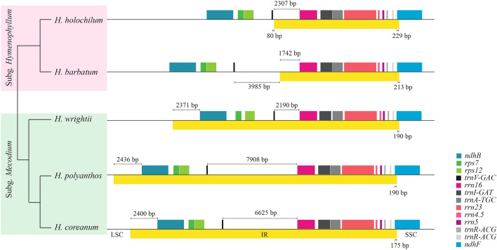
The newly sequenced plastomes of the four Hymenophyllum species were from 144,112 bp to 160,865 bp in length with a GC content of 37.5–38.6% (Table 1). The large single copy (LSC) region in the subgenus Hymenophyllum was 10 kb longer than that in the subgenus Mecodium, due to inverted repeat (IR) expansion of the LSC region (Fig. 2). The length difference of the small single copy (SSC) region between the two subgenera mainly resulted from the insertions at the rpl32-trnP and ndhA introns in the subgenus Mecodium. The IR-SSC boundary was slightly different among species but positioned near the 5ʹ end of ndhF. There were 85 coding genes, 8 rRNA genes, 33 tRNA genes, and one pseudogene (trnL-CAA) in the subgenus Hymenophyllum. However, three species in the subgenus Mecodium had one more rps7, ndhB and trnV due to IR expansion. Gene order remained stable among species in the genus Hymenophyllum except the IR expansion of the subgenus Mecodium.
Table 1.
Summary of plastome sequences of Hymenophyllum.
| Subgenus | Species | Total length (bp) | LSC (bp) | SSC (bp) | IR (bp) | GC content (%) | Coverage depth (X) |
|---|---|---|---|---|---|---|---|
| Hymenophyllum | H. barbatum | 144,112 | 102,259 | 20,789 | 10,532 | 37.6 | 227.9 |
| H. holochilum a | 142,214 | 99,299 | 20,765 | 11,075 | 37.6 | ||
| Mecodium | H. coreanum | 157,967 | 89,616 | 21,277 | 23,537 | 38.2 | 367.3 |
| H. polyanthos | 160,865 | 89,811 | 21,290 | 24,882 | 38.6 | 313.7 | |
| H. wrightii | 149,309 | 90,003 | 21,266 | 19,020 | 37.5 | 325.3 |
aDownloaded from Kuo et al. (2018).
Figure 2.
IR boundaries in the plastomes of Hymenophyllum. The phylogeny of Hymenophyllum species is extracted from the phylogeny of Hymenophyllaceae using 85 genes in this study. The boxes above the black line refer to the genes and the yellow box below the line refers to the region.
Length variation hotspots in Hymenophyllum plastomes
The intergenic spacer (IGS) between trnV and rrn16 was the most variable region in length (Fig. 2). Because coverage depths between three plant genomes in a NGS data were significantly different41, stable read depths throughout the plastomes in this study confirmed that these length variations did not result from mitochondrial plastome sequences or nuclear plastome sequences.
In subgenus Hymenophyllum, trnV-rrn16 of H. holochilum was 2387 bp in length and mostly belonged to the IR region. However, that of H. barbatum was expanded 3.4 kb more than H. holochilum and two-thirds of this region was part of the LSC region (Supplementary Table 1). In the subgenus Mecodium, trnV-rrn16 of H. wrightii was 2190 bp in length and 5.7 kb and 4.4 kb expansions were confirmed at the plastomes of H. polyanthos and H. coreanum, respectively. In total, 20 ORFs having more than 100 amino acid (aa) sequences in length were also found in this length-variable region of five Hymenophyllum species (Supplementary Fig. 1). These ORFs ranged from 101 aa to 1274 aa in length and blastp search showed that half of them had similar amino acid sequences to ORFs that were embedded in the plastome of Mankyua chejuense3 (Table 2). Based on the DNA sequences, trnV-rrn16 of five Hymenophyllum species contained at least a part of MORFFO. In addition, H. wrightii and H. holochilum contained a part of MORFFO at trnE-trnG and rrn16-ycf2, respectively.
Table 2.
Blastp results for ORFs within trnV-rrn16 in Hymenophyllum plastomes.
| Query sequence | Blastp result | |||||||
|---|---|---|---|---|---|---|---|---|
| Sequence Name | Name | Length (aaa) | Organism | Gene | Query cover (%) | E-value | Identity | Accession |
| H. holochilum | ORF_H1 | 103 | Pinus koraiensis | ORF46h | 20 | 3.40E-02 | 85.71 | YP_001152247.1 |
| ORF_H2 | 363 | Mankyua chejuense | ORF295 | 73 | 3.00E-76 | 46.99 | YP_005352949.1 | |
| Roya anglica | hypothetical protein | 44 | 3.00E-07 | 26.95 | YP_009033761.1 | |||
| ORF_H3 | 150 | –b | ||||||
| H. barbatum | ORF_B1 | 1,274 | Mankyua chejuense | ORF531 | 41 | 4.00E-130 | 42.45 | ADZ47985.2 |
| Mankyua chejuense | ORF295 | 22 | 3.00E-84 | 48.12 | YP_005352949.1 | |||
| Beggiatoa leptomitoformis | AL038_02335 | 33 | 2.00E-24 | 28.01 | ALG66761.1 | |||
| ORF_B2 | 101 | – | ||||||
| ORF_B3 | 280 | – | ||||||
| H. wrightii | ORF_W1 | 216 | – | |||||
| ORF_W2 | 132 | – | ||||||
| H. polyanthos | ORF_P1 | 664 | Mankyua chejuense | ORF295 | 44 | 3.00E-86 | 48.12 | YP_005352949.1 |
| Mankyua chejuense | ORF187 | 18 | 3.00E-22 | 41.94 | YP_005352954.1 | |||
| ORF_P2 | 222 | – | ||||||
| ORF_P3 | 107 | – | ||||||
| ORF_P4 | 147 | Mankyua chejuense | ORF531 | 97 | 9.00E-14 | 37.67 | YP_005352953.1 | |
| Bacteroidetes bacterium | hypothetical protein | 59 | 1.50E-01 | 33.33 | HAV23789.1 | |||
| ORF_P5 | 572 | Mankyua chejuense | ORF295 | 52 | 5.00E-77 | 45.97 | YP_005352949.1 | |
| ORF_P6 | 129 | Mankyua chejuense | ORF531 | 82 | 5.00E-14 | 37.04 | YP_005352953.1 | |
| ORF_P7 | 107 | Thiobaca trueperi | peptide chain release factor 3 | 58 | 3.10E-01 | 43.75 | WP_132977102.1 | |
| H. coreanum | ORF_C1 | 1,064 | Mankyua chejuense | ORF531 | 47 | 1.00E-123 | 43.24 | YP_005352953.1 |
| Beggiatoa leptomitoformis | hypothetical protein | 46 | 4.00E-25 | 25.54 | WP_083991398.1 | |||
| Thiotrichales bacterium HS_08 | hypothetical protein | 43 | 3.00E-18 | 24.7 | WP_103918394.1 | |||
| ORF_C2 | 218 | – | ||||||
| ORF_C3 | 359 | – | ||||||
| ORF_C4 | 133 | – | ||||||
| ORF_C5 | 200 | – | ||||||
aAmino acid.
bNo significant similarity found.
Enlarged noncoding regions in fern plastomes
To investigate the relationship between MORFFO and inserted loci, expanded noncoding regions having a part of MORFFO in available fern plastomes (138 plastomes, Supplementary Table 2) were found using blastn search. In total, 32 loci were matched to MORFFO with at least 100 bp in length (Supplementary table 3). Among them, 22 loci (75%) were flanked by tRNA genes. The most frequent locus, including MORFFO, throughout the plastomes was rrn16-(trnV-GAC)-rps12 in which trnV-GAC was generally intact in eusporangiate ferns and early-diverging leptosporangiate ferns but was deleted or pseudogenized in most core leptosporangiate ferns.
In addition, all expanded regions had palindrome sequences at both ends with a minimum stem length of 5 bp, a minimum loop length of 5 bp, and a minimum stem-loop sequence of 20 bp.
Phylogenetic relationships of Hymenophyllum species
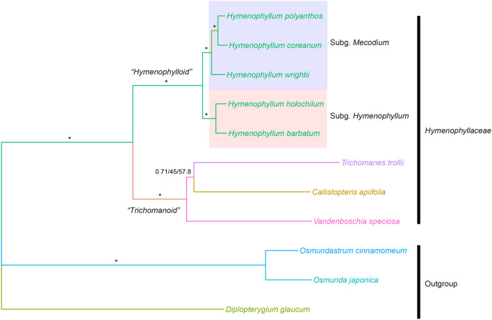
The concatenated alignment using 85 coding genes was 74,933 bp in length. Of these characters, 45,301 were constant, 11,129 variable characters were parsimonious uninformative, and 18,503 were parsimonious informative. Tree topologies among maximum parsimony (MP), maximum likelihood (ML) and Bayesian inference were completely identical with maximum support on all branches, with the exception of the moderate support on the clade Callistopteris apiifolia + Trichomanes trollii (Fig. 3). Hymenophyllaceae species were monophyletic and two traditional major clades, Hymenophylloid and Trichomanoid, were also strongly supported. H. coreanum was sister to H. polyanthos and they formed a clade with H. wrightii. As a result, two subgenera in Hymenophyllum were well resolved.
Figure 3.
Phylogeny of the family Hymenophyllaceae using Bayesian inference with 85 genes. Numbers on the branches refer to Bayesian posterior probability/bootstrap support of ML/bootstrap support of MP. *On the branches stands for the supporting values of 1/100/100.
Genome size variation between H. polyanthos and H. coreanum
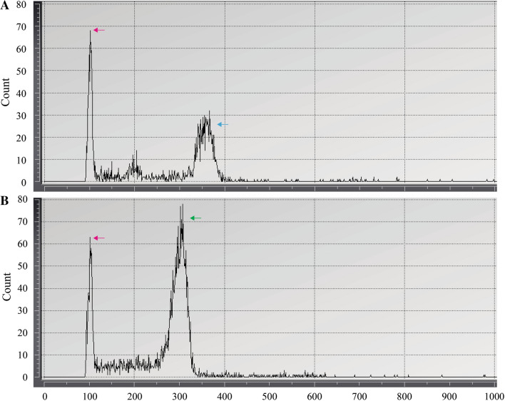
Because living materials of H. coreanum and H. polyanthos were very restricted in Korea, the genome size measurements for both taxa were carried out with two populations from two distant collecting sites and three populations from two distant sites, respectively. The measurement was repeated over three times for each sample. Compared to the calibration standard (1C-value of N. tabacum = 5.1842), the 1C-values of H. polyanthos and H. coreanum were 16.16 ± 0.17 and 14.85 ± 0, respectively (Fig. 4).
Figure 4.
The results of the genome size measurement using the flow cytometer. (A) H. polyanthos (Blue arrow) (B) H. coreanum (Green arrow). Red arrow refers to Nicotiana tabacum as reference.
Discussion
Taxonomical position of H. coreanum and relationship with H. polyanthos
The genus Hymenophyllum, delimited by Morton30 and Iwatsuki35, was recently reclassified into 10 subgenera by Ebihara et al.29 based on morphological characters and molecular evidence36. Among the 10 subgenera, sensu Ebihara et al.29, within Hymenophyllum, eight subgenera have the basic chromosome number of x = 36. However, the subgenus Mecodium has a chromosome number of x = 28 and the subgenus Hymenophyllum has chromosome numbers of x = 11 to 2829. Because the latter two subgenera seemed to be the most recently derived taxa within the genus36, the ancestor of Hymenophyllales was suggested to have the chromosome number 2n = 7243, and it is assumed that the reduction in chromosome number is a synapomorphy of these two subgenera in the genus Hymenophyllum44.
According to Ebihara et al.29, the subgenus Mecodium consisted of H. polyanthos and its local derivatives. On the other hand, it was noted that H. polyanthos shows variation in its chromosome number45,46 (n = 28 Japanese taxa or 27 taxa of other country outside of Japan), and more recently, the polyphyly of this cosmopolitan species, including several regional taxa, was discovered36,40. Therefore, the n = 27 population of H. polyanthos mentioned by Iwatsuki46 is likely to be the result of a transition from n = 28 to n = 27.
In this study, the chromosome numbers of H. polyanthos and H. coreanum were not directly counted. However, the result of flow cytometry implied that the genome of H. coreanum is downsized compared with that of H. polyanthos. It is not clear whether H. coreanum has been adopted to the n = 27 population of H. polyanthos, mentioned by Iwatsuki46, based on the data at hand. However, it is clear that H. coreanum is distinct from H. polyanthos based on morphological characteristics and genome size. Therefore, H. coreanum should be considered as an independent species not one of the synonymous taxa of H. polyanthos.
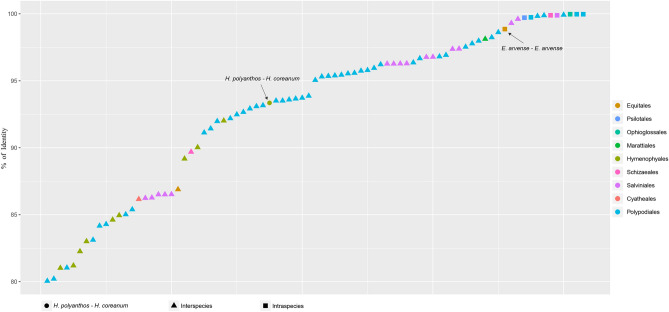
The divergence of plastome sequences between both species also strongly supports this taxonomical treatment. So far, it was rare to report more than two plastome sequences in the same species of ferns (Fig. 5 and supplementary table 4). The percentage of sequence identity between two plastomes in a species ranges from 98.86 (Equisetum arvense) to 99.97% (Mankyua chejuense). Except for the case of E. arvense in which the copy number variations frequently occurred15, sequence identities within the same species are higher than 99.7% in ferns. In contrast, the sequence identities between the plastomes of two different species in a genus, with the exception of the genera Cyrtomium and Azolla, are less than 98.7% (Fig. 5 and supplementary table 4). Sequence identity between plastomes of H. polyanthos and H. coreanum is quite low at 93.35% in the whole sequence and 98.13% even if the variation in trnV-rrn16 IGS was ignored. Consequently, plastome sequences of these two species revealed that they are quite distinct from each other and there is no reason to consider them as an intraspecific variation.
Figure 5.
Percentage of sequence identity between two plastomes. Circle refers to the sequence identity between H. polyanthos and H. coreanum. Triangle and square refer to the sequence identity between two plastomes of interspecific level in the same genus and that of infraspecific level in the same species, respectively.
In this study, we only investigated the Korean population of H. coreanum and it is not known whether the H. coreanum distributed in China, Taiwan and Japan38 also has the same genome size and plastome sequences as the Korean population. In Korea, the species mainly inhabits high mountains, however information for this species is very poor because it has previously been considered a synonymous taxon of H. polyanthos. Further study of this species facilitate better understanding of the speciation of the subgenus Mecodium because many species within the subgenus are derivatives of H. polyanthos, and H. coreanum, at least in Korea, seems to be recently diverged from H. polyanthos.
MORFFO of ferns prefer stem-loop structures
In previous studies25,47, the physical positions of MORFFO were considered to be related to inversions or IR borders even though it is not clear whether MORFFO are a cause or a consequence of inversions. In the present study, we made a new finding that the positions of MORFFO were between palindrome sequences, especially near tRNA genes (Supplementary table 3). Transfer RNA genes are very important in structural variation in fern plastomes. For instance, inversion between trnfM and trnE is specific to moninophytes48 and a number of inversions occurring near tRNA genes have been found in fern plastomes3,19,26,27. In addition, length variations in noncoding regions of plastomes owing to tRNA gene repeats have been suggested19,27. Transfer RNA-related genome rearrangements are not unique characteristic of fern plastomes. Intermolecular recombination between distinct tRNA genes has been found in cereals49, and extensive rearrangements were also suggested due to repeats or tRNA genes in Trachelium caeruleum50. Consequently, tRNA genes in plastomes of ferns seem to be strongly associated with genome instability events which can lead to insertion of MORFFO using double-strand breaks51.
In bacterial genome evolution, the most transposable elements have short terminal inverted repeats52. The insertion of some specific sequences related to repetitive extragenic palindromic (REP) elements have been reported53,54. Therefore, the mobility of MORFFO within the plastome is likely to be associated to the palindrome sequences at both ends of insertion regions. Two MORFFO in distant positions in a plastome of certain species in Polypodiales47 and Hymenophyllales in this study also seem to be caused by the palindrome sequences.
Correctively, it is likely that the mobile elements, consisting of palindrome sequences, lead to the insertion of MORFFO in the plastomes of ferns, and genome instability caused by hairpin structures at the ends of MORFFO influence genome rearrangements on the plastome. As a result, inversions seem to frequently occur in the loci having MORFFO.
Degradation after insertion of long DNA contig including MORFFO
So far, more than one hundred plastome sequences in ferns have been reported, and MORFFO have been found within at least one species in Marattiales, Ophioglossales, Hymenophyllales, Gleicheniales, Schizaeales, Cyatheales, and Polypodiales (Supplementary Table 2). The MORFFO status (present or absent) varies at the family level in Polypodiales47 or at the genus level in Ophioglossaceae, Schizaeaceae, Blechnaceae, Dryopteridaceae, Lomariopsidaceae, Polypodiaceae, Pteridaceae, and Thelypteridaceae. In addition, the inserted region of MORFFO also differs in certain genera25 (i.e., Asplenium, Cheilanthes, Myriopteris). It is uncertain exactly when MORFFO were inserted in the fern plastomes. However, if MORFFO were independent events at different lineages of ferns, it is difficult to explain (1) why expanded regions, including MORFFO, have high sequence similarities throughout the fern lineages25 and (2) how MORFFO were frequently transferred into the highly conserved genome, in terms of gene transfer55. In addition, MORFFO-like sequences found in cyanobacteria or green algae imply that MORFFO come from a single origin that was started from nuclear genomes after endosymbiotic or horizontal gene transfer3,25.
Even though MORFFO are supposed to be under selective pressure25, they are not assumed to play a vital role for fern plastomes because there was no vestige of MORFFO in various fern families (Supplementary Table 2). It is not surprising to degrade unnecessary genes in a plastome of land plants56,57. Especially, the degradation of ndh genes in Orchidaceae showed how easy it is to delete genes in plastomes among closely related taxa58. MORFFO in the plastome of Mankyua and that in the mitochondrial genome of Helminthostachys3 also suggest MORFFO degradation through intracellular gene transfer.
In Hymenophyllaceae, the length of rrn16-trnV of H. wrightii is slightly expanded similar to previously reported plastomes20,59, except Vandenboschia60, owing to MORFFO. Interestingly, rrn16-trnV is almost tripled in H. barbatum and H. coreanum or quadrupled in H. polyanthos compared with H. wrightii (Fig. 2). As mentioned earlier, MORFFO seems to have existed in the ancestor of Hymenophyllum, the length variation of rrn16-trnV in Hymenophyllum is able to be interpreted into a consequence of degradation.
Similar to the plastomes in Ophioglossaceae and Hymenophyllum, MORFFO survived or disappeared during the evolution of fern plastomes. Partial sequences of MORFFO in most core leptosporangiate ferns except Pteridaceae25 are consistent with the degradation hypothesis. MORFFO-lacking lineages may come from an ancestor that has undergone an independent MORFFO degradation event. However, it is not clear why MORFFO remain in relatively recently diverged families in the fern phylogeny17, Pteridaceae of Polypodiales25, if they do not have any function. One hypothesis is that morffo1, morffo2, and morffo3, which are found in Pteridaceae, are not intact genes but partial genes of a large ORF. Robison et al.25 described that these three ORFs cluster immediately adjacent to one another when they are present, and morffo1 and morffo2 often form a larger ORF. ORF_B1 in H. barbatum is highly similar to morffo1 and morffo2 consistent with their observation. If insertion of MORFFO contig was the consequence of genomic instability by stem-loop structures and then rearrangements including insertion/deletion and inversion were caused by genomic instability at the inserted region, a larger ORF can be fragmented and shuffling of smaller fragments can make novel chimeric ORFs. Actually, these novel chimeric ORFs have been reported in the mitochondrial genomes of land plants61,62.
Plastomes of eusporangiate and basal leptosporangiate ferns will shed light on the prototype of extant MORFFO, and this information will lead us to understand the evolution of MORFFO in fern plastomes.
Materials and Methods
Plant materials and DNA extraction
Four species, H. barbatum (CBNU-2018-0021), H. polyanthos (CBNU-2018-0187), H. wrightii (CBNU-2018-0330), and H. coreanum (CBNU-2018-0190), were sampled at Cheju Island and Jirisan in Korea. All specimens and living materials were deposited at the herbarium and greenhouse of Chungbuk National University. Using a fresh leaf, genomic DNAs were extracted by means of DNeasy Plant Mini Kit (Qiagen, Inc.) following the protocol proposed by the manufacturer.
Sequencing and assembling
Four high-quality DNAs were sequenced by Illimina HSeq X Ten. The raw reads were trimmed by trimmomatic 0.3663 with the options of leading:10, trailing:10, slidingwindow:4:20, and minlen:50. The assembly method was identical to Kim et al.64 using Hymenophyllum hollochilum (NC_039753) as a reference sequence.
Gene annotation
Genes in four plastome sequences were annotated compared with the genes of H. hollochilum using Geneious 10.2.6. Transfer RNA genes were verified using tRNAscan-SE65 and ORFs with > 303 bp in length were also annotated using Geneious 10.2.6.
Analyses of ORFs in Hymenophyllum and expanded noncoding regions in fern plastomes
To investigate the ORFs located in the rrn16-trnV IGS having high length variation in the genus, we extracted that the region from four plastome sequences. First, rrn16-trnV IGS was compared to other nucleotides using blastn66 to find out the homologous sequences. Thereafter, ORFs were searched using blastp to confirm the origin of these ORFs. To explore the physical positions of MORFFO, 126 plastome sequences were downloaded from NCBI and eight plastome sequences were received from Lehtonen and Cárdenas47 (Supplementary Table 2).
Phylogenetic relationships among four Hymenophyllum species
A total of 85 coding genes, except duplicated genes, were extracted from eight Hymenophyllaceae species and two Osmundales and one Gleicheniales species. To obtain the best partitioning scheme for each gene position in a single concatenated sequence, ModelFinder67 and PartitionFinder68 were used for ML analysis and BI analysis, respectively. MP and ML analyses were conducted using PAUP69 and IQ-TREE70 with 1000 bootstrap replications. BI analysis was inferred by Mrbayes71 under selected models in PartitionFinder with ngen = 1,000,000, samplefreq = 500, burninfrac = 0.25.
Estimation of DNA amounts
The genome size of H. polyanthos and H. coreanum was measured using living materials. For H. polyanthos, three populations were collected from Mt. Jiri-san in mainland of Korean peninsula and Mt. Halla-san of Jeju Island located at the most southern region of Korea. Two populations of H. coreanum were also collected from Mt. Jiri-san and Mt. Gariwang-san of southern and northern part of Korea, respectively. For a calibration standard, we grew Nicotiana tabacum in a greenhouse for weeks and used only 1 × 1 cm2 of young leaf. The reference and two samples were separately chopped with 500 μl of Nuclei extraction buffer in CyStain UV Precise P kit (Sysmex Partec, Germany) in a Petri dish on ice. The extraction time for samples was extended for eight minutes because the numbers of events less than that time were very low to visualize the graph. The debris was filtered using non-sterile CellTrics filter Green 30 μm (Sysmex Partec, Germany) and nuclei were stained by Staining Buffer in CyStain UV Precise P kit (Sysmex Partec, Germany). Particles of each sample were measured using CyFlow Cube 6 flow cytometer (Sysmex Partec, Germany).
Supplementary information
Acknowledgments
This work was supported by the National Research Foundation of Korea (NRF) under grant no. 2018R1D1A3B07048213. We really appreciate Sang Hee Park and Kanghyup Lee for helping in the sampling of Hymenophyllum species. We also thank Tanner A. Robison and Glenda G. Cárdenas for giving information about fern plastome analysis.
Author contributions
H.T.K. performed research design, writing manuscript, labwork, and data analysis. J.S.K. contributed to research design and writing manuscript.
Data availability
The complete sequence data used in the current study are available in the NCBI GenBank repository. All data generated or analyzed during this study are included in this published article and its Supplementary Information files.
Competing interests
The authors declare no competing interests
Footnotes
Publisher's note
Springer Nature remains neutral with regard to jurisdictional claims in published maps and institutional affiliations.
Supplementary information
is available for this paper at 10.1038/s41598-020-68000-7.
References
- 1.Ruhlman TA, Jansen RK. The plastid genomes of flowering plants. Methods Mol. Biol. 2014;1132:3–38. doi: 10.1007/978-1-62703-995-6_1. [DOI] [PubMed] [Google Scholar]
- 2.Viruel J, et al. Phylogenetics, classification and typification of extant horsetails (Equisetum, Equisetaceae) Bot J Linn Soc. 2019;189:311–352. doi: 10.1093/botlinnean/boz002. [DOI] [Google Scholar]
- 3.Kim HT, Kim KJ. Evolution of six novel ORFs in the plastome of Mankyua chejuense and phylogeny of eusporangiate ferns. Sci. Rep. 2018;8:16466. doi: 10.1038/s41598-018-34825-6. [DOI] [PMC free article] [PubMed] [Google Scholar]
- 4.Smith DR. Unparalleled GC content in the plastid DNA of Selaginella. Plant Mol. Biol. 2009;71:627–639. doi: 10.1007/s11103-009-9545-3. [DOI] [PubMed] [Google Scholar]
- 5.Wolf PG, et al. The first complete chloroplast genome sequence of a lycophyte, Huperzia lucidula (Lycopodiaceae) Gene. 2005;350:117–128. doi: 10.1016/j.gene.2005.01.018. [DOI] [PubMed] [Google Scholar]
- 6.Funk HT, Berg S, Krupinska K, Maier UG, Krause K. Complete DNA sequences of the plastid genomes of two parasitic flowering plant species, Cuscuta reflexa and Cuscuta gronovii. BMC Plant Biol. 2007;7:45. doi: 10.1186/1471-2229-7-45. [DOI] [PMC free article] [PubMed] [Google Scholar]
- 7.McNeal JR, Kuehl JV, Boore JL, de Pamphilis CW. Complete plastid genome sequences suggest strong selection for retention of photosynthetic genes in the parasitic plant genus Cuscuta. BMC Plant Biol. 2007;7:57. doi: 10.1186/1471-2229-7-57. [DOI] [PMC free article] [PubMed] [Google Scholar]
- 8.Guisinger MM, Kuehl JV, Boore JL, Jansen RK. Extreme reconfiguration of plastid genomes in the angiosperm family Geraniaceae: rearrangements, repeats, and codon usage. Mol. Biol. Evol. 2011;28:583–600. doi: 10.1093/molbev/msq229. [DOI] [PubMed] [Google Scholar]
- 9.Cai Z, et al. Extensive reorganization of the plastid genome of Trifolium subterraneum (Fabaceae) is associated with numerous repeated sequences and novel DNA insertions. J. Mol. Evol. 2008;67:696–704. doi: 10.1007/s00239-008-9180-7. [DOI] [PubMed] [Google Scholar]
- 10.Knox EB. The dynamic history of plastid genomes in the Campanulaceae sensu lato is unique among angiosperms. Proc. Natl. Acad. Sci. U. S. A. 2014;111:11097–11102. doi: 10.1073/pnas.1403363111. [DOI] [PMC free article] [PubMed] [Google Scholar]
- 11.Young HA, Lanzatella CL, Sarath G, Tobias CM. Chloroplast genome variation in upland and lowland switchgrass. PLoS ONE. 2011;6:e23980. doi: 10.1371/journal.pone.0023980. [DOI] [PMC free article] [PubMed] [Google Scholar]
- 12.Mariotti R, Cultrera NG, Diez CM, Baldoni L, Rubini A. Identification of new polymorphic regions and differentiation of cultivated olives (Olea europaea L.) through plastome sequence comparison. BMC Plant Biol. 2010;10:211. doi: 10.1186/1471-2229-10-211. [DOI] [PMC free article] [PubMed] [Google Scholar]
- 13.Tang J, et al. A comparison of rice chloroplast genomes. Plant Physiol. 2004;135:412–420. doi: 10.1104/pp.103.031245. [DOI] [PMC free article] [PubMed] [Google Scholar]
- 14.Doorduin L, et al. The complete chloroplast genome of 17 individuals of pest species Jacobaea vulgaris: SNPs, microsatellites and barcoding markers for population and phylogenetic studies. DNA Res. 2011;18:93–105. doi: 10.1093/dnares/dsr002. [DOI] [PMC free article] [PubMed] [Google Scholar]
- 15.Kim HT, Kim KJ. Chloroplast genome differences between Asian and American Equisetum arvense (Equisetaceae) and the origin of the hypervariable trnY-trnE intergenic spacer. PLoS ONE. 2014;9:e103898. doi: 10.1371/journal.pone.0103898. [DOI] [PMC free article] [PubMed] [Google Scholar]
- 16.Wolf PG, Rowe CA, Sinclair RB, Hasebe M. Complete nucleotide sequence of the chloroplast genome from a leptosporangiate fern Adiantum capillus-veneris L. DNA Res. 2003;10:59–65. doi: 10.1093/dnares/10.2.59. [DOI] [PubMed] [Google Scholar]
- 17.Smith AR, et al. A classification for extant ferns. Taxon. 2006;55:705–731. doi: 10.2307/25065646. [DOI] [Google Scholar]
- 18.Grewe F, Guo W, Gubbels EA, Hansen AK, Mower JP. Complete plastid genomes from Ophioglossum californicum, Psilotum nudum, and Equisetum hyemale reveal an ancestral land plant genome structure and resolve the position of Equisetales among monilophytes. BMC Evol. Biol. 2013;13:8. doi: 10.1186/1471-2148-13-8. [DOI] [PMC free article] [PubMed] [Google Scholar]
- 19.Kim HT, Chung MG, Kim KJ. Chloroplast genome evolution in early diverged leptosporangiate ferns. Mol. Cells. 2014;37:372–382. doi: 10.14348/molcells.2014.2296. [DOI] [PMC free article] [PubMed] [Google Scholar]
- 20.Kuo LY, Qi X, Ma H, Li FW. Order-level fern plastome phylogenomics: new insights from Hymenophyllales. Am. J. Bot. 2018;105:1545–1555. doi: 10.1002/ajb2.1152. [DOI] [PubMed] [Google Scholar]
- 21.Gao L, et al. Plastome sequences of Lygodium japonicum and Marsilea crenata reveal the genome organization transformation from basal ferns to core leptosporangiates. Genome Biol. Evol. 2013;5:1403–1407. doi: 10.1093/gbe/evt099. [DOI] [PMC free article] [PubMed] [Google Scholar]
- 22.Zhong B, Fong R, Collins LJ, McLenachan PA, Penny D. Two new fern chloroplasts and decelerated evolution linked to the long generation time in tree ferns. Genome Biol. Evol. 2014;6:1166–1173. doi: 10.1093/gbe/evu087. [DOI] [PMC free article] [PubMed] [Google Scholar]
- 23.Lu J-M, Zhang N, Du X-Y, Wen J, Li D-Z. Chloroplast phylogenomics resolves key relationships in ferns. J. System. Evol. 2015;53:448–457. doi: 10.1111/jse.12180. [DOI] [Google Scholar]
- 24.Wei R, et al. Plastid phylogenomics resolve deep relationships among Eupolypod II Ferns with rapid radiation and rate heterogeneity. Genome Biol. Evol. 2017;9:1646–1657. doi: 10.1093/gbe/evx107. [DOI] [PMC free article] [PubMed] [Google Scholar]
- 25.Robison TA, et al. Mobile elements shape plastome evolution in ferns. Genome Biol. Evol. 2018;10:2558–2571. doi: 10.1093/gbe/evy189. [DOI] [PMC free article] [PubMed] [Google Scholar]
- 26.Wolf PG, Roper JM, Duffy AM. The evolution of chloroplast genome structure in ferns. Genome. 2010;53:731–738. doi: 10.1139/g10-061. [DOI] [PubMed] [Google Scholar]
- 27.Gao L, Zhou Y, Wang ZW, Su YJ, Wang T. Evolution of the rpoB-psbZ region in fern plastid genomes: notable structural rearrangements and highly variable intergenic spacers. BMC Plant Biol. 2011;11:64. doi: 10.1186/1471-2229-11-64. [DOI] [PMC free article] [PubMed] [Google Scholar]
- 28.Pryer KM, Smith AR, Hunt JS, Dubuisson JY. rbcL data reveal two monophyletic groups of filmy ferns (Filicopsida: Hymenophyllaceae) Am. J. Bot. 2001;88:1118–1130. doi: 10.2307/2657095. [DOI] [PubMed] [Google Scholar]
- 29.Ebihara A, Dubuisson J-Y, Iwatsuki K, Hennequin S, Ito M. A taxonomic revision of Hymenophyllaceae. Blumea-Biodivers Evol Biogeogr Plants. 2006;51:221–280. doi: 10.3767/000651906X622210. [DOI] [Google Scholar]
- 30.Morton CV. The Genera, Subgenera, and sections of the Hymemophyllaceae. Contrib. U.S. Natl. Herb. 1968;38:153–214. [Google Scholar]
- 31.Iwatsuki K. The Hymenophyllaceae of Asia, excluding Malesia. J. Fac. Sci. U. Tokyo. 1985;III(13):501–551. [Google Scholar]
- 32.Copeland EB. Genera Filicum: the genera of ferns. Waltham: Chronica Botanica Co.; 1947. [Google Scholar]
- 33.Copeland EB. Genera Hymenophyllacearum. Philos. J. Sci. 1938;67:1–110. [Google Scholar]
- 34.contribution of morphology and cytology Hennequin, S. Phylogenetic relationships within the fern genus Hymenophyllum s.l. (Hymenophyllaceae, Filicopsida) C.R. Biol. 2003;326:599–611. doi: 10.1016/s1631-0691(03)00146-x. [DOI] [PubMed] [Google Scholar]
- 35.Iwatsuki K. Studies in the systematics of filmy ferns: VII. A scheme of classification based chiefly on the Asiatic species. Acta Phytotaxonomica et Geobotanica. 1984;35:165–179. [Google Scholar]
- 36.Hennequin S, Ebihara A, Ito M, Iwatsuki K, Dubuisson J-Y. New insights into the phylogeny of the genus Hymenophyllum sl (Hymenophyllaceae): revealing the polyphyly of Mecodium. Syst. Bot. 2006;31:271–284. doi: 10.1600/036364406777585775. [DOI] [Google Scholar]
- 37.Nakai T. Notes on Japanese Ferns III. Shokubutsugaku Zasshi. 1926;40:239–275. doi: 10.15281/jplantres1887.40.239. [DOI] [Google Scholar]
- 38.Ebihara A. The Standard of Ferns and Lycophytes in Japan. Tokyo: Gakken Plus; 2016. [Google Scholar]
- 39.Ebihara A, Nitta JH, Iwatsuki K. The Hymenophyllaceae of the Pacific area. 2. Hymenophyllum (excluding subgen. Hymenophyllum) Bull. Natl. Museum Nat. Sci. Ser. B. 2010;36:43–59. [Google Scholar]
- 40.Vasques DT, Ebihara A, Hirai RY, Prado J, Motomi I. Phylogeny of Hymenophyllum subg. Mecodium (Hymenophyllaceae), with special focus on the diversity of the Hymenophyllum polyanthos species complex. Plant System. Evol. 2019 doi: 10.1007/s00606-019-01609-y. [DOI] [Google Scholar]
- 41.Hoang NV, Furtado A, McQualter RB, Henry RJ. Next generation sequencing of total DNA from sugarcane provides no evidence for chloroplast heteroplasmy. New Negatives Plant Sci. 2015;1:33–45. doi: 10.1016/j.neps.2015.10.001. [DOI] [Google Scholar]
- 42.Leitch IJ, et al. The ups and downs of genome size evolution in polyploid species of Nicotiana (Solanaceae) Ann. Bot. 2008;101:805–814. doi: 10.1093/aob/mcm326. [DOI] [PMC free article] [PubMed] [Google Scholar]
- 43.Clark J, et al. Genome evolution of ferns: evidence for relative stasis of genome size across the fern phylogeny. New Phytol. 2016;210:1072–1082. doi: 10.1111/nph.13833. [DOI] [PubMed] [Google Scholar]
- 44.Hennequin S, Ebihara A, Dubuisson J-Y, Schneider H. Chromosome number evolution in Hymenophyllum (Hymenophyllaceae), with special reference to the subgenus Hymenophyllum. Mol. Phylogenet. Evol. 2010;55:47–59. doi: 10.1016/j.ympev.2010.01.001. [DOI] [PubMed] [Google Scholar]
- 45.Brownsey P, Perrie L. Hymenophyllaceae. In: Breitwieser I, Heenan PB, Wilton AD, editors. Flora of New Zealand—Ferns and Lycophytes. Fascicle 15. Lincoln: Manaaki Whenua Press; 2016. [Google Scholar]
- 46.Iwatsuki K. Pteridophytes and Gymnosperms. Berlin: Springer; 1990. pp. 157–163. [Google Scholar]
- 47.Lehtonen S, Cárdenas GG. Dynamism in plastome structure observed across the phylogenetic tree of ferns. Bot J Linn Soc. 2019;190:229–241. doi: 10.1093/botlinnean/boz020. [DOI] [Google Scholar]
- 48.Karol KG, et al. Complete plastome sequences of Equisetum arvense and Isoetes flaccida: implications for phylogeny and plastid genome evolution of early land plant lineages. BMC Evol. Biol. 2010;10:321. doi: 10.1186/1471-2148-10-321. [DOI] [PMC free article] [PubMed] [Google Scholar]
- 49.Hiratsuka J, et al. The complete sequence of the rice (Oryza sativa) chloroplast genome: intermolecular recombination between distinct tRNA genes accounts for a major plastid DNA inversion during the evolution of the cereals. Mol. Gen. Genet. MGG. 1989;217:185–194. doi: 10.1007/BF02464880. [DOI] [PubMed] [Google Scholar]
- 50.Haberle RC, Fourcade HM, Boore JL, Jansen RK. Extensive rearrangements in the chloroplast genome of Trachelium caeruleum are associated with repeats and tRNA genes. J. Mol. Evol. 2008;66:350–361. doi: 10.1007/s00239-008-9086-4. [DOI] [PubMed] [Google Scholar]
- 51.Aguilera A, Gómez-González B. Genome instability: a mechanistic view of its causes and consequences. Nat. Rev. Genet. 2008;9:204. doi: 10.1038/nrg2268. [DOI] [PubMed] [Google Scholar]
- 52.Darmon E, Leach DRF. Bacterial genome instability. Microbiol. Mol. Biol. Rev. 2014;78:1–39. doi: 10.1128/mmbr.00035-13. [DOI] [PMC free article] [PubMed] [Google Scholar]
- 53.Clément J-M, Wilde C, Bachellier S, Lambert P, Hofnung M. IS1397 is active for transposition into the chromosome of Escherichia coli K-12 and inserts specifically into palindromic units of bacterial interspersed mosaic elements. J. Bacteriol. 1999;181:6929–6936. doi: 10.1128/JB.181.22.6929-6936.1999. [DOI] [PMC free article] [PubMed] [Google Scholar]
- 54.Tobes R, Pareja E. Bacterial repetitive extragenic palindromic sequences are DNA targets for insertion sequence elements. BMC Genom. 2006;7:62. doi: 10.1186/1471-2164-7-62. [DOI] [PMC free article] [PubMed] [Google Scholar]
- 55.Leister D. Origin, evolution and genetic effects of nuclear insertions of organelle DNA. Trends Genet. 2005;21:655–663. doi: 10.1016/j.tig.2005.09.004. [DOI] [PubMed] [Google Scholar]
- 56.Logacheva MD, Schelkunov MI, Nuraliev MS, Samigullin TH, Penin AA. The plastid genome of mycoheterotrophic monocot Petrosavia stellaris exhibits both gene losses and multiple rearrangements. Genome Biol. Evol. 2014;6:238–246. doi: 10.1093/gbe/evu001. [DOI] [PMC free article] [PubMed] [Google Scholar]
- 57.Revill MJ, Stanley S, Hibberd JM. Plastid genome structure and loss of photosynthetic ability in the parasitic genus Cuscuta. J. Exp. Bot. 2005;56:2477–2486. doi: 10.1093/jxb/eri240. [DOI] [PubMed] [Google Scholar]
- 58.Kim HT, Chase MW. Independent degradation in genes of the plastid ndh gene family in species of the orchid genus Cymbidium (Orchidaceae; Epidendroideae) PLoS ONE. 2017;12:e0187318. doi: 10.1371/journal.pone.0187318. [DOI] [PMC free article] [PubMed] [Google Scholar]
- 59.Lehtonen S. The complete plastid genome sequence of Trichomanes trollii (Hymenophyllaceae) Nordic J. Bot. 2018;36:e02072. doi: 10.1111/njb.02072. [DOI] [Google Scholar]
- 60.Ruiz-Ruano FJ, Navarro-Domínguez B, Camacho JPM, Garrido-Ramos MA. Full plastome sequence of the fern Vandenboschia speciosa (Hymenophyllales): structural singularities and evolutionary insights. J. Plant. Res. 2019;132:3–17. doi: 10.1007/s10265-018-1077-y. [DOI] [PubMed] [Google Scholar]
- 61.Tanaka Y, Tsuda M, Yasumoto K, Yamagishi H, Terachi T. A complete mitochondrial genome sequence of Ogura-type male-sterile cytoplasm and its comparative analysis with that of normal cytoplasm in radish (Raphanus sativus L.) BMC Genom. 2012;13:352. doi: 10.1186/1471-2164-13-352. [DOI] [PMC free article] [PubMed] [Google Scholar]
- 62.Jo YD, Choi Y, Kim DH, Kim BD, Kang BC. Extensive structural variations between mitochondrial genomes of CMS and normal peppers (Capsicum annuum L.) revealed by complete nucleotide sequencing. BMC Genom. 2014;15:561. doi: 10.1186/1471-2164-15-561. [DOI] [PMC free article] [PubMed] [Google Scholar]
- 63.Bolger AM, Lohse M, Usadel B. Trimmomatic: a flexible trimmer for Illumina sequence data. Bioinformatics. 2014;30:2114–2120. doi: 10.1093/bioinformatics/btu170. [DOI] [PMC free article] [PubMed] [Google Scholar]
- 64.Kim HT, et al. Seven New Complete Plastome Sequences Reveal Rampant Independent Loss of the ndh Gene Family across Orchids and Associated Instability of the Inverted Repeat/Small Single-Copy Region Boundaries. PLoS ONE. 2015;10:e0142215. doi: 10.1371/journal.pone.0142215. [DOI] [PMC free article] [PubMed] [Google Scholar]
- 65.Lowe TM, Eddy SR. tRNAscan-SE: a program for improved detection of transfer RNA genes in genomic sequence. Nucl. Acids Res. 1997;25:955–964. doi: 10.1093/nar/25.5.955. [DOI] [PMC free article] [PubMed] [Google Scholar]
- 66.Altschul SF, Gish W, Miller W, Myers EW, Lipman DJ. Basic local alignment search tool. J. Mol. Biol. 1990;215:403–410. doi: 10.1016/S0022-2836(05)80360-2. [DOI] [PubMed] [Google Scholar]
- 67.Kalyaanamoorthy S, Minh BQ, Wong TK, von Haeseler A, Jermiin LS. ModelFinder: fast model selection for accurate phylogenetic estimates. Nat. Methods. 2017;14:587. doi: 10.1038/nmeth.4285. [DOI] [PMC free article] [PubMed] [Google Scholar]
- 68.Lanfear R, Frandsen PB, Wright AM, Senfeld T, Calcott B. PartitionFinder 2: new methods for selecting partitioned models of evolution for molecular and morphological phylogenetic analyses. Mol. Biol. Evol. 2016;34:772–773. doi: 10.1093/molbev/msw260. [DOI] [PubMed] [Google Scholar]
- 69.Swofford D. PAUP 4.0: phylogenetic analysis using parsimony. Washington: Smithsonian Institution; 1998. [Google Scholar]
- 70.Nguyen L-T, Schmidt HA, von Haeseler A, Minh BQ. IQ-TREE: a fast and effective stochastic algorithm for estimating maximum-likelihood phylogenies. Mol. Biol. Evol. 2014;32:268–274. doi: 10.1093/molbev/msu300. [DOI] [PMC free article] [PubMed] [Google Scholar]
- 71.Ronquist F, et al. MrBayes 3.2: efficient Bayesian phylogenetic inference and model choice across a large model space. Syst. Biol. 2012;61:539–542. doi: 10.1093/sysbio/sys029. [DOI] [PMC free article] [PubMed] [Google Scholar]
Associated Data
This section collects any data citations, data availability statements, or supplementary materials included in this article.
Supplementary Materials
Data Availability Statement
The complete sequence data used in the current study are available in the NCBI GenBank repository. All data generated or analyzed during this study are included in this published article and its Supplementary Information files.


