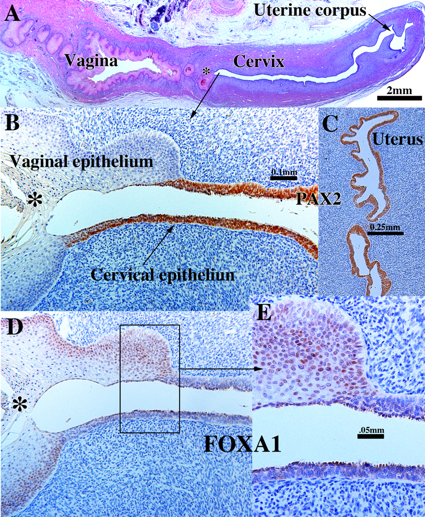
Figure 7.

Sagittal sections of a 21-week female reproductive tract. (A) depicts the upper portion of the vagina, cervix and uterus (H&E stain). The vaginal epithelium is many layers thick due to endogenous estrogenic stimulation. Note the abrupt transition in epithelial differentiation at the vaginal/cervical border. (B) PAX2 staining of the vaginal/cervical border shows prominent PAX2 immunostaining (indicative of Müllerian duct origin) of the pseudostratified stratified relatively thin cervical epithelium and PAX2-negative stratified squamous vaginal epithelium. (C) Epithelium of the uterine corpus is strongly PAX2-reactive as expected for a Müllerian epithelium. (D & E) FOXA1 nuclear immunostaining was seen uniformly throughout the entire vagina, and FOXA1 nuclear immunostaining abruptly stopped at the vaginal/cervical border (D-E) in mirror image to PAX2 immunostaining (B). (modified from Robboy et al., 2017 with permission). (B & D) are serial sections of the same specimen.
