Summary:
Wounds from orthopedic limb reconstruction are often difficult to heal due to the surgery, patient comorbidities, or a combination of these factors. The role of negative pressure wound therapy (NPWT) modalities in the perioperative management of patients with complex lower extremity wounds is evolving. Here, we present a case study using adjunctive NPWT with instillation and a dwell time, standard NPWT, and closed-incision negative pressure therapy (ciNPT) to manage a complex lower extremity wound. The patient was a 51-year-old man who presented with severe scarring of the lower extremity and infection following plate osteosynthesis of a tibial shaft fracture. Following lower extremity reconstruction, the patient received 5 days of NPWT with instillation and a dwell time with cycles that consisted of instilling normal saline with a 1-second dwell time, followed by 2 hours of continuous negative pressure at −125 mm Hg. The wound is then covered with an adjacent local tissue flap, which showed signs of vascular complication. ciNPT is applied over the flap incision for 7 days, which resulted in restored normal coloration; ciNPT is continued for another 7 days. A skin substitute is applied over the flap donor site, followed by NPWT using a silver foam dressing. Dressing changes are performed weekly for 4 weeks. At 8 weeks postsurgery, a skin graft is applied over the donor site. In this case, adjunctive use of multiple NPWT modalities resulted in a completely healed wound within 12 months with no complications.
Wounds from orthopedic limb reconstruction of tibial fractures are often difficult to resolve due to the risk of complications, such as disunion, infection, or soft tissue ischemia. Wound closure, often using a local flap, is an essential step in protecting the wound from infection, restoring vascular supply, and providing an environment conducive to wound healing.1 However, local flaps of the distal third leg are compromised by the limited availability of soft tissue, a high propensity for edema, and an unreliable subdermal blood supply. Negative pressure wound therapy (NPWT) has been reported to promote perfusion2 and assist with lower extremity flap resolution without complications.3 Here, we present a case using NPWT with instillation and dwell time (NPWTi-d), closed-incision negative pressure therapy (ciNPT), and standard NPWT to reverse signs of flap failure and support closure in a complex lower leg wound following limb reconstruction surgery.
CASE PRESENTATION
A 51-year-old man with a history of smoking and vascular disease was presented with an infected lower extremity wound with an exposed plate. Twelve months before, the patient had undergone open reduction internal fixation surgery for a tibial shaft fracture. Six months later, the patient was treated for an infection of a contralateral wound on the same leg that was closed after hardware removal using a local flap.
Presently, the patient was admitted to the hospital, and a peripherally inserted central catheter was used to administer a 6-week course of intravenous antibiotics. Because the fracture was fully healed, orthopedic services removed the plate, and the patient was left with an open wound measuring 8.9 × 2.8 cm2, exposing the medial distal tibia. A plastic surgery was consulted, and wound cleansing was initiated using NPWTi-d (V.A.C. VERAFLO Therapy; KCI, San Antonio, Tex.) and a reticulated open cell foam (ROCF) dressing (V.A.C. VERAFLO Dressing; KCI). Normal saline was instilled until the foam was filled with a 1-second dwell time, followed by 2 hours of continuous negative pressure at −125 mm Hg. While awaiting cleansing of the wound, imaging of the lower extremity revealed that the patient had a single inflow vessel to the distal extremity.
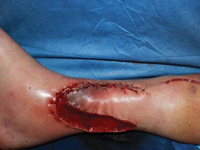
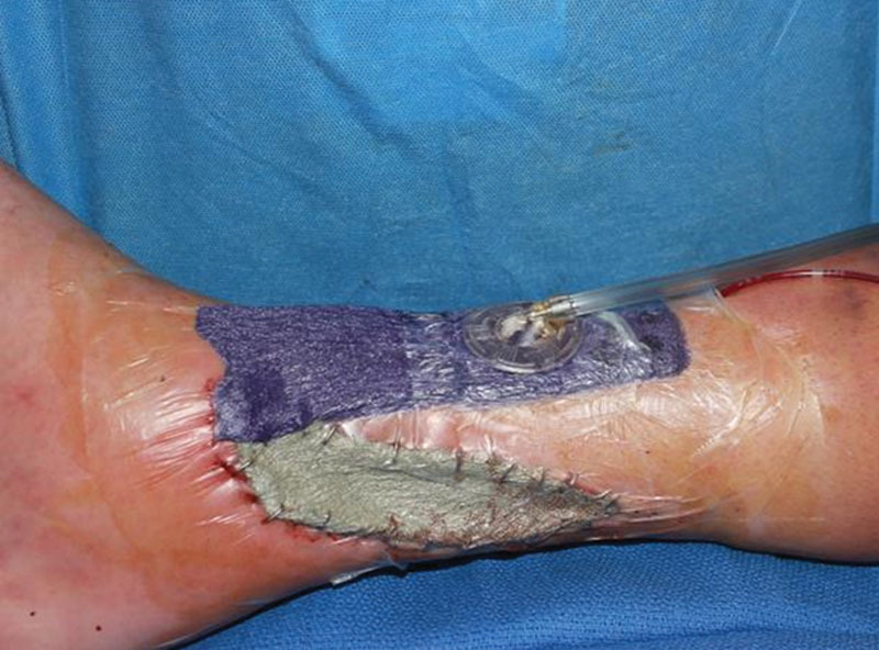
After 5 days, NPWTi-d was discontinued, and the decision was made to close the wound with a local flap rather than a free flap, given the concern regarding the single-vessel inflow. The wound was covered with an adjacent local flap after some perforator veins were identified, which was secured in place with sutures and surgical staples (Fig. 1). The distal end of the flap exhibited discoloration indicative of vascular compromise, at which point the decision was made to use ciNPT (PREVENA Therapy; KCI) at −125 mm Hg over the flap incision (Fig. 2). The goal of the therapy was to assist venous outflow. Over the donor site, a bilaminate skin substitute (Integra Dermal Regeneration Template; Integra LifeSciences, Plainsboro, N.J.) was placed, followed by application of a silver ROCF dressing (ROCF-silver; V.A.C. GRANUFOAM SILVER Dressing; KCI). Continuous NPWT (V.A.C. Therapy; KCI) was applied at −125 mm Hg over both dressings. The patient was sent home the following day.
Fig. 1.

Initial coverage of infected medial left leg wound with a lateral local flap. Progressive discoloration was visible at the distal end of the flap.
Fig. 2.

Application of ciNPT over the flap incision and NPWT with ROCF-silver dressing over the donor site.
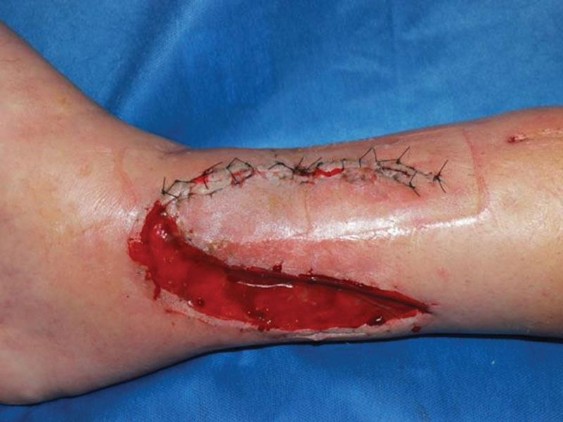
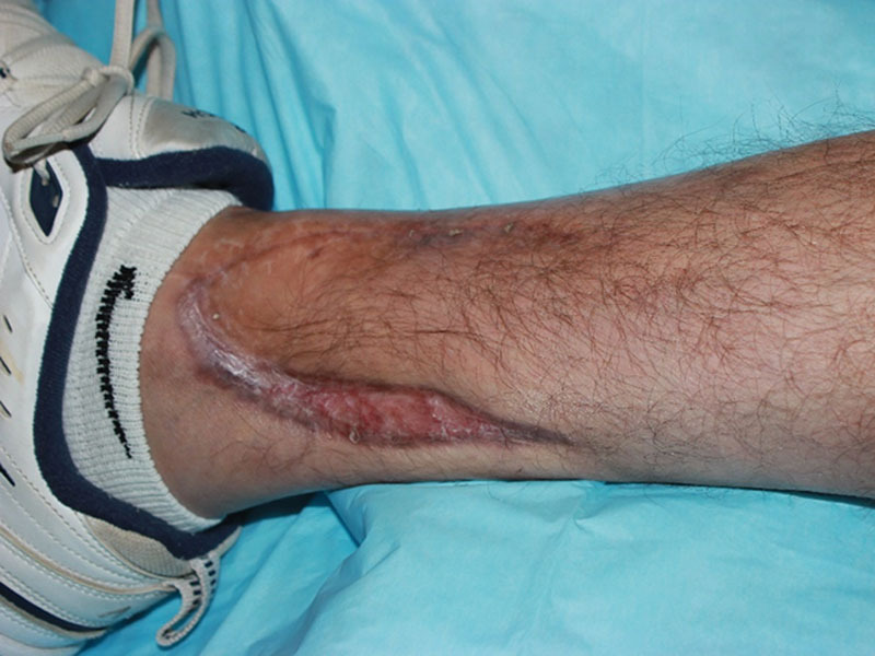
After 7 days of ciNPT, the patient returned to the operating room with the expectation that the flap would require debridement and reassessment. Once the dressings were removed, the flap appeared to be completely viable (Fig. 3). Because both sites appeared to be healing, ciNPT was reapplied for an additional 7 days, upon which the flap incision remained closed. The sutures over the incision were removed on postoperative day 14, and a nonadherent silicone dressing (ADAPTIC TOUCH Non-Adhering Silicone Dressing; Systagenix Wound Management, Gatwick, United Kingdom) was placed over the donor site wound. After 8 weeks, the flap had healed, and the bilaminate skin substitute had matured. The patient then underwent a split thickness skin graft (STSG), which was bolstered by NPWT using ROCF-silver dressing for 6 days. NPWT was discontinued, and the STSG was covered with a nonadherent silicone dressing. At 6 weeks post STSG placement, the wounds were closed with normal scabbing. At the 12-month follow-up, both wounds remained completely healed (Fig. 4).
Fig. 3.

Wound after 7 days of ciNPT and NPWT with ROCF-silver dressing.
Fig. 4.

Wound at 12-month follow-up.
DISCUSSION
The infection risk for injuries requiring orthopedic limb reconstruction surgery is particularly high during the initial fracture and upon installation or removal of fixation hardware.4 Although internal plate osteosynthesis carries the lowest risk of infection among fixation methods, it still has an infection rate of 22%, which can delay wound closure and elevate risk of more serious complications.5 Under such circumstances, closure with a soft tissue flap can assist wound healing, and ensuring that survival of the repositioned tissue is critical.1 Unfortunately, a lack of soft tissue, risk of edema, and limited blood supply in the lower leg can compromise flap survival. In this case, the single-vessel inflow combined with the patient’s history of smoking, vascular disease, and previous surgeries led us to choose a local flap to close the wound, rather than to use a free flap. Once the distal end of the local flap showed significant discoloration, a sign of vascular failure, and imminent loss of vitality, we proactively applied the combination of ciNPT and NPWT over both the flap incision and the open donor site to support inflow and stimulate wound healing. The previous use of a flap on the contralateral side of the patient’s leg left little tissue for use if the present flap failed. Loss of the local flap would have necessitated a more invasive method of soft tissue replacement for successful reconstruction.
Despite the application of ciNPT and NPWT, the ultimate flap prognosis remained uncertain until the patient returned on postoperative day 7. To our surprise, the flap was viable, and the sutured incision remained closed, negating the need for revision or debridement. This positive outcome after the use of negative pressure devices is supported by previously published clinical and preclinical studies. In a porcine wound model, NPWT at −125 mm Hg decreased discoloration in local flaps and increased survival compared with controls.6 In human subjects, NPWT has been used to bolster local flaps in the ankle region3 and resolve venous congestion in lower limb reconstruction.7 In our case study, we additionally supported flap adherence by cleansing the wound bed with NPWTi-d, which can improve healing even in infected wounds.8 This was followed by the use of ciNPT to ensure closure of the flap incision, with adjunctive support of the donor site with standard NPWT, which has shown success in resolving donor site wounds associated with other flap types.9,10
In this case, the use of a mobile NPWT device also enabled our patient to remain ambulatory and be discharged home for recovery in the 2 weeks following flap closure. We note that according to the manufacturer’s instructions, dressing changes for NPWT over an open wound should be performed every 2–3 days. However, we did not observe any adverse effects of keeping the dressings in place for up to 7 days. In our experience, the combination of NPWTi-d, ciNPT, and NPWT to synchronously improve the overall health of the flap, incision, and donor site presents a useful strategy for successful flap closure of a complex wound.
Footnotes
Published online 18 June 2020.
Disclosure: Dr. Gabriel has a consulting agreement with KCI, San Antonio, Tex. The other authors have no financial interest to declare. The Article Processing Charge was paid by the authors.
Statement of Conformity: This case report complies with the ethical principles outlined in the Declaration of Helsinki.
REFERENCES
- 1.Chan JK, Harry L, Williams G, et al. Soft-tissue reconstruction of open fractures of the lower limb: muscle versus fasciocutaneous flaps. Plast Reconstr Surg. 2012;130:284e–295e. [DOI] [PMC free article] [PubMed] [Google Scholar]
- 2.Ma Z, Li Z, Shou K, et al. Negative pressure wound therapy: regulating blood flow perfusion and microvessel maturation through microvascular pericytes. Int J Mol Med. 2017;40:1415–1425. [DOI] [PMC free article] [PubMed] [Google Scholar]
- 3.Goldstein JA, Iorio ML, Brown B, et al. The use of negative pressure wound therapy for random local flaps at the ankle region. J Foot Ankle Surg. 2010;49:513–516. [DOI] [PubMed] [Google Scholar]
- 4.Chan JK, Gardiner MD, Pearse M, et al. Farhadieh RD, Bulstrode NW, Cugno S. Lower limb reconstruction. In: Plastic and Reconstructive Surgery: Approaches and Techniques. 2015;1st ed John Wiley & Sons, Ltd, Hoboken, New Jersey; 607–627. [Google Scholar]
- 5.Harris AM, Althausen PL, Kellam J, et al. Complications following limb-threatening lower extremity trauma. J Orthop Trauma. 2009;23:1–6. [DOI] [PubMed] [Google Scholar]
- 6.Morykwas MJ, Argenta LC, Shelton-Brown EI, et al. Vacuum-assisted closure: a new method for wound control and treatment: animal studies and basic foundation. Ann Plast Surg. 1997;38:553–562. [DOI] [PubMed] [Google Scholar]
- 7.Vaienti L, Gazzola R, Benanti E, et al. Failure by congestion of pedicled and free flaps for reconstruction of lower limbs after trauma: the role of negative-pressure wound therapy. J Orthop Traumatol. 2013;14:213–217. [DOI] [PMC free article] [PubMed] [Google Scholar]
- 8.Brinkert D, Ali M, Naud M, et al. Negative pressure wound therapy with saline instillation: 131 patient case series. Int Wound J. 2013;10suppl 156–60. [DOI] [PMC free article] [PubMed] [Google Scholar]
- 9.Harris BN, Bewley AF. Minimizing free flap donor-site morbidity. Curr Opin Otolaryngol Head Neck Surg. 2016;24:447–452. [DOI] [PubMed] [Google Scholar]
- 10.Schmedes GW, Banks CA, Malin BT, et al. Massive flap donor sites and the role of negative pressure wound therapy. Otolaryngol Head Neck Surg. 2012;147:1049–1053. [DOI] [PubMed] [Google Scholar]


