Abstract
Objectives
Ischemic postconditioning (PC) with “stuttering” reintroduction of blood flow after prolonged ischemia has been shown to offer protection from ischemia reperfusion injury to the myocardium and brain. We hypothesized that four 20-s pauses during the first 3 min of standard CPR would improve post resuscitation cardiac and neurological function, in a porcine model of prolonged untreated cardiac arrest.
Methods
18 female farm pigs, intubated and isoflurane anesthetized had 15 min of untreated ventricular fibrillation followed by standard CPR (SCPR). Nine animals were randomized to receive PC with four, controlled, 20-s pauses, during the first 3 min of CPR (SCPR + PC). Resuscitated animals had echocardiographic evaluation of their ejection fraction after 1 and 4 h and a blinded neurological assessment with a cerebral performance category (CPC) score assigned at 24 and 48 h. All animals received 12 h of post resuscitation mild therapeutic hypothermia.
Results
SCPR + PC animals had significant improvement in left ventricular ejection fraction at 1 and 4 h compared to SCPR (59 ± 11% vs 35 ± 7% and 55 ± 8% vs 31 ± 13% respectively, p < 0.01). Neurological function at 24 h significantly improved with SCPR + PC compared to SCPR alone (CPC: 2.7 ± 0.4 vs 3.8 ± 0.4 respectively, p = 0.003). Neurological function significantly improved in the SCPR + PC group at 48 h and the mean CPC score of that group decreased from 2.7 ± 0.4 to 1.7 ± 0.4 (p < 0.00001).
Conclusions
Ischemic postconditioning with four 20-s pauses during the first 3 min of SCPR improved post resuscitation cardiac function and facilitated neurological recovery after 15 min of untreated cardiac arrest in pigs.
Keywords: Post conditioning, Cardiopulmonary resuscitation, Survival, Neurological function, Left ventricular function
1. Introduction
Cardiopulmonary resuscitation (CPR) survival rates have remained poor over the past half-century with only minimal if any improvements in neurologically intact survival.1 In humans, when ventricular fibrillation (VF) is left untreated for more than 10 min, short and long term survival is severely reduced.2 In animals, current non-invasive methods of CPR are unable to provide short and long-term neurological recovery when ventricular fibrillation is left untreated longer than 12–13 min.3–5 Furthermore, successful resuscitation following 12–15 min of untreated VF has been used as the model to evaluate post resuscitation left ventricular dysfunction.6
Re-introduction of blood flow with “controlled pauses” has been shown to protect the myocardium and the brain from ischemia-reperfusion injury in clinical scenarios of regional ischemia during ST elevation myocardial infarction and stroke both in animals and humans.7–10 This concept has been called “ischemic postconditioning” and describes a method of reperfusion injury protection.9,11
We sought to introduce ischemic postconditioning at the initiation of CPR efforts by providing four, 20-s pauses in chest compressions and ventilations over the first 3 min of reperfusion. We hypothesize that the implementation of a simple ischemic postconditioning (PC) strategy during the first 3 min of CPR, will improve cardiac and cerebral function and 48-h survival rates after 15 min of untreated ventricular fibrillation.
2. Materials and methods
All studies were performed by an experienced research team in Yorkshire female farm pigs weighing 32 ± 2 kg. A certified and licensed veterinarian provided a blinded neurological assessment at 24 and 48 h. The protocol was approved by the Institutional Animal Care Committee of the Minneapolis Medical Research Foundation of Hennepin County Medical Center. All animal care was compliant with the National Research Council’s 1996 Guidelines for the Care and Use of Laboratory Animals.
2.1. Preparatory phase
The anesthesia, surgical preparation, data monitoring, and recording procedures used in this study have been described previously.12 Briefly, we employed aseptic surgical conditions using initial sedation with intramuscular ketamine (7 mL of 100 mg/mL, Ketaset, Fort Dodge Animal Health, Fort Dodge, Iowa) followed by inhaled isoflurane at a dose of 0.8–1.2%. Pigs were intubated with a size 7.0 endotracheal tube. The animal’s temperature was maintained at 37.5 ± 0.5 °C, with a warming blanket (Bair Hugger, Augustine Medical, Eden Prairie, Minnesota). Central aortic blood pressure was recorded continuously with a micromanometer-tipped (Mikro-Tip Transducer, Millar Instruments, Houston, Texas) catheter placed at the beginning of the descending thoracic aorta. A second Millar catheter was inserted in the right atrium via the right external jugular vein. All animals received an intravenous heparin bolus (100 units/kg) and 500 units of heparin every hour until surgical repair was completed. An ultrasound flow probe (Transonic 420 series multichannel, Transonic Systems, Ithaca, New York) was placed to the right internal carotid artery to record blood flow (mL/min). The animals were then ventilated with room air, using a volume-control ventilator (Narcomed, Telford, Pennsylvania), with a tidal volume of 10 mL/kg and a respiratory rate adjusted to continually maintain a PaCO2 of 40 mmHg and PaO2 of 80 mmHg (blood oxygen saturation > 95%), as measured from arterial blood (Gem 3000, Instrumentation Laboratory, Lexington, Massachusetts) to adjust the ventilator as needed. Surface electrocardiographic tracings were continuously recorded. All data were recorded with a digital recording system (BIOPAC MP 150, BIOPAC Systems, Inc., CA, USA). End tidal CO2 (ETCO2), tidal volume, minute ventilation, and blood oxygen saturation were continuously measured with a respiratory monitor (CO2SMO Plus, Novametrix Medical Systems, Wallingford, Connecticut).
2.2. Measurements and recording
Thoracic aortic pressure, right atrial pressure, ETCO2, and carotid blood flow were continuously recorded. Coronary perfusion pressure (CPP) during CPR was calculated from the mean arithmetic difference between right-atrial pressure and aortic pressure during the decompression phase. Carotid artery blood flow was reported in mL/s.
2.3. Experimental protocol
After the surgical preparation was complete, oxygen saturation on room air was greater than 95%, and ETCO2 was stable between 35 and 42 mmHg for 5 min, ventricular fibrillation was induced by delivering direct intracardiac current via a temporary pacing wire (St. Jude Medical, Minnetonka, Minnesota). The ventilator was disconnected from the endotracheal tube. Standard CPR was performed with a pneumatically driven automatic piston device (Pneumatic Compression Controller, Ambu International, Glostrup, Denmark) as previously described.13 During S-CPR, uninterrupted chest compressions were performed at a rate of 100 compressions/min, with a 50% duty cycle and a compression depth of 25% of the anterior–posterior chest diameter. Asynchronous positive-pressure ventilations were delivered with room air (FiO2 of 0.21) with a manual resuscitator bag. The tidal volume was maintained at ∼10 mL/kg and the respiratory rate was 10 breaths/min. The investigators were blinded to hemodynamics during CPR.
2.4. Protocol
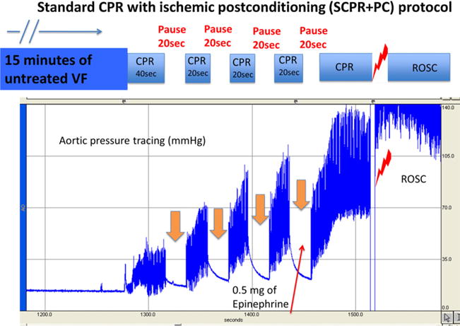
Following 15 min of untreated ventricular fibrillation, 18 pigs were randomized to receive standard CPR (SCPR) or SCPR plus postconditioning (SCPR + PC). The SCPR + PC group received initially 40 s of SCPR followed by a 20-s pause of compressions and ventilations followed by another 20 s of SCPR and the cycle was repeated for a total of 4 pauses (Fig. 1). Epinephrine was administered in both groups in 0.5 mg (∼15 mcg/kg) bolus at minute three and was repeated every 3 min until return of spontaneous circulation (ROSC). Resuscitation efforts were continued until ROSC was achieved or a total of 15 min of CPR had occurred. The first defibrillation effort was delivered with 200-J biphasic shocks after 4 min of CPR in both groups. If ROSC was not achieved, defibrillation was delivered every 2 min thereafter during CPR.
Fig. 1.

Ischemic postconditioning protocol during standard cardiopulmonary resuscitation In the SCPR + PC group during the first 3 min of CPR, animals received four 20-s pauses and each pause was followed by 20 s of SCPR. The “stuttering” introduction of reperfusion is called “ischemic postconditioning”. SCPR: standard CPR; VF: ventricular fibrillation; ROSC: return of spontaneous circulation.
2.5. Post ROSC care
After ROSC was achieved, animals were connected to the mechanical ventilator. Supplemental oxygen was added only if arterial saturation was lower than 90%. Animals were observed under general anesthesia with isoflurane until hemodynamically stable. Hemodynamic stability was defined as a mean aortic pressure > 55 mmHg without pharmacological support for 30 min and normalization of ETCO2 and acidosis. Animals that had a stable post-ROSC rhythm but were hypotensive (mean arterial pressure < 50 mmHg) received increments of 0.1–0.2 mg intravenous epinephrine every 5 min until MAP rose above 50 mmHg. If pH was lower than 7.2, 100 mEq of NaHCO3 was given intravenously. Amiodarone 20 mg intravenously was given in all animals if there was recurrence of ventricular fibrillation after initial ROSC. At that point vascular repair of the internal jugular vein and the left common femoral artery were then performed. Arterial blood gases were obtained at baseline, at 5 min after ROSC and every 30 min thereafter.
Both groups received post resuscitation therapeutic hypothermia as per American Heart Association recommendations for resuscitated comatose patients from ventricular fibrillation in order to simulate best practice and optimize the chances of the control group for neurological recovery. Our institutional animal care committee mandated therapeutic hypothermia for protocol approval. Immediately after ROSC all animals in both groups received 1.5 l of chilled Saline (8–10 °C) and surface cooling with towels soaked with ethanol. Target temperature was set at 34 °C and was maintained at that level for 12 h with the use of a cooling blanket device (Arctic sun, Medivance Inc., Louisville, CO, USA). Target temperature was reached within 29 ± 8 and 31 ± 6 min in the control and IPC groups respectively (p = 0.56). Central temperature was measured at the bladder of the animals. Animals were re-warmed at 0.5 °C/h to 36 °C. Subsequently, animals were weaned off anesthesia and were extubated once they could maintain normal respiratory pattern and blood gasses for 30 min off the ventilator.
Survivors were given intramuscular analgesic injections of non-steroidal anti-inflammatory medication as previously described and had free access to water and food14. No other post-ROSC medical care was provided after the vascular repair. Animals were returned to their runs and were observed every 2 h for the next 6 h for signs of distress or accelerated deterioration of their function. If animals met predetermined criteria or if the veterinarian judged that they were in severe distress they were euthanized per IACUC protocol.
2.6. Neurological assessment
Twenty-four and 48-h after ROSC, a certified veterinarian, blinded to the intervention, assessed the pigs’ neurological function based upon a cerebral performance category (CPC) scoring system modified for pigs. The veterinarian used clinical signs such as response to opening the cage door, response to noxious stimuli if unresponsive, response to trying to lift the pig, whether the animal could stand, move all four limbs, walk, eat, urinate, defecate, and respond appropriately to the presence of a person walking into the cage. The following scoring system was used: 1 = normal; 2 = slightly disabled; 3 = moderately disabled but conscious; 4 = vegetative state.14 For each group, mean CPC score (excluding deaths) was calculated at 24 and 48 h.
2.7. Echocardiographic evaluation of left ventricular function
A transthoracic echocardiogram was obtained on all survivors 1 and 4 h post ROSC. Images were obtained from the right parasternal window that provides similar views as the long and short parasternal windows in humans.15 Ejection fraction was assessed using Simpson’s method of volumetric analysis by an independent clinical echocardiographer blinded to the treatments.16 Before echocardiographic evaluation, any inotropic support was stopped for at least 20 min and, if needed, was restarted immediately after the echocardiographic evaluation.
2.8. Statistical analysis
Values were expressed as mean ± standard deviation. Baseline data, hemodynamic parameters, blood gases and left ventricular ejection fraction measurements between groups were analyzed with unpaired t-test. A 2-tailed Fischer exact test was used to compare 24 and 48-h survival rate. Unpaired t-test was used to evaluate mean CPC scores between the two groups at 24 h. A paired t-test was used for comparison between 24 and 48-h mean CPC score of the SCPR + PC. The primary study endpoint was the mean CPC score at 24 h and left ventricular ejection fraction at 4 h. Secondary endpoint was survival at 48 h after ROSC. A p-value of <0.05 was considered statistically significant.
3. Results
There were no significant baseline differences between treatment groups in any hemodynamic or respiratory parameters (Tables 1 and 2).
Table 1.
Hemodynamics, resuscitation rates.
| CPR method | Measurement | Baseline | 2 min CPR | 4 min CPR (after 0.5 mg EPI) | 1 h ROSC | 4 h ROSC | Number of shocks to initial ROSC | Total EPI dose (mg) | ROSC | 24 h survival | 48 h survival |
|---|---|---|---|---|---|---|---|---|---|---|---|
| SCPR + PC | SBP | 94 ± 11 | 69 ± 11 | 112 ± 10* | 95 ± 5 | 83 ± 4 | 2.7 ± 2 | 0.8 ± 0.3* | 9/9 | 8/9 | 8/9* |
| DBP | 67 ± 8 | 31 ± 5 | 53 ± 4* | 59 ± 4 | 57 ± 4 | ||||||
| RA | 2 ± 1 | 4 ± 0 | 4 ± 1 | 6 ± 1 | 3 ± 3 | ||||||
| CPP | 75 ± 9 | 27 ± 5 | 49 ± 7* | 67 ± 5 | 55 ± 4 | ||||||
| CBF | 184 ± 45 | 47 ± 7 | 38 ± 4* | 217 ± 24 | 181 ± 37 | ||||||
| LVEF % | 64 ± 6 | N/A | N/A | 59 ± 11* | 55 ± 8* | ||||||
| SCPR | SBP | 91 ± 8 | 66 ± 8 | 88 ± 16 | 91 ± 7 | 88 ± 8 | 3 ± 4 | 1.5 ± 0.5 | 8/9 | 5/9 | 1/9 |
| DBP | 62 ± 9 | 29 ± 6 | 41 ± 8 | 56 ± 9 | 61 ± 9 | ||||||
| RA | 3 ± 2 | 3 ± 1 | 2 ± 3 | 0.5 ± 2 | 4 ± 2 | ||||||
| CPP | 59 ± 4 | 26 ± 4 | 39 ± 6 | 56 ± 8 | 57 ± 4 | ||||||
| CBF | 181 ± 41 | 39 ± 15 | 18 ± 7 | 167 ± 46 | 172 ± 33 | ||||||
| LVEF % | 66 ± 5 | N/A | N/A | 35 ± 7 | 31 ± 13 |
Values are shown as mean ± SD. CPR was performed with either SCPR or SCPR + PC. All pressures in mmHg, all flows in mL/min. SBP = systolic blood pressure, DBP = diastolic blood pressure, RA = right atrial pressure, CPP = coronary perfusion pressure, CBF = carotid blood flow, LVEF: left ventricular ejection fraction (%).
Mean statistically significant difference between groups with p < 0.05 SCPR: standard CPR; PC: postconditioning.
Table 2.
Arterial blood gasses during cardiopulmonary resuscitation and after return of spontaneous circulation.
| Arterial blood gasses | Baseline | 5 min ROSC | 30 min ROSC | 120 min ROSC | |
|---|---|---|---|---|---|
| SCPR + PC | pH | 7.45 ± 0.04 | 7.26 ± 0.08* | 7.35 ± 0.03 | 7.42 ± 0.05 |
| pCO2 | 40 ± 5 | 47 ± 3* | 38 ± 4 | 40 ± 4 | |
| pO2 | 90 ± 9 | 90 ± 13 | 84 ± 5 | 92 ± 11 | |
| HCO3 | 23.4 ± 0.5 | 21 ± 3* | 21 ± 2 | 25 ± 2* | |
| ETCO2 | 38 ± 3 | 44 ± 7* | 37 ± 2 | 33 ± 3 | |
| SCPR | pH | 7.42 ± 0.02 | 7.16 ± 0.04 | 7.34 ± 0.02 | 7.38 ± 0.06 |
| pCO2 | 41 ± 6 | 38 ± 1 | 37 ± 2 | 35 ± 6 | |
| pO2 | 94 ± 7 | 85 ± 11 | 90 ± 5 | 92 ± 6 | |
| HCO3 | 29.3 ± 0.6 | 15 ± 2 | 19 ± 3 | 18.1 ± 0.7 | |
| ETCO2 | 41 ± 1 | 33 ± 6 | 38 ± 1 | 35 ± 2 |
Mean ± SD. Arterial blood gas measurements at baseline and after ROSC. Partial pressures in torr. SaO2: percent oxygen saturation; HCO3: bicarbonate; ETCO2: end-tidal CO2.
Means statistically significant difference between groups.
At the end of 15 min of untreated fibrillation before CPR initiation, the core body temperature of the animals was 36.9 ± 0.3 °C and 37.1 ± 0.2 °C in the SCPR and SCPR + PC groups respectively.
3.1. CPR hemodynamics
Both groups had similar aortic and right atrial pressures with similar pre epinephrine coronary perfusion pressures. The SCPR + PC group demonstrated significantly higher post epinephrine aortic and coronary perfusion pressures compared to SCPR alone (Table 1).
3.2. Return of spontaneous circulation and survival
There were no significant differences in ROSC and 24 h survival between groups (Table 1). In the S-CPR group, 8/9 animals achieved ROSC, and 5/9 animals survived 24 h. Only 1/9 animal survived to 48 h. In the SCPR + PC group, 9/9 animal had initial ROSC and 8/9 survived to 24 and 48 h (p = 0.003 for 48 h survival rate). Animals in the SCPR + PC group were significantly more stable and received significantly less epinephrine than the control animals during the recovery period (Table 1). Three of the five animals treated with SCPR that had ROSC, died during the first night. Animals that had a CPC score of 4 (coma) at 24 h died before the 48 h evaluation.
3.3. Left ventricular function
Echocardiographic evaluation at 1 h revealed that animals receiving SCPR alone had a significantly lower left ventricular ejection fraction than the animals treated with SCPR + PC who appeared to have normal function (35 ± 7%, vs 59 ± 11%, p < 0.01). The effect was maintained at 4 h (31 ± 13% vs 55 ± 8%, p < 0.01) (Table 1).
3.4. Neurological function at 24 and 48 h
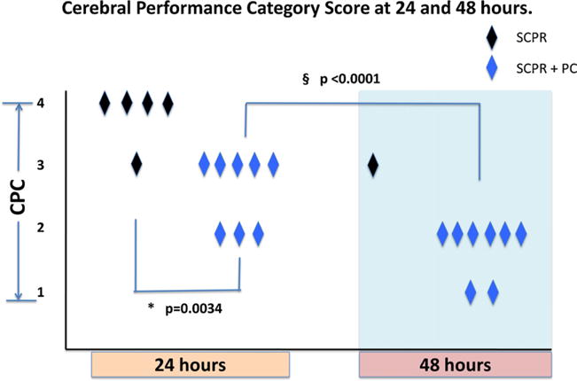
Mean CPC score at 24 h was significantly lower (better neurological function) in the animals that received SCPR + PC compared to SCPR alone (2.7 ± 0.4 vs 3.8 ± 0.4 respectively, p = 0.003). Neurological function in SCPR + PC group significantly improved in all but one animals at 48 h and the mean CPC score of the group decreased from 2.7 ± 0.4 to 1.7 ± 0.4 (p < 0.001). The one SCPR animal that survived 48 h had the same CPC score of 3 at 24 and 48 h (Fig. 2).
Fig. 2.

24 and 48-h neurological assessment. Addition of controlled pauses during SCPR significantly improved neurological function compared to SCPR alone at 24 h. In the SCPR + PC neurological function improved in all but one animal at 48 h. Mean CPC at 24 h was significant lower in the SCPR + PC compared to SCPR alone (CPC: 2.7 ± 0.4 vs 3.8 ± 0.4 respectively, p = 0.003) cerebral performance category score: (1 = normal, 2 = mild deficit, 3 = moderate deficit but conscious, 4 = coma); SCPR: standard CPR. PC: postconditioning. *Means statistically significant difference between groups at 24 h. §Means statistically significant difference between the SCPR + PC group at 24 vs 48 h.
3.5. Blood gasses, end tidal CO2 and lactate
There were no significant differences in blood gas values at baseline between groups. Immediately after ROSC, pH and HCO3 and ETCO2 were significantly higher in the SCPR + PC group a finding that can be explained by higher circulation at the last few minutes of CPR and immediately after ROSC (Table 2).
4. Discussion
Our study, for the first time, shows that a simple strategy of ischemic postconditioning introduced early during standard-CPR with four 20-s pauses can significantly improve cardio-cerebral outcomes in a porcine model of very prolonged cardiac arrest and global ischemia. When good quality SCPR was coupled with controlled pauses at the initiation of reperfusion, the resuscitated animals documented normal left ventricular function post resuscitation in the absence of inotropic support and improved neurologic outcome. Furthermore, to our knowledge, this is the only study that has demonstrated complete neurological recovery is possible after 15 min of untreated cardiac arrest with standard CPR and a non-invasive intervention.
It is important to emphasize that unintentional pauses in chest compressions spread throughout resuscitative efforts have been associated with worse outcomes by adding to the injury that has accumulated from the no-flow period.17–19 The effects of pauses on coronary perfusion pressure and carotid blood flow were exactly as previously described by Berg et al.18 Pauses caused elimination of the trans-coronary pressure gradient and carotid flow. The type of intentional pauses described in this report is thought to harness endogenous protective processes associated with specific mitochondrial protective mechanics and should not be confused with the poor outcomes known to be associated with poor CPR quality that includes prolonged intervals of interrupted chest compressions.20
The fact that there were no differences in ROSC rates between groups and that the S-CPR (control) group had such high ROSC rate is a testament to the high quality of CPR performed. While introduction of CPR pauses at the initiation of resuscitation efforts may appear to be contrary to some beliefs that continuous uninterrupted chest compressions are essential, four pauses of 20 s duration during the first 3 min after CPR initiation followed by continuous chest compressions with asynchronous ventilations for the remainder of the resuscitation appeared to positively impact neurological outcome after very prolonged global cerebral ischemia.21
The animals that were treated with controlled pauses showed absence of post resuscitation left ventricular dysfunction and they were hemodynamically more stable post ROSC requiring less epinephrine. There is very strong evidence that post conditioning is advantageous for cardiac muscle protection after ischemia.11,22 Three to four pauses during reperfusion of acute myocardial infarction have been shown to significantly decrease infarct size in human studies.7,8 Our results show that a similar strategy of controlled reperfusion after prolonged global ischemia in cardiac arrest exhibits the same benefits for the myocardium as a whole and mitigates post ROSC cardiac dysfunction that contributes heavily to post resuscitation morbidity and mortality. Recently animal studies have described strategies to alleviate ischemia reperfusion injury and promote cardiac recovery after cardiac arrest with inhaled anesthetics and hypothermia. It is possible that a strategy combining pauses during CPR and pharmacological postconditioning could demonstrate further benefits.23,24
The most striking observation in our study was that the brain demonstrated the potential for full recovery after 15 min of global ischemia with no flow. Shaffner et al. showed that cerebral recovery was not feasible after 12 min of untreated arrest because regeneration of ATP was not possible despite high cerebral perfusion pressures.25 Our data suggest otherwise. To our knowledge, this is the first time that survival rates with consistently favorable neurological outcomes have been reported after 15 min of untreated cardiac arrest. Controlled reperfusion with short pauses at the initiation of reperfusion of stroke has been shown to significantly decrease injury in a rat model.10 Furthermore, 15–30 s cycles of on/off flow in the same model with 10 min of global ischemic cerebral insult have provided significant cerebral preservation and recovery.10 The latter model is relevant to cardiac arrest where the ischemic insult is systemic and global. For the above mentioned reasons we combined the duration (15–30 s) and number of cycles (3–4) of different ischemic postconditioning strategies to create our tested protocol of four 20-s pauses during the first 3 min of CPR.
The mechanisms for protection of both the heart and brain have been well-studied and are currently considered to be mediated by direct and indirect modulation of mitochondrial permeability transition pore state.20,26–31 We used this strategy exactly because of previously documented protection of vital organs in stroke and myocardial infarction. Based on our data, it appears that postconditioning with pauses in CPR efforts at the initiation of reperfusion, offer significant protection and facilitates functional cardiac and cerebral recovery.8,11,32,33
Postconditioning with controlled reperfusion has also been shown to be beneficial in preventing ischemia reperfusion injury in most of the organs such as liver, kidney, retina, and small intestine.34–40 That could explain why the animals with SCPR + CP had an overall improvement in their status, survived and continued to improve to 48 h.
This study has limitations. First, we did not perform a dosing study and therefore we cannot comment what is the best combination of cycles and duration of pauses. We used the specific combination based on the literature targeting neurological protection.10 Second we do not know if the same benefits could be realized if resuscitation efforts are prolonged and we cannot exclude the possibility of synergy between mild therapeutic hypothermia and ischemic postconditioning. Third, our study was not designed to address the mechanism of protection offered by postconditioning. Nonetheless, we have no reason to believe that the mechanism should be different than the one described extensively in the cardiac and cerebral literature.8,11,32,33 We also did not assess biomarkers of injury for the heart and brain. Although the clinical endpoints reported here (echocardiographic LV function and blinded neurological assessment), in our opinion, represent higher quality preclinical endpoints than biomarkers, we cannot claim there was tissue protection despite the observed improvement in myocardial and cerebral functional outcomes. A histopathology/brain MRI study is underway to correlate the clinical endpoints observed in this study with brain pathology. However, we tested those interventions exactly as described for acute myocardial infarction and stroke and found them to be extremely effective in our model of cardiac arrest.8,32,33 Finally, it is unknown if the benefits demonstrated in this study would be seen with coexisting myocardial ischemia or can be translated to humans.
5. Conclusion
A simple strategy of ischemic postconditioning with four 20-s pauses during the first 3 min of SCPR mitigates post resuscitation cardiac dysfunction and facilitates neurological recovery after 15 min of untreated cardiac arrest in pigs.
Acknowledgments
Funding source
The study was funded by an Institutional, Division of Cardiology grant at the University of Minnesota and an R01 HL108926-01 NIH grant to Dr. Yannopoulos.
Footnotes
A Spanish translated version of the summary of this article appears as Appendix in the final online version at http://dx.doi.org/10.1016/j.resuscitation.2012.04.005.
Authors’ note
All authors have participated to the conception, design and writing of this manuscript.
Conflict of interest
Demetris Yannopoulos is the Medical Director of the Minnesota Resuscitation Consortium, a state wide initiative to improve survival in the state of MN from cardiac arrest. This initiative is sponsored by the Medtronic Foundation and is part of the Heart Rescue Program. There are no conflicts related to this investigation.
Keith G. Lurie is the founder of Advanced Circulatory Systems Incorporated (ACSI), and co-inventor of the inspiratory impedance threshold device and ACD CPR technique but he has no conflicts to this study. Tom P. Aufderheide has board membership for Take Heart America and Citizen CPR Foundation, has consulted for JoLife Medical and Medtronic Foundation, and has received grants/grants pending from the NHLBI Immediate Trial, NHLBI Resuscitation Outcomes Consortium and NINDS Neurological Emergency Treatment Trials Network.
Henry R Halperin MD is a consultant to Zoll Medical and has received grants from NHLBI.
Menekhem Zviman is a consultant to Zoll Medical.
The other authors have no conflicts of interest with the present study.
Ethical approval
The study was approved by the Institutional Animal Care Committee of the Minneapolis Medical Research Foundation of Hennepin County Medical Center, and all animals received treatment in compliance with the National Research Council’s 1996 Guide for the Care and Use of Laboratory Animals.
References
- 1.Nichol G, Aufderheide TP, Eigel B, et al. Regional systems of care for out-of-hospital cardiac arrest: a policy statement from the American Heart Association. Circulation. 2010;121:709–29. doi: 10.1161/CIR.0b013e3181cdb7db. [DOI] [PubMed] [Google Scholar]
- 2.Cummins RO. From concept to standard-of-care? Review of the clinical experience with automated external defibrillators. Ann Emerg Med. 1989;18:1269–75. doi: 10.1016/s0196-0644(89)80257-4. [DOI] [PubMed] [Google Scholar]
- 3.Hogler S, Sterz F, Sipos W, et al. Distribution of neuropathological lesions in pig brains after different durations of cardiac arrest. Resuscitation. 2010;81:1577–83. doi: 10.1016/j.resuscitation.2010.07.005. [DOI] [PubMed] [Google Scholar]
- 4.Janata A, Bayegan K, Sterz F, et al. Limits of conventional therapies after prolonged normovolemic cardiac arrest in swine. Resuscitation. 2008;79:133–8. doi: 10.1016/j.resuscitation.2008.04.005. [DOI] [PubMed] [Google Scholar]
- 5.Janata A, Weihs W, Schratter A, et al. Cold aortic flush and chest compressions enable good neurologic outcome after 15 min of ventricular fibrillation in cardiac arrest in pigs. Crit Care Med. 2010;38:1637–43. doi: 10.1097/CCM.0b013e3181e78b9a. [DOI] [PubMed] [Google Scholar]
- 6.Kern KB, Hilwig RW, Berg RA, et al. Postresuscitation left ventricular systolic and diastolic dysfunction. Treatment with dobutamine. Circulation. 1997;95:2610–3. doi: 10.1161/01.cir.95.12.2610. [DOI] [PubMed] [Google Scholar]
- 7.Garcia S, Henry TD, Wang YL, et al. Long-term follow-up of patients undergoing postconditioning during ST-elevation myocardial infarction. J Cardiovasc Transl Res. 2011;4:92–8. doi: 10.1007/s12265-010-9252-0. [DOI] [PubMed] [Google Scholar]
- 8.Ovize M, Baxter GF, Di Lisa F, et al. Postconditioning and protection from reperfusion injury: where do we stand? Position paper from the Working Group of Cellular Biology of the Heart of the European Society of Cardiology. Cardiovasc Res. 2010;87:406–23. doi: 10.1093/cvr/cvq129. [DOI] [PubMed] [Google Scholar]
- 9.Zhao ZQ, Corvera JS, Halkos ME, et al. Inhibition of myocardial injury by ischemic postconditioning during reperfusion: comparison with ischemic preconditioning. Am J Physiol Heart Circ Physiol. 2003;285:H579–88. doi: 10.1152/ajpheart.01064.2002. [DOI] [PubMed] [Google Scholar]
- 10.Wang JY, Shen J, Gao Q, et al. Ischemic postconditioning protects against global cerebral ischemia/reperfusion-induced injury in rats. Stroke. 2008;39:983–90. doi: 10.1161/STROKEAHA.107.499079. [DOI] [PubMed] [Google Scholar]
- 11.Yellon DM, Hausenloy DJ. Myocardial reperfusion injury. N Engl J Med. 2007;357:1121–35. doi: 10.1056/NEJMra071667. [DOI] [PubMed] [Google Scholar]
- 12.Yannopoulos D, Matsuura T, McKnite S, et al. No assisted ventilation cardiopulmonary resuscitation and 24-h neurological outcomes in a porcine model of cardiac arrest. Crit Care Med. 2010;38:254–60. doi: 10.1097/CCM.0b013e3181b42f6c. [DOI] [PubMed] [Google Scholar]
- 13.Shultz JJ, Coffeen P, Sweeney M, et al. Evaluation of standard and active compression–decompression CPR in an acute human model of ventricular fibrillation. Circulation. 1994;89:684–93. doi: 10.1161/01.cir.89.2.684. [DOI] [PubMed] [Google Scholar]
- 14.Yannopoulos D, Matsuura T, Schultz J, Rudser K, Halperin HR, Lurie KG. Sodium nitroprusside enhanced cardiopulmonary resuscitation improves survival with good neurological function in a porcine model of prolonged cardiac arrest. Crit Care Med. 2011;39:1269–74. doi: 10.1097/CCM.0b013e31820ed8a6. [DOI] [PMC free article] [PubMed] [Google Scholar]
- 15.Marino BS, Yannopoulos D, Sigurdsson G, et al. Spontaneous breathing through an inspiratory impedance threshold device augments cardiac index and stroke volume index in a pediatric porcine model of hemorrhagic hypovolemia. Crit Care Med. 2004;32:S398–405. doi: 10.1097/01.ccm.0000139950.39972.68. [DOI] [PubMed] [Google Scholar]
- 16.Quinones MA, Waggoner AD, Reduto LA, et al. A new, simplified and accurate method for determining ejection fraction with two-dimensional echocardiography. Circulation. 1981;64:744–53. doi: 10.1161/01.cir.64.4.744. [DOI] [PubMed] [Google Scholar]
- 17.Yu T, Weil MH, Tang W, et al. Adverse outcomes of interrupted pre-cordial compression during automated defibrillation. Circulation. 2002;106:368–72. doi: 10.1161/01.cir.0000021429.22005.2e. [DOI] [PubMed] [Google Scholar]
- 18.Berg RA, Sanders AB, Kern KB, et al. Adverse hemodynamic effects of interrupting chest compressions for rescue breathing during cardiopulmonary resuscitation for ventricular fibrillation cardiac arrest. Circulation. 2001;104:2465–70. doi: 10.1161/hc4501.098926. [DOI] [PubMed] [Google Scholar]
- 19.Christenson J, Andrusiek D, Everson-Stewart S, et al. Chest compression fraction determines survival in patients with out-of-hospital ventricular fibrillation. Circulation. 2009;120:1241–7. doi: 10.1161/CIRCULATIONAHA.109.852202. [DOI] [PMC free article] [PubMed] [Google Scholar]
- 20.Cour M, Loufouat J, Paillard M, et al. Inhibition of mitochondrial permeability transition to prevent the post-cardiac arrest syndrome: a pre-clinical study. Eur Heart J. 2011;32:226–35. doi: 10.1093/eurheartj/ehq112. [DOI] [PubMed] [Google Scholar]
- 21.Sayre MR, Koster RW, Botha M, et al. Part 5: adult basic life support: 2010 international consensus on cardiopulmonary resuscitation and emergency cardiovascular care science with treatment recommendations. Circulation. 2010;122:S298–324. doi: 10.1161/CIRCULATIONAHA.110.970996. [DOI] [PubMed] [Google Scholar]
- 22.Shinohara G, Morita K, Nagahori R, et al. Ischemic postconditioning promotes left ventricular functional recovery after cardioplegic arrest in an in vivo piglet model of global ischemia reperfusion injury on cardiopulmonary bypass. J Thorac Cardiovasc Surg. 2011;142:926–32. doi: 10.1016/j.jtcvs.2011.01.028. [DOI] [PubMed] [Google Scholar]
- 23.Albrecht M, Gruenewald M, Zitta K, et al. Hypothermia and anesthetic postconditioning influence the expression and activity of small intestinal proteins possibly involved in ischemia/reperfusion-mediated events following cardiopulmonary resuscitation. Resuscitation. 2012;83:113–8. doi: 10.1016/j.resuscitation.2011.06.038. [DOI] [PubMed] [Google Scholar]
- 24.Meybohm P, Gruenewald M, Albrecht M, et al. Pharmacological postconditioning with sevoflurane after cardiopulmonary resuscitation reduces myocardial dysfunction. Crit Care. 2011;15:R241. doi: 10.1186/cc10496. [DOI] [PMC free article] [PubMed] [Google Scholar]
- 25.Shaffner DH, Eleff SM, Brambrink AM, et al. Effect of arrest time and cerebral perfusion pressure during cardiopulmonary resuscitation on cerebral blood flow, metabolism, adenosine triphosphate recovery, and pH in dogs. Crit Care Med. 1999;27:1335–42. doi: 10.1097/00003246-199907000-00026. [DOI] [PubMed] [Google Scholar]
- 26.Ayoub IM, Radhakrishnan J, Gazmuri RJ. Targeting mitochondria for resuscitation from cardiac arrest. Crit Care Med. 2008;36:S440–6. doi: 10.1097/ccm.0b013e31818a89f4. [DOI] [PMC free article] [PubMed] [Google Scholar]
- 27.Crompton M. Mitochondrial intermembrane junctional complexes and their role in cell death. J Physiol. 2000;529:11–21. doi: 10.1111/j.1469-7793.2000.00011.x. [DOI] [PMC free article] [PubMed] [Google Scholar]
- 28.Halestrap AP, Clarke SJ, Javadov SA. Mitochondrial permeability transition pore opening during myocardial reperfusion—a target for cardioprotection. Cardiovasc Res. 2004;61:372–85. doi: 10.1016/S0008-6363(03)00533-9. [DOI] [PubMed] [Google Scholar]
- 29.Weiss JN, Korge P, Honda HM, Ping P. Role of the mitochondrial permeability transition in myocardial disease. Circ Res. 2003;93:292–301. doi: 10.1161/01.RES.0000087542.26971.D4. [DOI] [PubMed] [Google Scholar]
- 30.Argaud L, Gateau-Roesch O, Raisky O, Loufouat J, Robert D, Ovize M. Postconditioning inhibits mitochondrial permeability transition. Circulation. 2005;111:194–7. doi: 10.1161/01.CIR.0000151290.04952.3B. [DOI] [PubMed] [Google Scholar]
- 31.Argaud L, Gateau-Roesch O, Muntean D, et al. Specific inhibition of the mitochondrial permeability transition prevents lethal reperfusion injury. J Mol Cell Cardiol. 2005;38:367–74. doi: 10.1016/j.yjmcc.2004.12.001. [DOI] [PubMed] [Google Scholar]
- 32.Zhao H. Ischemic postconditioning as a novel avenue to protect against brain injury after stroke. J Cereb Blood Flow Metab. 2009;29:873–85. doi: 10.1038/jcbfm.2009.13. [DOI] [PMC free article] [PubMed] [Google Scholar]
- 33.Cour M, Gomez L, Mewton N, Ovize M, Argaud L. Postconditioning: from the bench to bedside. J Cardiovasc Pharmacol Ther. 2011;16:117–30. doi: 10.1177/1074248410383174. [DOI] [PubMed] [Google Scholar]
- 34.Zeng Z, Huang HF, Chen MQ, Song F, Zhang YJ. Postconditioning prevents ischemia/reperfusion injury in rat liver transplantation. Hepatogastroenterology. 2010;57:875–81. [PubMed] [Google Scholar]
- 35.Snoeijs MG, van Heurn LW, Buurman WA. Biological modulation of renal ischemia-reperfusion injury. Curr Opin Organ Transplant. 2010;15:190–9. doi: 10.1097/MOT.0b013e32833593eb. [DOI] [PubMed] [Google Scholar]
- 36.Gurusamy KS, Gonzalez HD, Davidson BR. Current protective strategies in liver surgery. World J Gastroenterol. 2010;16:6098–103. doi: 10.3748/wjg.v16.i48.6098. [DOI] [PMC free article] [PubMed] [Google Scholar]
- 37.Dreixler JC, Shaikh AR, Alexander M, Savoie B, Roth S. Post-ischemic conditioning in the rat retina is dependent upon ischemia duration and is not additive with ischemic pre-conditioning. Exp Eye Res. 2010;91:844–52. doi: 10.1016/j.exer.2010.06.015. [DOI] [PMC free article] [PubMed] [Google Scholar]
- 38.Ferencz A, Takacs I, Horvath S, et al. Examination of protective effect of ischemic postconditioning after small bowel autotransplantation. Transplant Proc. 2010;42:2287–9. doi: 10.1016/j.transproceed.2010.05.023. [DOI] [PubMed] [Google Scholar]
- 39.Zhao W, Che XM, Fan L, et al. Protective effect of ischemic preconditioning against cold ischemia and reperfusion injury of rat small intestinal graft. Nan Fang Yi Ke Da Xue Xue Bao. 2007;27:1764–6. [PubMed] [Google Scholar]
- 40.Lauzier B, Sicard P, Bouchot O, et al. After four hours of cold ischemia and cardioplegic protocol, the heart can still be rescued with postconditioning. Transplantation. 2007;84:1474–82. doi: 10.1097/01.tp.0000288637.18796.0e. [DOI] [PubMed] [Google Scholar]


