Abstract
This study uses national survey data to describe testosterone levels by age in youths aged 6 to 20 years and the onset and magnitude of divergence in levels between boys and girls.
Data on testosterone levels in children and adolescents segregated by sex are scarce and based on convenience samples or assays with limited sensitivity and accuracy. Such data would be useful in evaluating children with pubertal or androgen disorders and dichotomizing male and female youths participating in sport. Thus, we analyzed the timing of the onset and magnitude of the divergence in testosterone in youths aged 6 to 20 years by sex using a highly accurate assay.
Methods
Testosterone concentrations from separate cohorts of male and female youths collected during 2 periods of the National Health and Nutrition Examination Survey (NHANES; 2013-2014 and 2015-2016) were pooled into 1 data set for analyses. Briefly, NHANES uses a multistage probability design to randomly sample US residents from all 50 states. The overall response rate in the 2 data collection cycles was 70.4% in youths, and 80% of those responders elected to participate in the collection of biospecimens for testosterone analyses. All procedures accessed public, deidentified information and did not require ethical review as determined by the Mayo Clinic Institutional Review Board.
As described previously,1 testosterone was quantified via isotope dilution liquid chromatography tandem mass spectrometry, which demonstrates a broad analytical measurement range (0.75-1400 ng/dL), excellent precision across a wide range (<3% coefficient of variation) and high accuracy (–0.7% mean bias for a 2-year period), confirmed using reference materials from the National Institute for Standards and Technology.
Full factorial analysis of variance was used to examine the change in testosterone concentration from ages 6 to 20 years by sex, focusing on the age of divergence of testosterone and the overlap at the extremes. Two-tailed post hoc analyses (Scheffe test) were used to test for differences between pairs with Bonferroni-corrected P values (P < .025). For all other analyses, significance was determined at P < .05. All analyses were performed with R software, version 3.4.2 (R Foundation).
Results
The data set included 4495 youth samples—2293 male and 2202 female—with diverse racial representation including Hispanic (36%), white (26.6%), black (23.0%), Asian (8.8%), and multiracial (6.1%). No statistical differences of race (effects or interactions) were noted.
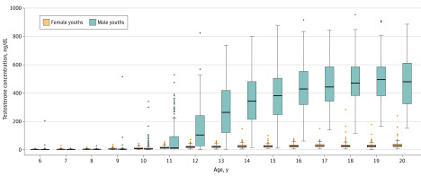
The median testosterone concentration increased for female youths from age 6 to 20 years from 2.4 ng/dL to 29.5 ng/dL (P < .001), with a plateau beginning at age 14 years (Table). Over the same age range, the median testosterone concentration increased considerably more for male youths compared with female youths (age × sex; P < .001), from 1.9 ng/dL at age 6 years to 516 ng/dL at age 20 years (P < .001), with a plateau beginning at age 17 years. Testosterone concentration was not different between the sexes from age 6 to 10 years; however, male youths had greater testosterone concentrations than female youths from age 11 to 20 years (Figure).
Table. Age-Adjusted Testosterone Concentration Percentiles.
| Age, y | Male youths | Female youths | P valuea | ||||||||||
|---|---|---|---|---|---|---|---|---|---|---|---|---|---|
| No. | Testosterone concentration, median, ng/dL, by percentile | No. | Testosterone concentration, median, ng/dL, by percentile | ||||||||||
| 5th | 25th | 50th | 75th | 95th | 5th | 25th | 50th | 75th | 95th | ||||
| 6 | 157 | 0.5 | 1.1 | 1.9 | 2.7 | 4.9 | 166 | 0.8 | 1.6 | 2.4 | 4.0 | 6.5 | .64 |
| 7 | 187 | 0.5 | 1.4 | 2.2 | 3.5 | 5.4 | 144 | 1.2 | 2.1 | 3.0 | 4.2 | 8.4 | .16 |
| 8 | 171 | 1.2 | 2.0 | 3.0 | 4.4 | 7.6 | 158 | 1.3 | 2.6 | 3.7 | 5.0 | 9.4 | .17 |
| 9 | 161 | 1.1 | 2.3 | 3.7 | 5.4 | 9.1 | 161 | 1.7 | 3.4 | 5.3 | 7.9 | 18.6 | .66 |
| 10 | 177 | 1.8 | 3.7 | 5.6 | 11.2 | 76.1 | 168 | 3.0 | 5.5 | 8.0 | 15.2 | 26.2 | .35 |
| 11 | 171 | 3.1 | 6.2 | 13.3 | 95.7 | 327 | 185 | 4.9 | 9.4 | 14.2 | 20.0 | 38.5 | <.001 |
| 12 | 148 | 7.9 | 27.5 | 105 | 250 | 497 | 144 | 7.1 | 13.3 | 19.5 | 26.6 | 40.3 | <.001 |
| 13 | 157 | 16.4 | 121 | 264 | 424 | 620 | 127 | 8.0 | 15.7 | 21.9 | 27.9 | 42.8 | <.001 |
| 14 | 165 | 64.0 | 216 | 343 | 482 | 699 | 157 | 9.5 | 17.1 | 23.4 | 33.0 | 47.0 | <.001 |
| 15 | 158 | 140 | 246 | 382 | 506 | 745 | 133 | 12.1 | 17.9 | 24.1 | 30.5 | 51.4 | <.001 |
| 16 | 151 | 149 | 319 | 429 | 554 | 695 | 178 | 11.4 | 18.3 | 25.5 | 34.6 | 56.5 | <.001 |
| 17 | 140 | 220 | 343 | 443 | 589 | 779 | 131 | 13.0 | 21.6 | 28.9 | 36.9 | 56.8 | <.001 |
| 18 | 142 | 265 | 380 | 470 | 587 | 737 | 145 | 12.1 | 18.6 | 26.3 | 33.2 | 62.0 | <.001 |
| 19 | 120 | 235 | 381 | 496 | 595 | 804 | 125 | 10.9 | 18.8 | 25.4 | 34.0 | 65.0 | <.001 |
| 20 | 88 | 188 | 326 | 516 | 632 | 862 | 80 | 10.2 | 21.8 | 29.5 | 40.0 | 98.1 | <.001 |
Comparing male youths vs female youths at the 50th percentile.
Figure. Total Testosterone Concentrations of the US Population Aged 6 to 20 Years.
The horizontal line in the middle of each box indicates the median; top and bottom borders, 75th and 25th percentiles; whiskers above and below the box, 90th and 10th percentiles; and circles beyond the whiskers, outliers beyond the 90th or 10th percentiles.
Among youths aged 12 years or older, there was no overlap of the interquartile range of testosterone between male and female youths. After cessation of the age-related increase in testosterone for female youths (at 14 years), there was an intersection of testosterone concentration distributions between the lowest (first) percentile of male youths and the uppermost (99th) percentile of female youths (≥100 ng/dL), which includes 8 of 949 samples (<1%) for female youths.
Discussion
These data demonstrated the following: (1) the sex-related divergence of testosterone initiated at 11 years of age on average; (2) clear and distinct distributions of serum testosterone between the sexes after 11 years of age; and (3) the distribution of testosterone within male youths was much larger in magnitude and spread than the distribution of testosterone within female youths. At the population level, serum testosterone created a clear dichotomy between male and female youths, and the presented age-adjusted distributions may be useful in evaluation of pubertal and androgenic disorders in youths.
A testosterone value of 100 ng/dL distinctly separated the sexes with minimal overlap, which may have broad implications for athletic competition, as serum testosterone has been demonstrated to be strongly associated with sex differences in athletic performance.2,3 Potential testosterone thresholds for eligibility in sports may need to be adjusted based on further information on outliers and direction of error accepted.
These analyses were limited by a lack of information on pubertal stages and history of androgenic disorders and by self- or parental report of male/female sex.
Section Editor: Jody W. Zylke, MD, Deputy Editor.
References
- 1.Zhou H, Wang Y, Gatcombe M, et al. Simultaneous measurement of total estradiol and testosterone in human serum by isotope dilution liquid chromatography tandem mass spectrometry. Anal Bioanal Chem. 2017;409(25):5943-5954. doi: 10.1007/s00216-017-0529-x [DOI] [PMC free article] [PubMed] [Google Scholar]
- 2.Senefeld JW, Clayburn AJ, Baker SE, Carter RE, Johnson PW, Joyner MJ. Sex differences in youth elite swimming. PLoS One. 2019;14(11):e0225724. doi: 10.1371/journal.pone.0225724 [DOI] [PMC free article] [PubMed] [Google Scholar]
- 3.Handelsman DJ. Sex differences in athletic performance emerge coinciding with the onset of male puberty. Clin Endocrinol (Oxf). 2017;87(1):68-72. doi: 10.1111/cen.13350 [DOI] [PubMed] [Google Scholar]


