Abstract
Background:
Isotonic exercise is commonly adopted for shoulder rehabilitation, but the efficacy of isokinetic exercise for rehabilitation has not been evaluated.
Purpose:
To evaluate the efficacy of isotonic and isokinetic external shoulder rotation exercises.
Study Design:
Controlled laboratory study.
Methods:
Using surface electromyography (EMG) and the Biodex system, we investigated the EMG amplitude of the infraspinatus (IS), total work (tWK), and EMG(IS)/tWK ratio and examined the relative IS and posterior deltoid (PD) contributions to all exercises. A total of 24 healthy participants without musculoskeletal injuries were included. Participants performed isotonic external shoulder rotation at 10%, 20%, 30%, 40%, and 50% of the maximum voluntary isometric contraction (MVIC) as well as isokinetic external shoulder rotation at angular velocities of 60, 120, 180, 240, and 300 deg/s. Levels of intensity were classified from 1 to 5: level 1 corresponded to 10% of the MVIC and a 300-deg/s angular velocity; level 2 corresponded to 20% MVIC and 240 deg/s; level 3 corresponded to 30% MVIC and 180 deg/s; level 4 corresponded to 40% MVIC and 120 deg/s; and level 5 corresponded to 50% MVIC and 60 deg/s. Normalized IS and tWK amplitudes were calculated for each exercise.
Results:
During isotonic exercise, the EMG(IS)/tWK ratio significantly decreased from level 5 to 3, 2, and 1; from level 4 to 2 and 1; and from level 3 to 1. During isokinetic exercise, the EMG(IS)/tWK ratio at level 3 was greater than that at all other levels except level 1. Statistical differences were found between isotonic and isokinetic modes at levels 1, 2, and 3. The IS/PD activation ratios were not significantly different between exercise modes at any level.
Conclusion:
Isokinetic resistance may provide more effective stimulation of the IS muscle compared with isotonic resistance.
Clinical Relevance:
Isokinetic exercise needs to be considered as a method of rehabilitation that effectively increases infraspinatus muscle activity.
Keywords: Isotonic, isokinetic, concentric, shoulder, external rotation, electromyography
The rotator cuff is known to contribute to dynamic stabilization of the glenohumeral joint.28 The subscapularis, infraspinatus (IS), and teres minor conjointly generate a force that acts as a counterforce to the pulling force of the deltoid and supraspinatus muscles during arm elevation.25 A fine balance between the IS, teres minor, and subscapularis is important to control anteroposterior translation of the humeral head and hence to provide a stable fulcrum. Therefore, dysfunction of the rotator cuff results in shoulder disabilities, such as subacromial impingement or rotator cuff tears on progression.22 Early-stage shoulder rehabilitation should focus on the improvement of rotator cuff balance and stabilizing ability.8 External rotation exercise is commonly performed to restore function of the IS, a major external rotator, which is activated first during external rotation and plays an important role in maintaining shoulder stability.1
Isotonic exercise involves movement under a constant load throughout the active range of motion, which is applied to all body parts, including the shoulder.4 However, isokinetic contraction is performed with accommodating resistance throughout the active range of motion at a constant speed and is usually adopted for muscle testing in the shoulder joint.3 There is a lack of evidence demonstrating the effectiveness of isokinetic exercise on rotator cuff rehabilitation. Malliou et al15 were the only group to report that isokinetic strengthening is a more effective method of altering rotator cuff muscle strength ratios compared with other kinds of exercise, including isotonic exercise. Therefore, isokinetic exercise has a potential value in rotator cuff rehabilitation.
Surface electromyography (EMG) has been used to evaluate muscle action potentials through surface electrodes located directly over an activated muscle.12 EMG amplitude is measured during muscle contraction and provides a quantification of muscle activation.23 Muscle activation, rather than muscle force production, may be more useful in reflecting training-induced adaptations of the nervous system in the early rehabilitation phase.5,20 Schmitz and Westwood20 proposed the EMG amplitude-to-work (EMG/WK) ratio to examine the efficacy of isokinetic and isotonic exercises during leg extension. By normalizing the EMG amplitude to the amount of work, those authors intended to determine the optimal type of resistance that results in the recruitment of more motor units or an increase in frequency. Purkayastha et al17 extended the study of Schmitz and Westwood by investigating EMG/WK ratios for concentric isokinetic muscle actions at 60, 120, 180, 240, and 300 deg/s as well as concentric isotonic muscle actions at 10%, 20%, 30%, 40%, and 50% of the maximum voluntary isometric contraction (MVIC) during leg extension exercises. An investigation of isotonic and isokinetic external shoulder rotation exercises using the methods of Purkayastha et al may help to find the ideal resistance to increase IS activation.
The primary purpose of this study was to evaluate the IS EMG/WK ratio for concentric isokinetic muscle actions at 60, 120, 180, 240, and 300 deg/s as well as concentric isotonic muscle actions at 10%, 20%, 30%, 40%, and 50% of the MVIC during external shoulder rotation exercises. The extraction of pure IS work from total work (tWK) during external rotation exercises was not feasible. Therefore, tWK was used as a denominator in calculating the EMG(IS)/tWK ratio. Second, to validate the use of tWK as a denominator, the IS-to–posterior deltoid (PD) activation ratios were also calculated. The IS/PD activation ratio has been used to examine the relative IS and PD contributions to external shoulder rotation exercises.1,19 Using the IS/PD activation ratio, we assessed whether the IS contributes to tWK at a similar rate for each exercise.
Methods
Participants
This study was approved by our institutional review board, and informed consent was obtained from all participants. All participants were recruited from the Sports Medical Center of our institute between June 2017 and January 2018. A total of 24 right-handed healthy male participants (mean age, 25.3 years; mean height, 175.4 cm; mean weight, 67.3 kg) were included in this study. Participants were excluded if they (1) had previous musculoskeletal injuries or underwent rehabilitation treatments for any injuries (n = 2) or (2) had difficulty in following the exercise protocols (n = 3).
EMG and Work Data
Surface EMG signals were recorded from the IS and PD using a TeleMyo DTS Desk Receiver (Noraxon USA), and all data processing was performed with MR3 software (version 3; Noraxon USA). The signals were amplified with a gain of 400, noise <1 µV, and common mode rejection ratio of 100 dB. They were sampled at 1500 Hz and filtered with a bandwidth of 10 to 500 Hz. To construct a linear envelope, full-wave rectification was performed. A Lancosh finite impulse response digital filter (Noraxon USA) was used to filter the raw signal. The bandpass filter frequency was between 10 and 350 Hz.
We processed the EMG data into the root mean square (RMS) value in 200-millisecond windows. For normalization, RMS values of the MVIC were measured 3 times for each IS and PD. The EMG amplitude of the IS recorded during each exercise was normalized as a percentage of the maximum EMG amplitude of the IS during the MVIC (% RMS). To minimize skin impedance, we prepared the skin surface by cleaning the area with an alcohol swab before placing the electrodes. Bipolar surface EMG electrodes with a distance of 2 cm between active recording sites were used. The IS electrode was placed 3 cm below the spine of the scapula laterally but not over the PD muscle.6 For the PD, the electrode was placed 2 cm below the posterolateral corner of the acromion over the PD muscle (Figure 1). The highest tWK value during each exercise was selected for analysis, which was derived from Biodex System 4 (Biodex). For the EMG(IS)/tWK ratio (% RMS/J), the % RMS values of the IS EMG amplitude were divided by the respective isotonic or isokinetic tWK values. The IS activation values were divided by the PD activation values (IS/PD activation ratios) to investigate the recruitment of the PD muscle during each external rotation exercise.
Figure 1.
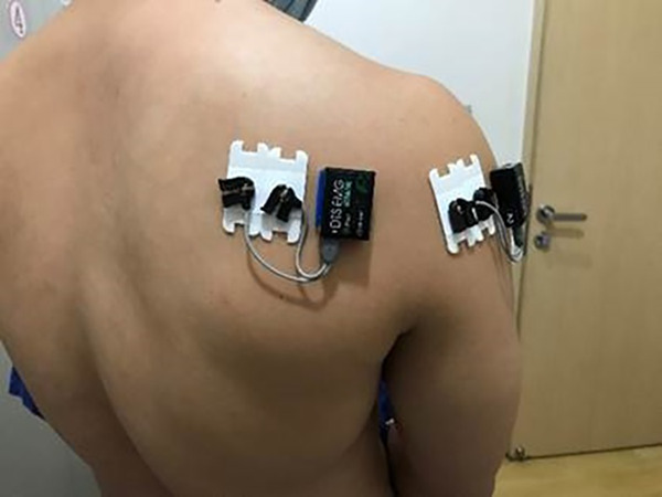
Electrode placements for the infraspinatus and posterior deltoid muscles.
Procedures
The participants underwent IS MVIC testing with the shoulder at 20° of abduction and forward flexion with neutral rotation while sitting on a dynamometer (Figure 2). MVIC testing was conducted in 3 sets of 5 seconds according to the guidelines of Kendall et al.13 Participants were provided with verbal encouragement and visual feedback of signal amplitudes to generate the maximum IS force. At least 2 minutes of rest between MVIC tests was given to minimize muscle fatigue. The maximum amplitude after 3 sets of contractions was used for normalization, and the highest peak torque during the MVIC was selected as a representative score.
Figure 2.
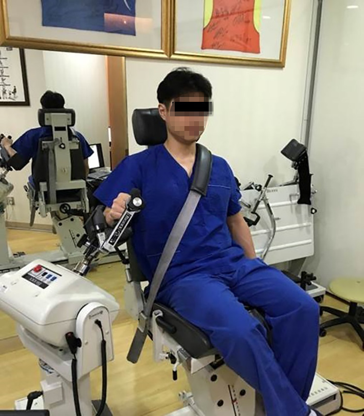
External rotation exercises were carried out with the shoulder at 20° of abduction and forward flexion. The participant sat on the dynamometer with the trunk fixed by a cross seat belt.
During the external rotation exercises, each participant rotated the arm under investigation externally 40° from neutral rotation with the shoulder at 20° of abduction. The isotonic or isokinetic exercise protocols were selected randomly, and concentric external rotation exercises in each protocol were performed 3 times in random order with 2 minutes of rest between the exercises. Each protocol consisted of a series of 5 exercises. The isotonic protocol comprised concentric external rotation exercises at the following 5 loads: 10%, 20%, 30%, 40%, and 50% of the previously determined MVIC. For the isokinetic protocol, the angular velocity was set sequentially at 60, 120, 180, 240, and 300 deg/s. According to the study of Purkayastha et al,17 the levels of intensity were categorized from 1 to 5: level l corresponded to an isotonic intensity of 10% of the MVIC and an isokinetic velocity of 300 deg/s; level 2 corresponded to 20% MVIC and 240 deg/s; level 3 corresponded to 30% MVIC and 180 deg/s; level 4 corresponded to 40% MVIC and 120 deg/s; and level 5 corresponded to 50% MVIC and 60 deg/s.
Statistical Analysis
The normality of variables was assessed using the Shapiro-Wilk test. Also, 1-way repeated-measures analysis of variance (ANOVA) (parametric) or the Kruskal-Wallis test (nonparametric) was performed to detect differences in the normalized IS, tWK, EMG(IS)/tWK ratio, and IS/PD activation ratio amplitudes for both exercise modes. A post hoc test was conducted using the Bonferroni method. A comparison between isotonic and isokinetic modes at the same level of intensity was performed using the independent Student t test (parametric) or Mann-Whitney U test (nonparametric). P < .05 was considered as statistically significant. SPSS (version 17.0; IBM) was used for all data analyses.
Results
Normalized EMG Amplitude of the IS Muscle
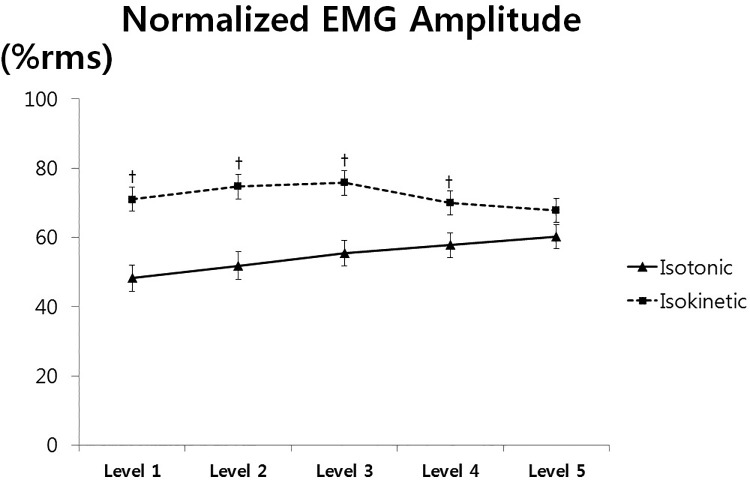
During isotonic exercise, 1-way repeated-measures ANOVA indicated that the normalized EMG amplitude of the IS at level 5 was greater than that at level 1 (P < .001) or 2 (P = .008). During isokinetic exercise, IS muscle activity at levels 2 and 3 was higher than that at level 4 or 5 (levels 2 vs 4: P = .020; levels 2 vs 5: P = .006; levels 3 vs 4 and 3 vs 5: P < .001 for both). Statistical differences were found between the isotonic and isokinetic modes at levels 1, 2, 3, and 4 (levels 1-3: P < .001; level 4: P = .003) (Figure 3).
Figure 3.
Normalized electromyographic amplitudes of the infraspinatus muscle versus loads (isotonic exercise) or velocities (isokinetic exercise). †Statistically significant difference between the exercises. EMG, electromyography.
Total Work
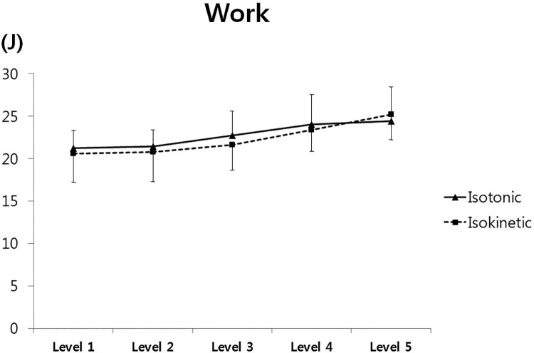
During isotonic exercise, tWK at level 5 was significantly greater than that at level 1 (P = .001), 2 (P < .001), or 3 (P = .008), and the tWK at level 4 was also greater than that at level 1, 2, or 3 (P < .001). The tWK at level 3 was greater than that at lower levels (P < .001). During isokinetic exercise, tWK decreased significantly from level 5 to 3 (P = .02), from level 5 to 2 or 1 (P < .001), from level 4 to 3 (P = .027), and from level 4 to 2 or 1 (P < .001). No statistical differences in tWK were detected between the isotonic and isokinetic modes at any level investigated (Figure 4).
Figure 4.
Work versus loads (isotonic exercise) or velocities (isokinetic exercise). No statistical difference was detected between the exercises at any level.
EMG(IS)/tWK Ratio
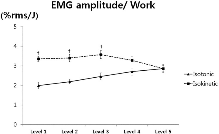
During isotonic exercise, the EMG(IS)/tWK ratio decreased significantly from level 5 to 3 (P = .003), 2 (P < .001), and 1 (P < .001); from level 4 to 2 (P < .001) and 1 (P < .001); and from level 3 to 1 (P < .001). During isokinetic exercise, the EMG(IS)/tWK ratio at level 3 was greater than that at any other level except level 1 (level 2: P = .044; level 4: P = .019; level 5: P = .008). The EMG(IS)/tWK ratio at level 5 was lower than that at any other level (level 1: P = .017; level 2: P = .012; level 3: P = .020; level 4: P = .029). Statistical differences were found between the isotonic and isokinetic modes at levels 1, 2, and 3 (level 1: P = .005; level 2: P = .006; level 3: P = .023) (Figure 5).
Figure 5.
Amplitude-to-work ratios versus loads (isotonic exercise) or velocities (isokinetic exercise) of the infraspinatus muscle. †Statistically significant difference between the exercises. EMG, electromyography; RMS, root mean square.
IS/PD Activation Ratio
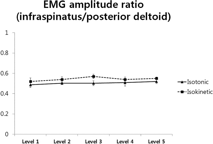
One-way repeated-measures ANOVA did not indicate any significant differences between the levels for both isotonic and isokinetic exercises. Similarly, no differences between the isotonic and isokinetic modes were detected at any level investigated (Figure 6).
Figure 6.
Activation ratios of the infraspinatus and posterior deltoid muscles versus loads (isotonic exercise) or velocities (isokinetic exercise). No significant difference was detected between the exercises at any level. EMG, electromyography.
Discussion
The primary purpose of this study was to evaluate the efficacy of external rotation exercises using surface EMG and a dynamometer. Our study showed that the highest IS amplitude was recorded at 180 and 240 deg/s during isokinetic exercise. On the other hand, isotonic exercise led to an increase in IS amplitude under higher intensity loads. Theoretically, isokinetic concentric exercise induces constant EMG amplitudes across velocities, as all available motor units are recruited and firing at their optimal frequencies.1 However, there are conflicting results about velocity-related EMG responses during concentric isokinetic muscle actions.17,18,21,27 In a study evaluating EMG activity of the quadriceps femoris muscle at 4 isokinetic speeds (30, 60, 90, and 120 deg/s), no significant differences in muscle activities across speeds were detected, and the authors concluded that the participants made equivalent efforts at all speeds.18 The induction of higher muscle activities at middle to high angular velocities compared with low angular velocities was noted in the current study as well as the study by Purkayastha et al,17 in which an increase of knee extensor activities from 60 to 180 and 240 deg/s was observed. An increase in EMG values to velocities between 45 and 180 deg/s has been reported during knee extension.21 A possible explanation of why low angular velocity induces lower muscle activation is that neural drive reduction may protect the musculoskeletal system from injuries under high-tension loading conditions.27 Based on our clinical experience, external shoulder rotation at low angular velocity becomes burdensome even in healthy participants; therefore, an unconscious neural response to protect the rotator cuff seems plausible.
Work is defined as the product of force × distance.7 In theory, if the distance is constant (40° external rotation in this study), work is purely dependent on force. There was a trend toward a tWK increase during both isotonic and isokinetic exercises as the intensity level increased from 1 to 5. However, no significant differences were detected in tWK between levels 1 and 2 or levels 4 and 5 in either isotonic or isokinetic exercises. This implies that force production at low or high levels fails to elaborately respond to the given external intensities. One possibility is that participants failed to complete the given distance at level 5. Although we did not measure the range of motion achieved during the exercises, a decrease in the range of motion and an increase in intensities have been reported in previous studies.10,17 Purkayastha et al17 demonstrated an 18% decrease in the range of motion during isotonic knee extension exercise with increased intensity, which was more prominent at the highest intensity. Sustained tWK during external rotation exercises was used in this study, as the isolation of work performed by the IS was not feasible. To investigate possible problems in adopting tWK, we examined the IS/PD activation ratio at each level, which reflects the IS and PD contributions to tWK. Throughout all levels, regardless of the exercise mode, no significant differences were observed in the IS/PD activation ratios. Constant IS and PD contributions to tWK at all levels would justify the adoption of tWK in evaluating the efficacy of exercises on IS activation.
Currently, there is a lack of evidence to support the effectiveness of isokinetic exercise for shoulder joint rehabilitation.2,16,24 A systematic review demonstrated the effect of isokinetic training; however, most of the studies could not identify the isolated effect of isokinetic training.26 In isokinetic exercise training, there is no established protocol for angular velocity. When considering the EMG(IS)/tWK ratio, isokinetic exercise at 180 deg/s seemed to be most effective in inducing higher IS muscle activation per unit of work. However, incorporating isokinetic training at 180 deg/s into the rehabilitation program is complicated. Although the early adoption of 180 deg/s may be relatively safe for patients with rotator cuff tendinopathy, this may not be the case for patients who undergo rotator cuff repair. Boissonnault et al2 recommended the postoperative incorporation of isokinetic exercise at 8 weeks in patients undergoing rotator cuff repair. Nevertheless, current evidence suggests that early motion increases the risk of rotator cuff retears.11 A careful investigation on the optimal timing for the adoption of isokinetic strengthening exercise is needed. In the early phase of rehabilitation, static exercises, such as inferior glide or low row, are safer for protecting repaired or inflamed rotator cuffs because of the limited range of motion during these exercises.
Various shoulder postures have been studied to identify the most beneficial for enhancing IS activation. External rotation with the shoulder at 0° of abduction has been reported to lead to the most effective IS isolation, maximizing IS activation and minimizing PD activation.18 Another study also demonstrated that the IS showed greatest activation during external rotation at 0° of abduction.14 In this study, we adopted 20° of abduction and forward flexion, as participants comfortably positioned their arms and performed external rotation while sitting in a Biodex dynamometer. Although experimental studies have reported 0° of abduction as the most effective angle to increase the IS/PD activation ratio, slight changes in posture should also be considered so the patient is comfortable during external shoulder rotation exercise using a dynamometer.
Limitations
There are several limitations that warrant further review. First, pure work generated by the IS was not used as a denominator in calculating EMG/WK ratios. Because the extraction of IS pure work from tWK was not feasible, tWK was used as a denominator. In this study, a consistency in the IS/PD activation ratio at all intensities was noted; therefore, the percentage of tWK contributed by the IS may be consistent. Second, this study adopted the classification of intensity levels from knee extension exercises,17 which has not been validated for the shoulder. For a comparison of isotonic and isokinetic exercises, standardization to equalize angular velocity and tWK is necessary.9 As tWK at each level was not significantly different between the exercises, the classification of intensity levels may be applicable to the shoulder. However, further research is needed to validate the method adopted in this study.
A third limitation was that the distribution of muscle activations was relatively uneven through our study, despite the fact that only male participants were included. Other factors besides sex may also affect muscle activations. Physical characteristics such as body fat percentage and muscle mass should be relatively uniform to minimize the standard deviation of muscle activations. We did not consider these factors in this study; hence, we could not provide the minimal clinically important difference for stimulating muscle adaptation to resistance using the distribution-based method. In addition, our methodological approach for shoulder rehabilitation was experimental, as little has been known for isokinetic exercise of the shoulder. Therefore, with limited data, we could not provide clinical relevance regarding the resistance levels adopted in this study or safe loads without damaging the rotator cuff. Future studies will be needed to justify the usefulness of isokinetic shoulder exercise in various aspects.
Conclusion
This study demonstrated statistical differences in the normalized IS amplitude between exercise modes at all investigated intensity levels except for level 5. Therefore, isokinetic resistance may provide more effective stimulation of the IS muscle compared with isotonic resistance. The normalized IS amplitude increased under higher intensity loads during isotonic exercise, while angular velocities of 180 and 240 deg/s induced higher IS activation than lower angular velocities during isokinetic exercise. Regarding exercise efficacy, as measured by the EMG/WK ratio, our data indicate that isokinetic exercise at an angular velocity of 180 deg/s induced higher IS activation per unit of work.
Footnotes
Final revision submitted February 13, 2020; accepted February 26, 2020.
The authors declared that there are no conflicts of interest in the authorship and publication of this contribution. AOSSM checks author disclosures against the Open Payments Database (OPD). AOSSM has not conducted an independent investigation on the OPD and disclaims any liability or responsibility relating thereto.
Ethical approval for this study was obtained from the Konkuk University Medical Center Institutional Review Board (No. KUH1060170).
References
- 1. Bitter NL, Clisby EF, Jones MA, Magarey ME, Jaberzadeh S, Sandow MJ. Relative contributions of infraspinatus and deltoid during external rotation in healthy shoulders. J Shoulder Elbow Surg. 2007;16(5):563–568. [DOI] [PubMed] [Google Scholar]
- 2. Boissonnault WG, Badke MB, Wooden MJ, Ekedahl S, Fly K. Patient outcome following rehabilitation for rotator cuff repair surgery: the impact of selected medical comorbidities. J Orthop Sports Phys Ther. 2007;37(6):312–319. [DOI] [PubMed] [Google Scholar]
- 3. Brown LE. Isokinetics in Human Performance. Human Kinetics; 2000. [Google Scholar]
- 4. Coffey TH. Delorme method of restoration of muscle power by heavy resistance exercises. Treat Serv Bull. 1946;1(2):8–11. [PubMed] [Google Scholar]
- 5. Cram JR, Kasman GS, Holtz J. Introduction to Surface Electromyography. Aspen Publishers; 1998. [Google Scholar]
- 6. Criswell E. Cram’s Introduction to Surface Electromyography. 2nd ed Jones and Bartlett Publishers; 2011. [Google Scholar]
- 7. Cutnell JD, Johnson KW. Physics. 8th ed John Wiley & Sons; 2009. [Google Scholar]
- 8. Edwards P, Ebert J, Joss B, Bhabra G, Ackland T, Wang A. Exercise rehabilitation in the non-operative management of rotator cuff tears: a review of the literature. Int J Sports Phys Ther. 2016;11(2):279–301. [PMC free article] [PubMed] [Google Scholar]
- 9. Guilhem G, Guevel A, Cornu C. A standardization method to compare isotonic vs. isokinetic eccentric exercises. J Electromyogr Kinesiol. 2010;20(5):1000–1006. [DOI] [PubMed] [Google Scholar]
- 10. Hinson M, Rosentswieg J. Comparative electromyographic values of isometric, isotonic, and isokinetic contraction. Res Q. 1973;44(1):71–78. [PubMed] [Google Scholar]
- 11. Houck DA, Kraeutler MJ, Schuette HB, McCarty EC, Bravman JT. Early versus delayed motion after rotator cuff repair: a systematic review of overlapping meta-analyses. Am J Sports Med. 2017;45(12):2911–2915. [DOI] [PubMed] [Google Scholar]
- 12. Keenan KG, Farina D, Maluf KS, Merletti R, Enoka RM. Influence of amplitude cancellation on the simulated surface electromyogram. J Appl Physiol (1985). 2005;98(1):120–131. [DOI] [PubMed] [Google Scholar]
- 13. Kendall FP, McCreary EK, Provance PG, Rodgers MM, Romani WA. Muscles: Testing and Function With Posture and Pain. 5th ed Lippincott Williams & Wilkins; 2005. [Google Scholar]
- 14. Kurokawa D, Sano H, Nagamoto H, et al. Muscle activity pattern of the shoulder external rotators differs in adduction and abduction: an analysis using positron emission tomography. J Shoulder Elbow Surg. 2014;23(5):658–664. [DOI] [PubMed] [Google Scholar]
- 15. Malliou PC, Giannakopoulos K, Beneka AG, Gioftsidou A, Godolias G. Effective ways of restoring muscular imbalances of the rotator cuff muscle group: a comparative study of various training methods. Br J Sports Med. 2004;38(6):766–772. [DOI] [PMC free article] [PubMed] [Google Scholar]
- 16. Martin DR, Garth WP., Jr Results of arthroscopic debridement of glenoid labral tears. Am J Sports Med. 1995;23(4):447–451. [DOI] [PubMed] [Google Scholar]
- 17. Purkayastha S, Cramer JT, Trowbridge CA, Fincher AL, Marek SM. Surface electromyographic amplitude-to-work ratios during isokinetic and isotonic muscle actions. J Athl Train. 2006;41(3):314–320. [PMC free article] [PubMed] [Google Scholar]
- 18. Rothstein JM, Delitto A, Sinacore DR, Rose SJ. Electromyographic, peak torque, and power relationships during isokinetic movement. Phys Ther. 1983;63(6):926–933. [DOI] [PubMed] [Google Scholar]
- 19. Ryan G, Johnston H, Moreside J. Infraspinatus isolation during external rotation exercise at varying degrees of abduction. J Sport Rehabil. 2018;27(4):334–339. [DOI] [PubMed] [Google Scholar]
- 20. Schmitz RJ, Westwood KC. Knee extensor electromyographic activity-to-work ratio is greater with isotonic than isokinetic contractions. J Athl Train. 2001;36(4):384–387. [PMC free article] [PubMed] [Google Scholar]
- 21. Seger JY, Thorstensson A. Muscle strength and myoelectric activity in prepubertal and adult males and females. Eur J Appl Physiol Occup Physiol. 1994;69(1):81–87. [DOI] [PubMed] [Google Scholar]
- 22. Singh B, Bakti N, Gulihar A. Current concepts in the diagnosis and treatment of shoulder impingement. Indian J Orthop. 2017;51(5):516–523. [DOI] [PMC free article] [PubMed] [Google Scholar]
- 23. Solomonow M, Baten C, Smit J, et al. Electromyogram power spectra frequencies associated with motor unit recruitment strategies. J Appl Physiol (1985). 1990;68(3):1177–1185. [DOI] [PubMed] [Google Scholar]
- 24. Thomas M, Grunert J, Standtke S, Busse MW. [Rope pulley isokinetic system in shoulder rehabilitation: initial results]. Z Orthop Ihre Grenzgeb. 2001;139(1):80–86. [DOI] [PubMed] [Google Scholar]
- 25. Thompson WO, Debski RE, Boardman ND, 3rd, et al. A biomechanical analysis of rotator cuff deficiency in a cadaveric model. Am J Sports Med. 1996;24(3):286–292. [DOI] [PubMed] [Google Scholar]
- 26. Wang TL, Fu BM, Ngai G, Yung P. Effect of isokinetic training on shoulder impingement. Genet Mol Res. 2014;13(1):744–757. [DOI] [PubMed] [Google Scholar]
- 27. Westing SH, Cresswell AG, Thorstensson A. Muscle activation during maximal voluntary eccentric and concentric knee extension. Eur J Appl Physiol Occup Physiol. 1991;62(2):104–108. [DOI] [PubMed] [Google Scholar]
- 28. Wilk KE, Reinold MM, Dugas JR, Arrigo CA, Moser MW, Andrews JR. Current concepts in the recognition and treatment of superior labral (SLAP) lesions. J Orthop Sports Phys Ther. 2005;35(5):273–291. [DOI] [PubMed] [Google Scholar]


