Figure 1.
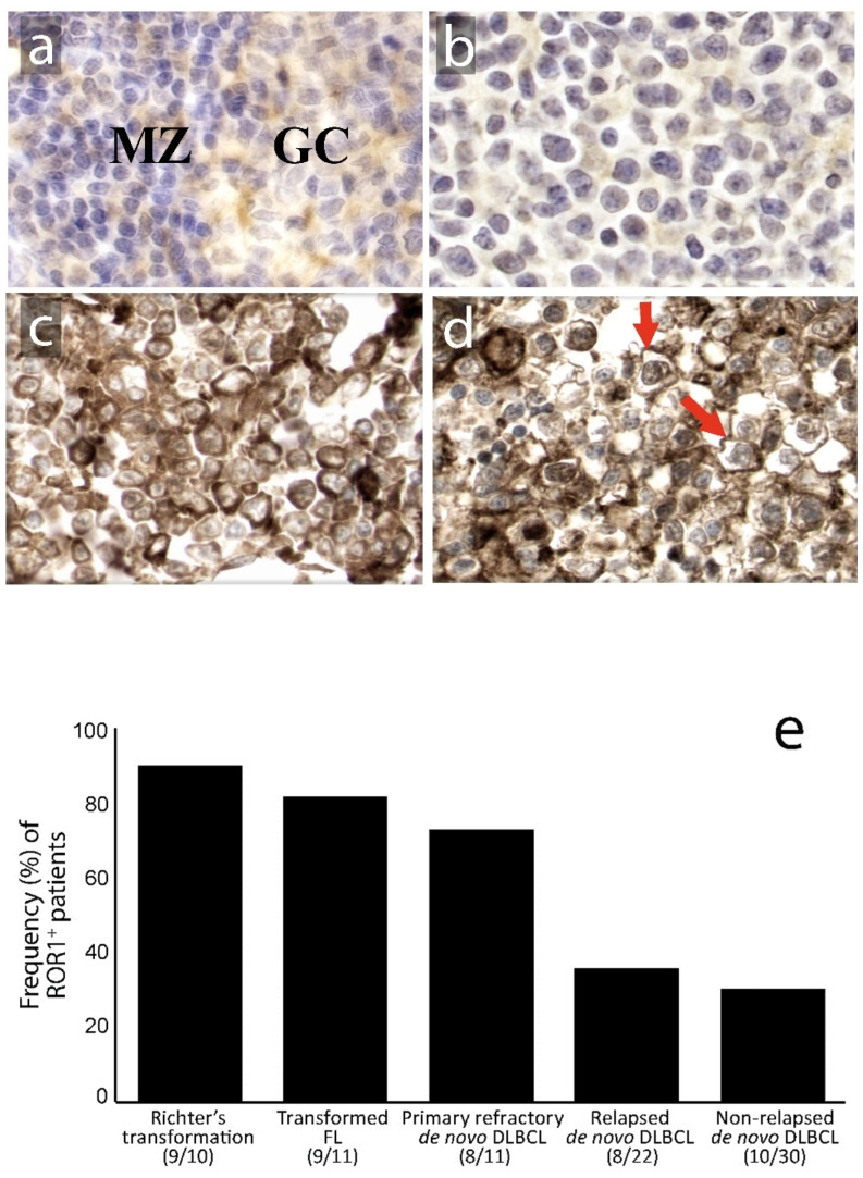
Expression (Immunohistochemical (IHC)) of the ROR1 protein in reactive lymphoid tissue and diffuse large B-cell lymphoma (DLBCL) lymphoma tissues. (magnification × 400; 3,3’-Diaminobenzidine (DAB) as chromogen, hematoxylin as counterstain). ROR1 was expressed (a) weakly in the cytoplasm of a subset of centroblasts in the germinal center (GC) and rare small lymphocytes in the mantle zone (MZ) of reactive lymph nodes; (b) representative case of DLBCL negative for ROR1; (c) DLBCL positive for ROR1 with predominantly cytoplasmic staining and (d) with a predominantly membranous staining pattern (red arrows); (e) ROR1 expression in tumor cells was more often observed in primary refractory DLBCL, Richter’s syndrome and transformed follicular lymphoma than in relapsed and non-relapsed DLBCL patients (p < 0.001, Chi-Square test). Numbers in brackets represent the number of ROR1-positive cases compared to the total number of cases in each group.

