Abstract
Background:
Leonardo de Vinci contributed several observations and drawings on facial proportion and the lower one third of the face. Many facial and body measurements to determine vertical dimension at occlusion. These facial measurements can be implemented in construction of complete denture patients.
Aim:
This study aims to correlate the vertical dimension at occlusion to 13 anthropometric measurements. Then correlating, which measurement is more accurate to the vertical dimension at occlusion.
Methodology:
20 male and female subjects were selected. Vertical dimension at occlusion and 12 anthropometric parameters were measured.
Results and Conclusion:
Twice the length of the eye and distance between the tip of the thumb and tip of the index finger is closest to the vertical dimension at occlusion in male patients and that vertical distance from the pupil to corner of the mouth, vertical height of the ear is closest to the vertical dimension at occlusion in female patients.
Keywords: Anthropometric parameters, divine proportion, vertical dimension
Introduction
Although advances in techniques and materials are being made in prosthodontics, still no accurate method of assessing the vertical dimension of occlusion in edentulous patients is available to dentists.[1] Determining the vertical dimension of occlusion is a critical procedure for a totally or partially edentulous patient. Many edentulous patients have adapted to a vertical dimension which has decreased due to bone resorption and posterior tooth wear.[2] While the considerable importance of a proper vertical dimension of occlusion is generally accepted,[3] the means of obtaining it are considered unreliable by some investigators,[4] and some authors recommend the use of “clinical judgment.“ The importance of the problem seems incompatible with the great diversity of methods for obtaining the vertical dimension of occlusion and the reliance on something as vague and incommunicable as “clinical judgment.“ Many factors have been suggested as responsible for the ambiguities associated with such measurements and calculations, which include difficulties in obtaining measurements on the skin of the face and the range of variability in physiologic and pathologic states.[5]
A variety of techniques have been proposed to determine measurements for the correct vertical dimension of occlusion.[3,4,5,6] When selecting the best method to use, criteria to be considered are accuracy and repeatabilityof the measurement, adaptability of the technique, type and complexity of the equipment needed, and the length of time required to secure the measurement.[5]
Irving M. Sheppard and Stephen M. Sheppard did a study to determine some of the characteristics of the described changes in the vertical dimension of mandibular rest position. In addition, it includes a comparison of facial and skeletal measurements and a comparison of conventionally obtained rest space with that of existing functioning dentures.[5]
Though there are many methods to establish vertical dimension at rest position but in geriatric patients where there is difficulty in establishing physiologic rest position, many facial and body measurements determine vertical dimension at occlusion. These facial measurements can be implemented in construction of complete denture patients.[6]
The current study aims at establishing the vertical dimension so that after the replacement of teeth the facial form and contour can be maintained. Since these markings, facial landmarks and dimensions are easily reproducible, therefore these are of utmost importance for the general public to maintain their facial form and contour and as well as important for the general dentist to reproduce the same dimensions again and again.
Materials and Method
Subjects
Thirty dentulous male subjects and 20 dentulous female were selected for this study, the subjects were screened and selected from the outpatient and students of Awadh dental college and hospital, Jamshedpur.
Inclusion criteria
Full complement of teeth (third molar excluded)
Age group of subjects between 18 and 25 years
Exclusion criteria
No history of orthodontic treatment
Malocclusion
Attrited teeth
Facial asymmetry
Armamentarium required
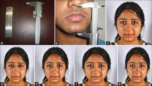
Vernier calliper [Figure 1a]
Figure 1.
(a) Vernier calliper. (b and c) The vertical dimension at occlusion. (d) Horizontal distance between pupils. (e) Vertical height of the eyebrow to the ala of the nose (f) Vertical distance from pupil of the eye to the corner of mouth. (g) Vertical height of the nose
Metal scale
The Study
This study aims to correlate the vertical dimension at occlusion to 13 anthropometric measurements. Then correlating, which measurement is more accurate to the vertical dimension at occlusion.
The vertical dimension at occlusion was established by two points—subnasion to gnathion, when subjects were in centric occlusion [Figure 1b].
The anthropometry parameters to which vertical dimension at occlusion was compared were:
Horizontal distance between pupils [Figure 1c].
Vertical distance from the external corner of the eye to the corner of mouth [Figure 1d].
Vertical height of the eyebrow to the ala of the nose [Figure 1e].
Vertical height of the nose (subnasion to glabella) [Figure 1f].
Distance from one corner of the lip to the other, following curvature of the mouth [Figure 1g].
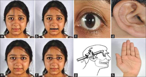
Distance from the eyebrow line to the hair line (in female) [Figure 2a].
Distance from the outer corner of the eye to the inner corner [Figure 2b].
Vertical height of the ear [Figure 2c].
Distance between the tip of the thumb and the tip of the index finger when the hand lays flat [Figure 2d].
Twice the length of one eye [Figure 2e].
Twice the distance between the inner canthus of the eye to the other [Figure 2f].
Distance between the outer canthus and ear [Figure 2g].
Figure 2.
(a) Distance from the eyebrow line to the hair line (in female). (b) Distance from the one corner of the lip to the other. (c) the length of one eye. (d) Vertical height of the ear. (e) distance between the inner canthus of the eye to the other. (f) Distance from the outer corner of the eye to the inner corner. (g) Distance between the outer canthus and ear. (h) Distance between the tip of the thumb and the tip of the index finger when the hand lays flat
Results
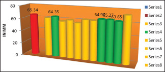
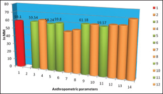
Statistical analysis was done to compare the relationship of vertical dimension at occlusion to 12 anthropometric parameters using unpaired sample t-test. The results for male patients are tabulated in Table 1 and graphically represented in Graph 1 and the results obtained for female patients are tabulated in Table 2 and graphically represented in Graph 2.
Table 1.
Statistical analysis of male patients
| Mean | SD | P | |
|---|---|---|---|
| Vertical dimension on occlusion | 65.34 | 40.14 | |
| Horizontal distance between pupils | 61.68 | 2.68 | 0.000** |
| Vertical distance from the external corner of the eye to the corner of mouth | 64.35 | 3.48 | 0.238 |
| Vertical height of the eyebrow to the ala of the nose | 57.79 | 2.73 | 0.000** |
| Vertical height of the nose (subnasion to glabella) | 60.45 | 3.93 | 0.000** |
| Distance from the one corner of the lip to the other, following curvature of the mouth | 56.91 | 4.13 | 0.000** |
| Distance from the eyebrow line to the hair line (in female) | |||
| Distance from the outer corner of the eye to the inner corner | 62.70 | 2.76 | 0.004* |
| Vertical height of the ear | 62.42 | 2.81 | 0.004* |
| Distance between the tip of the thumb and the tip of the index finger when the hand lays flat | 64.97 | 7.00 | 0.791 |
| Twice the length of one eye | 65.27 | 3.86 | 0.944 |
| Twice the distance between the inner canthus of the eye to the other | 63.65 | 3.83 | 0.198 |
| Distance between the outer canthus and ear | 73.52 | 2.92 | 0.000** |
Note: **Denotes significant at <1% level * Denotes significant at <5% level no star- not significant
Graph 1.

Anthropometric parameters for male patients
Table 2.
Statistical analysis of female patients
| Mean | SD | P | |
|---|---|---|---|
| Vertical dimension on occlusion | 59.10 | 4.77 | |
| Horizontal distance between pupils | 59.54 | 3.46 | 0.754 |
| Vertical distance from the external corner of the eye to the corner of mouth | 62.26 | 2.41 | 0.211 |
| Vertical height of the eyebrow to the ala of the nose | 58.24 | 3.01 | 0.468 |
| Vertical height of the nose (subnasion to glabella) | 59.80 | 3.54 | 0.530 |
| Distance from the one corner of the lip to the other, following curvature of the mouth | 50.20 | 2.81 | 0.000** |
| Distance from the eyebrow line to the hair line (in female) | 53.17 | 4.61 | 0.003* |
| Distance from the outer corner of the eye to the inner corner | 61.18 | 4.17 | 0.169 |
| Vertical height of the ear | 61.23 | 3.49 | 0.111 |
| Distance between the tip of the thumb and the tip of the index finger when the hand lays flat | 59.57 | 4.73 | 0.722 |
| Twice the length of one eye | 62.43 | 2.77 | 0.008 |
| Twice the distance between the inner canthus of the eye to the other | 62.56 | 7.35 | 0.090 |
| Distance between the outer canthus and ear | 70.41 | 3.47 | 0.000** |
Note: **Denotes significant at <1% level *Denotes significant at <5% level no star- not significant
Graph 2.

Anthropometric parameters for female patients
The statistical analysis for male patients showed that twice the distance between the inner canthus of the eye to the other was most close to vertical dimension at occlusion followed by vertical distance from the external corner of the eye to the corner of mouth, distance between the tip of the thumb and the tip of the index finger when the hand lays flat and twice the length of one eye as they were not significant statistically [Table 1].
The statistical analysis for female patients showed that vertical height of the eyebrow to the ala of the nose was most close to vertical dimension at occlusion followed by vertical height of the ear, horizontal distance between pupils, vertical height of the nose (subnasion to glabella), and distance between the tip of the thumb and the tip of the index finger when the hand lays flat [Table 2].
Discussion
Leonardo de Vinci contributed several observations and drawings on facial proportion and the lower one-third of the face. Many facial and body measurements to determine vertical dimension at occlusion. These facial measurements can be implemented in construction of complete denture patients.[6]
Facial measurements are used in establishing vertical dimension. Ivy, according to Bowman and Chick, mentioned the use of facial measurements to determine vertical dimension for the edentulous patient. A good friend suggested that the distance from the pupil of the eye to the junction of the lips equalled that from the subnasion to the gnathion. However, Willishas has been given the credit for popularizing these measurements.[1,3]
McGee correlated the known vertical dimension of occlusion with three facial measurements which he claimed remain constant throughout life. The three measurements are: the distance from the center of the pupil of the eye to a line projected laterally from the median line of the lips; the distance from the glabella to the subnasion; and the distance between the angles of the mouth with the lips in repose. McGee stated that two of these three measurements will be invariably equal, and occasionally all three will be equal to one another. He claimed that, in 95% of his subjects with natural teeth, two or three of these measurements corresponded to the vertical dimension of occlusion.[1]
Ivy, a good friend, Willis suggested that the distance from the pupil of the eye to the junction of the lips equaled that from the subnasion to gnathion.
Knebleman in 1988 patented a method to determine the VDO. In his method, he observed that distance between subnasion to gnathion (vertical dimension at occlusion) is equal to the length between the outer canthus of the eye to ear.[7,8]
Facial measurements can be very useful in establishing vertical dimension where physiologic rest position is difficult to achieve in geriatric patients and patients with poor neuromuscular coordination.
In this study, we observed that vertical distance from the pupil to corner of the mouth, vertical height of the ear is closest to the vertical dimension at occlusion in female patients and twice the length of the eye and distance between the tip of the thumb and tip of the index finger is closest to the vertical dimension at occlusion in male patients.
Conclusion
From this study we can conclude that anthropometric measurements can help us in determining vertical dimension while constructing complete denture in geriatric patients, suffering from neuromuscular disorder.
Financial support and sponsorship
Nil.
Conflicts of interest
There are no conflicts of interest.
References
- 1.Turrell JW. Clinical assessment of vertical dimension. J Prosthet Dent. 2006;96:79–84. doi: 10.1016/j.prosdent.2006.05.015. [DOI] [PubMed] [Google Scholar]
- 2.Brian Toolson L, Smith DE. Clinical measurement and evaluation of vertical dimension. J Prosthet Dent. 2006;95:335–40. doi: 10.1016/j.prosdent.2006.03.013. [DOI] [PubMed] [Google Scholar]
- 3.Shanahan TEJ. Physiologic vertical dimension and centric relation. J Prosthet Dent. 2004;91:1–4. doi: 10.1016/j.prosdent.2003.09.002. [DOI] [PubMed] [Google Scholar]
- 4.Sheppard IM, Sheppard SM. Vertical dimension measurements. J Prosthet Dent. 2006;95:175–81. doi: 10.1016/j.prosdent.2006.01.004. [DOI] [PubMed] [Google Scholar]
- 5.Use of facial measurements in determining vertical dimension. J Am Dent Assoc. 1947;35:342–50. doi: 10.14219/jada.archive.1947.0361. [DOI] [PubMed] [Google Scholar]
- 6.Misch CE. Vertical occlusal dimension by facial measurements Continuum. Misch Implant Institute Newsletter. 1997 [Google Scholar]
- 7.Toolson LB, Smith DE. Clinical measurement and evaluation of vertical dimension. J Prosthet Dent. 1982;47:236–41. doi: 10.1016/0022-3913(82)90147-0. [DOI] [PubMed] [Google Scholar]
- 8.Majeed MI, Haralur SB, Khan MF. An anthropometric study of cranio-facial measurements and their correlation with vertical dimension of occlusion among Saudi Arabian subpopulations. Open Access Maced J Med Sci. 2018;6:680–6. doi: 10.3889/oamjms.2018.082. [DOI] [PMC free article] [PubMed] [Google Scholar]


