Abstract
Patient: Female, 38-year-old
Final Diagnosis: Peritonitis
Symptoms: Abdominal and/or epigastric pain
Medication:—
Clinical Procedure: —
Specialty: Infectious Diseases
Objective:
Rare disease
Background:
Mycobacterium abscessus is one of the most important mycobacteria, but its associated peritonitis in patients on continuous ambulatory peritoneal dialysis (CAPD) appears relative rare, and the treatment regimen of the antibiotics are still unclear.
Case Report:
A 38-year-old female with chronic glomerulonephritis on CAPD who was diagnosed with M. abscessus-associated peritonitis. Symptoms exacerbated despite treatment with a 3-antibiotic regimen combining clarithromycin, imipenem/cilastatin (IPM/CS), and minocycline (MINO). However, after changing IPM/CS and MINO to linezolid (LZD), her condition and inflammation improved, and she was able to be maintained on oral tedizolid (TZD).
Conclusions:
Oxazolidinones such as LZD and TZD might be candidate antibiotics for the treatment of M. abscessus-associated diseases with chronic renal failure due to their immunomodulatory effects and non-renal excretion.
MeSH Keywords: Antibiotics, Antitubercular; Immunocompromised Host; Mycobacterium Infections, Nontuberculous; Renal Insufficiency, Chronic
Background
Non-tuberculous mycobacteria (NTM)-associated peritonitis during continuous ambulatory peritoneal dialysis (CAPD) is considered relative rare although peritonitis is one of the most serious complications associated with peritoneal dialysis [1].
Peritonitis is a frequent complication of CAPD and most of the episodes of peritonitis are caused by touch contamination of the dialysis tubing or by extension of the catheter exit site or tunnel infection [2]. Coagulase-negative and coagulase-positive staphylococci are the 2 most common organisms, accounting for 50% or more of all CAPD peritonitis, and other Gram-positive and Gram-negative bacteria and fungi account for the rest. Intraperitoneal antibiotic treatments are usually effective in eradicating the infection, and the choice of antibiotics depends on organisms isolated from cultured dialysate.
Furthermore, Tian et al. reported that they had 31 patients die among 2917 CAPD patients during the study period from 2002 to 2014 and that the most common causes of CAPD-related death were cerebrovascular disease (29.0%) and infection (19.4%) [3]. They also found similar common pathogens, such as Gram-positive bacteria followed by Gram-negative bacteria in CAPD peritonitis
Here, we report a case of definite Mycobacterium abscessus associated peritonitis in a woman on CAPD treated by linezolid (LZD) and tedizolid (TZD) in addition to clarithromycin (CAM). This case and related study were approved by the Committee for Clinical Scientific Research of Tohoku Medical and Pharmaceutical University Hospital in October 9, 2015 as No. ID2015-2-023 and the patients whose specimens were used provided written informed consent to have the case details and any accompanying images published.
Case Report
A 38-year-old female developed renal failure and glomerulonephritis and mainly received immunosuppressive agents such as prednisolone, tacrolimus, and cyclophosphamide. However, extensive chronic renal failure subsequently developed.
During the 2 years following onset of chronic kidney disease, she experienced repeated infectious diseases, including cytomegalovirus and aspergillosis. Kidney function was gradually worsened due to long-term administration of nephrotoxic agents, including tacrolimus (controlled concentration range: 5–10 ng/mL), voriconazole, and valganciclovir. CAPD was started after 2 years of follow-up on an outpatient basis.
The patient experienced bacterial peritonitis several times mainly due to Staphylococcus aureus including methicillin resistant S. aureus (MRSA) and received some antibiotics, such as cefazolin 1 g/day, levofloxacin 0.5 g/day, and vancomycin 1 g/day in the first year of CAPD. Each antibiotic was used 2 to 4 weeks every day or every other day, and her condition had improved. However, her peritoneal tube has not removed because the patient requested peritoneal dialysis and rejected the change to hemodialysis.
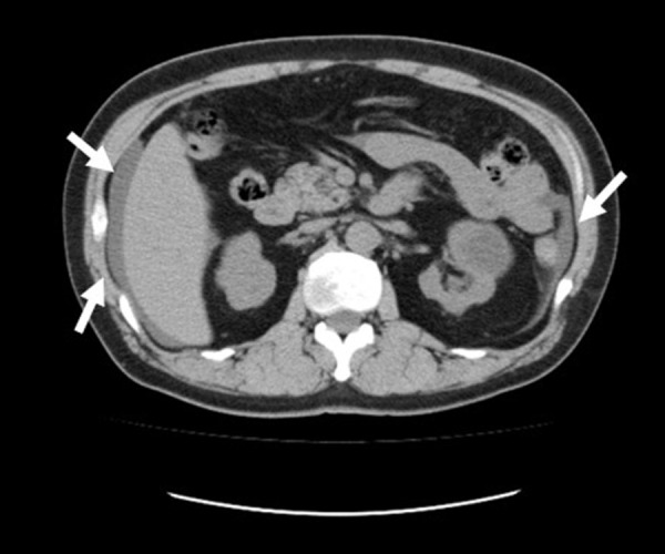
She was admitted to our hospital because of turbidity in the peritoneal drainage fluid with fever and abdominal pain after 18 months on CAPD administration although levofloxacin was received orally in the outpatient clinic for 1 week. Physical examination showed slight tenderness around the umbilicus in abdomen. Her blood pressure was 120/70 mmHg, temperature of 37.2°C, respiratory rate of 18 breaths per minute, and consciousness level of E4V5M6 on the Glasgow coma scale (GCS). Laboratory data were as follows: white blood cell (WBC) count, 6.1×10³/µL with 76.5% neutrophils, 11.3% lymphocytes, 9.3% monocytes, 0.6% basophils, and 2.3% eosinophils; platelet count, 224×103/µL; hemoglobin, 7.6 g/dL; serum creatinine, 7.85 mg/dL; blood urea nitrogen, 51 g/L; uric acid, 5.1 mg/dL; and C-reactive protein, 2.16 mg/dL. Peritoneal fluid contained WBCs (2234/µL; 4% mononuclear cells, 96% polymorphonuclear cells). We found slight pus and erythema at the peritoneal dialysis catheter exit site and the ascites and thickening of the peritoneum with edema were shown in the computed tomography of the abdomen (Figure 1).
Figure 1.

Abdominal computed tomography of the patients. Slight ascites and thickening of the peritoneum with edema were found (arrows).
We did not find the other pathological changes, including the intra-abdominal abscess and the perforation of intestines, therefore, CAPD-associated peritonitis was diagnosed and peritoneal fluid from the peritoneal tube and blood were collected as culture specimens. Treated with 1 g of intravenous meropenem on alternate days in combination with intraperitoneal administration of cefazolin 1 g/day and ceftazidime 1 g/day were started empirically. Although these antibiotic were continued for a week, WBC in the peritoneal fluid were increased to 2796/µL and the condition of the patient deteriorated with vomiting and abdominal pain, and high fever of 38.3°C was found, although her blood pressure of 116/60 mmHg, respiratory rate of 19 breaths per minute, and consciousness level was E4V5M6 of GCS had not worsen. Peritoneal fluid showed WBC counts in her peritoneal fluid and serum C-reactive protein were increased to 3463/µL and 3.72 mg/dL, respectively.
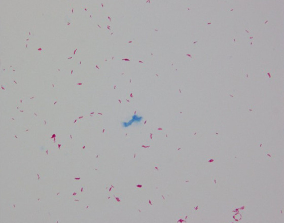
Bacterial cultures, including blood and sputum, which are collected on admission routinely to rule out the bacteremia and pneumonia due to resistant pathogens, yielded negative results on day 3 post-admission, but acid-fast bacilli were identified from smears of peritoneal fluid (Figure 2). M. abscessus was collected after culture of peritoneal fluid and was confirmed by amino acid analysis using Time-of-Flight Mass Spectrometer (TOF-MS), leading to definitive diagnosis of M. abscessus associated CAPD peritonitis.
Figure 2.

Light microscopy showing Ziehl-Nelsen staining in the peritoneal fluid. Red and thin stained acid-fast bacilli were detected (×1000).
Because a severe condition of the patient was suspected, the peritoneal dialysis catheter was removed, and she was started on hemodialysis. In addition, the combination antibiotic therapy comprising oral CAM at 800 mg, intravenous imipenem/cilastatin (IPM/CS) 1 g, and minocycline (MINO) 200 mg was started and continued for more 1 week. However, her condition and laboratory data worsened, and intra-abdominal fluid cell numbers were still high at 3014/µL. We then changed 2 of the 3 antibiotics, removing IPM/CS and MINO, and adding intravenous LZD at 1.2 g/day because the agent was known as immunomodulation, and non-dose adjustment necessary even in patients with renal failure.
Her condition subsequently improved, and WBC in the perito-neal fluid decreased to 1876/µL and C-reactive protein in the serum returned to 1.26 mg/dL. More than 2 weeks later, we found decreased of platelet counts in the blood from 2.2×10⁵/µL to 1.0×10⁵/µL, which is a well- known side effect of LZD, therefore, we changed intravenous LZD to oral TZD, an oxazolidinone similar to LZD but not associated with thrombocytopenia, and found that her condition remained stable and her WBC in the peritoneal fluid and C-reactive protein in the serum decreased to 1072/µL and 0.17 mg/dL, respectively, although the drug sensitivity of M. abscessus had shown susceptibility to CAM, but not LZD (Table 1).
Table 1.
Drug susceptibilities of Mycobacterium abscessus isolated from peritoneal fluid.
| No. | Drugs | MIC | S/I/R |
|---|---|---|---|
| 1 | SM | 32 | R |
| 2 | EB | 32 | R |
| 3 | KM | 16 | R |
| 4 | RIP | >32 | R |
| 5 | LVFX | 2 | I |
| 6 | CAM | 0.125 | S |
| 7 | TH | >16 | R |
| 8 | AMK | 16 | R |
| 9 | IPM/CS | >32 | |
| 10 | MINO | >256 | |
| 11 | DAP | >256 | |
| 12 | LZD | >256 |
MIC – minimal inhibitory concentrations; S – susceptible; I – intermediate; R – resistance; SM – streptomycin; EB – ethambutol; KM – kanamycin; RIP – rifampicin; LVFX – levofloxacin; CAM – clarithromycin; TH – ethionamide; AMK – amikacin; IPM/CS – imipenem/cilastatin; MINO – minocycline; DAP – daptomycin; LZD – linezolid.
M. abscessus treatment was planned to be continued for half a year. She was discharged on day 40 because improvement of her condition was found. Her fever and WBC count in her peritoneal fluids were decreased to 36.8°C and 84/µL, respectively, although thickening of the peritoneum on computed tomography (CT) images remained; she continued the hemo-dialysis to avoid the recurrence of the peritonitis. One year after discharge, the patient remains well without recurrence of M. abscessus infection.
Discussion
M. abscessus is the most important rapidly growing mycobacteria, and 80% of chronic pulmonary diseases were caused by this rapid growth-type of mycobacterium. However, mycobacteria are also responsible for extrapulmonary disease, especially in terms of cutaneous and surgery-related infections [1,4,5].
We previously reported M. avium complex-associated peritonitis in patients on CAPD, but this condition appears to be relative rare [6]. It has been reported that most cases of NTM peritonitis occur in patients with immunosuppression underlying disease, such as systemic lupus erythematosus, diabetes, human immunodeficiency virus infection, and CAPD among the cases of peritoneal dialysis associated NTM peritonitis [7]. In our case, peritoneal dialysis may have been the most important risk factor of M. abscessus infection, and our patient’s low-immunological condition might be related to her severe infection.
Abdominal M. abscessus infections related with CAPD are extremely rare and few cases have been reported [8–10] (Table 2). Ding et al. analyzed 11 patients with abdominal NTM infections identified during a 7-year period, and found immuno-compromised status (liver cirrhosis, malignancy, acquired immunodeficiency syndrome) was noted in 10 patients (91%); however, none of the patients who developed NTM peritonitis had received CAPD although M. abscessus played a major role (27% of cases) [8].
Table 2.
Reported peritonitis cases where Mycobacterium abscessus was isolated.
| Study | Patients (n) | Male/Female | Age (yrs) | Underlying diseases | Major pathogen | Antibiotics | Survived/death |
|---|---|---|---|---|---|---|---|
| Ding et al. [8] | 11 | 6/5 | 64.5 | Malignancy (73%) | M. abscessus (27%) | Unknown | 1 patient survived more than 1 year |
| Kleinpeter and Krane [9] | 2 | 1/1 | 63/28 | Diabetes/nephritis | M. fortumtum/M. abscessus | CAM and/or AMK | Both survived |
| Yoshimura et al. [10] | 1 | 1/0 | 56 | Diabetes | M. abscessus | CAM and IPM/CS | Survived |
yrs – years; M. abscessus – Mycobacterium abscessus; M. fortumtum – Mycobacterium fortumtum; CAM – clarithromycin; AMK – amikacin; IMP/CS – imipenem/cilastatin.
M. abscessus is considered the most pathogenic, antibiotic-resistant of the mycobacteria [1,5]. Among the various antibiotics, macrolides such as CAM and azithromycin have usually been considered highly effective against M. abscessus. However, recent studies have suggested that more than 75% of isolates demonstrate inducible resistance to macrolides, including CAM [1,5]. IPM/CS data have varied markedly among studies and depend on the countries/regions [5]. Amikacin, an aminoglycoside, has consistently demonstrated good activity against M. abscessus, with susceptibilities usually greater than 90% [1,5]. However, amikacin is known for its renal side effects, and little is known about its in vivo efficacy and whether myco-bacterial minimal inhibitory concentrations (MICs) are reached within peritoneal fluid [7]. Ruth et al. found no clear role for MINO in the treatment of M. abscessus infection, because of the high MIC, rapid acquisition of drug resistance, and lack of synergistic effects with other antibiotics [11]. Fluoroquinolones have been the most widely available antibiotics, but increasing evidence suggests resistance to most representative drugs of this class after repeated use [5]. We therefore did not use levofloxacin although we found intermediate susceptibility of M. abscessus to levofloxacin later (Table 1).
Moxifloxacin, azithromycin, and cefoxitin might be good alterative candidates, however, we had already use levofloxacin, which was similar to moxifloxacin, and found less improvement. In addition, azithromycin is a macrolide, as is CAM which was already used, and cefoxitin is a beta-lactams as is IPM/CS which was already used, too. Therefore, we selected LZD, which was very different from the other agents we had already used that had poor effects clinically, although LZD showed less susceptibility in vivo.
LZD has been used as an emerging treatment option for systemic NTM infections. Susceptibility of M. abscessus to LZD varies from about 40% to 100% across various studies although the M. abscessus isolates showed multiple resistances to the other conventional drugs [4,12]. In our in vitro data, the isolated M. abscessus showed resistance to LZD. However, clinical improvement was seen after administration of LZD, which was started in place of IPM/CS and MINO.
LZD, one of the oxazolidinone antibiotics, is known as an immunomodulatory drug, and inhibitory effects on cytokines and inflammation have been reported [13]. Decreased levels of chemokines KC and MIP-2, and proinflammatory cytokine IFN-γ, TNFα, and IL-1β were found in the lungs of influenza-related community-acquired MRSA pneumonia mice models after treatment with LZD as compared to vancomycin [8].
These effects might have been sufficient in the present case, in addition to macrolides antibiotics such as CAM and azithromycin which are also known to exert immunomodulatory effects [14,15]. Furthermore, LZD is known to show very good penetration into most tissues in humans, and a full dose can be used even in patients with renal failure, including patients on CAPD as in the present case [16,17].
In addition, TZD has become available as an oxazolidinone with immunomodulatory effects similar to LZD but offering greater effectiveness at only half the dose of LZD [18,19]. We might be able to treat M. abscessus-associated peritonitis more successfully with CAM and LZD/TZD instead of conventional regimens comprising CAM, IMP/CS, and MINO combinations, because TZD has shown effectiveness against clinical isolates of M. abscessus [20].
Conclusions
We report a M. abscessus-associated peritonitis in a CAPD patient. We achieved improvement in this patient using treatment with LZD and TZD, even though drug susceptibility testing had shown resistance in vitro, while IPM/CS and MINO had resulted in worsening of her condition. Oxazolidinone antibiotics, especially TZD, might have immunomodulatory effects in addition to better tissue penetration and anti-bacterial activities, and longer half reduction time than LZD. Thus, these agents represent strong candidates for the treatment of M. abscessus-associated diseases in patients with chronic renal failure due to independence from kidney function.
Footnotes
Conflict of interest
None.
References:
- 1.Chen J, Zhao L, Mao Y, et al. Clinical efficacy and adverse effects of antibiotics used to treat Mycobacterium abscessus pulmonary disease. Front Microbiol. 2019;10:1977. doi: 10.3389/fmicb.2019.01977. [DOI] [PMC free article] [PubMed] [Google Scholar]
- 2.Saktayen MG. CAPD peritonitis. Incidence, pathogens, diagnosis, and management. Med Clin North Am. 1990;74:997–1010. doi: 10.1016/s0025-7125(16)30532-6. [DOI] [PubMed] [Google Scholar]
- 3.Tian Y, Xie X, Xiang S, et al. Risk factors and outcomes of high peritonitis rate in continuous ambulatory peritoneal dialysis patients: A retrospective study. Medicine (Baltimore) 2016;(2):e5569. doi: 10.1097/MD.0000000000005569. 95. [DOI] [PMC free article] [PubMed] [Google Scholar]
- 4.Bostan C, Slim E, Choremis J, et al. Successful management of severe post-LASIK Mycobacterium abscessus keratitis with topical amikacin and linezolid, flap ablation, and topical corticosteroids. J Cataract Refract Surg. 2019;45:1032–35. doi: 10.1016/j.jcrs.2019.03.001. [DOI] [PubMed] [Google Scholar]
- 5.Shen Y, Wang X, Jin J, et al. In vitro susceptibility of Mycobacterium abscessus and Mycobacterium fortuitum isolates to 30 antibiotics. Biomed Res Int. 2018;2018:4902921. doi: 10.1155/2018/4902941. [DOI] [PMC free article] [PubMed] [Google Scholar]
- 6.Miyashita E, Yoshida H, Mori D, et al. Mycobacterium avium complex-associated peritonitis with CAPD after unrelated bone marrow transplantation. Pediatr Int. 2014;56:e96–98. doi: 10.1111/ped.12463. [DOI] [PubMed] [Google Scholar]
- 7.Song Y, Wu J, Yan H, Chen J. Peritoneal dialysis-associated nontuberculous mycobacterium peritonitis: A systematic review of reported cases. Nephrol Dial Transplant. 2012;27:1639–44. doi: 10.1093/ndt/gfr504. [DOI] [PubMed] [Google Scholar]
- 8.Ding LW, Lai C, Lee LN, Hsueh PR. Abdominal nontuberculous mycobacterial infection in a university hospital in Taiwan from 1997 to 2003. J Formos Med Assoc. 2006;105:370–76. doi: 10.1016/S0929-6646(09)60132-7. [DOI] [PubMed] [Google Scholar]
- 9.Kleinpeter MA, Krane N. Treatment of mycobacterial exit-site infections in patients on continuous ambulatory peritoneal dialysis. Adv Perit Dial. 2001;17:172–75. [PubMed] [Google Scholar]
- 10.Yoshimura R, Kawanishi M, Fujii S, et al. Peritoneal dialysis-associated infection caused by Mycobacterium abscessus: A case report. BMC Nephrol. 2018;19(1):341. doi: 10.1186/s12882-018-1148-2. [DOI] [PMC free article] [PubMed] [Google Scholar]
- 11.Ruth MM, Sangen J, Pennings LJ, et al. Minocycline has no clear role in the treatment of Mycobacterium abscessus disease. Antimicrob Agents Chemother. 2018;62:e01208–18. doi: 10.1128/AAC.01208-18. pii: [DOI] [PMC free article] [PubMed] [Google Scholar]
- 12.Kim SY, Jhun B, Moon SM, et al. Genetic mutations in linezolid-resistant Mycobacterium avium complex and Mycobacterium abscessus clinical isolates. Diagn Microbiol Infect Dis. 2019;94:38–40. doi: 10.1016/j.diagmicrobio.2018.10.022. [DOI] [PubMed] [Google Scholar]
- 13.Bhan U, Podstad A, Kovach MA, et al. Linezolid has unique immunomodulatory effects in post-influenza community acquired MRSA pneumonia. PLoS One. 2015;10(1):e114574. doi: 10.1371/journal.pone.0114574. [DOI] [PMC free article] [PubMed] [Google Scholar]
- 14.Seki M, Sakata T, Toyokawa M, et al. A chronic respiratory Pasteurella multocida infection is well-controlled by long-term macrolide therapy. Intern Med. 2016;55:307–10. doi: 10.2169/internalmedicine.55.4929. [DOI] [PubMed] [Google Scholar]
- 15.Kakeya H, Seki M, Izumikawa K, et al. Efficacy of combination therapy with oseltamivir phosphate and azithromycin for influenza: A multicenter, open-label, randomized study. PLoS One. 2014;14:e91293. doi: 10.1371/journal.pone.0091293. [DOI] [PMC free article] [PubMed] [Google Scholar]
- 16.Bogard KN, Peterson N, Plumb TJ, et al. Antibiotic dosing during sustained low-efficiency dialysis: Special considerations in adult critically ill patients. Crit Care Med. 2011;2(39):56–70. doi: 10.1097/CCM.0b013e318206c3b2. [DOI] [PubMed] [Google Scholar]
- 17.El-Naggari M, El Nour I, Al-Nabhani D, et al. Nocardia asteroides peritoneal dialysis-related peritonitis: First case in pediatrics, treated with protracted linezolid. J Infect Public Health. 2016;9(2):192–97. doi: 10.1016/j.jiph.2015.11.003. [DOI] [PubMed] [Google Scholar]
- 18.Housman ST, Pope J, Russomanno J, et al. Pulmonary disposition of tedizolid following administration of once-daily oral 200-milligram tedizolid phosphate in healthy adult volunteers. Antimicrob Agents Chemother. 2012;56:2627–34. doi: 10.1128/AAC.05354-11. [DOI] [PMC free article] [PubMed] [Google Scholar]
- 19.Lemaire S, Van Bambeke F, Appelbaum PC, Tulkens PM. Cellular pharmacokinetics and intracellular activity of torezolid (TR-700): studies with human macrophage (THP-1) and endothelial (HUVEC) cell lines. J Antimicrob Chemother. 2009;64:1035–43. doi: 10.1093/jac/dkp267. [DOI] [PubMed] [Google Scholar]
- 20.Tang YW, Cheng B, Yeoh SF, et al. Tedizolid activity against clinical Mycobacterium abscessus complex isolatesan in vitro characterization study. Front Microbiol. 2018;9:e20950. doi: 10.3389/fmicb.2018.02095. [DOI] [PMC free article] [PubMed] [Google Scholar]


