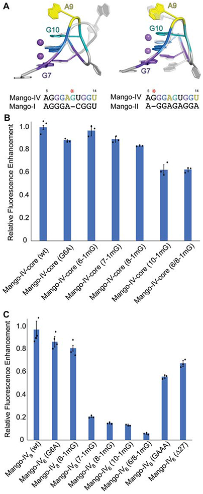
Figure 5.

Structural comparison and mutational analysis of the first guanine tract of Mango-IV. (A) Overlay of first guanine tract of Mango-IV (colored) with Mango-I (grey transparent, left) and Mango-II (grey, right). Sequence alignments of first guanine tract region are shown below each overlay. Red asterisk indicates location of guanine insertion for the Mango-IV sequence. (B) Fluorescence enhancement of the Mango-IVcore sequence and mutants to the first guanine tract. Residue numbering is consistent with nucleotide positions of crystalizing construct depicted in panel A. (C) Fluorescence enhancement of the Mango-IV8 aptamer and mutants to the first guanine tract and helical linker. Residue numbering is consistent with nucleotide positions of crystalizing construct depicted in panel A. Data are represented as the mean ± SEM.
