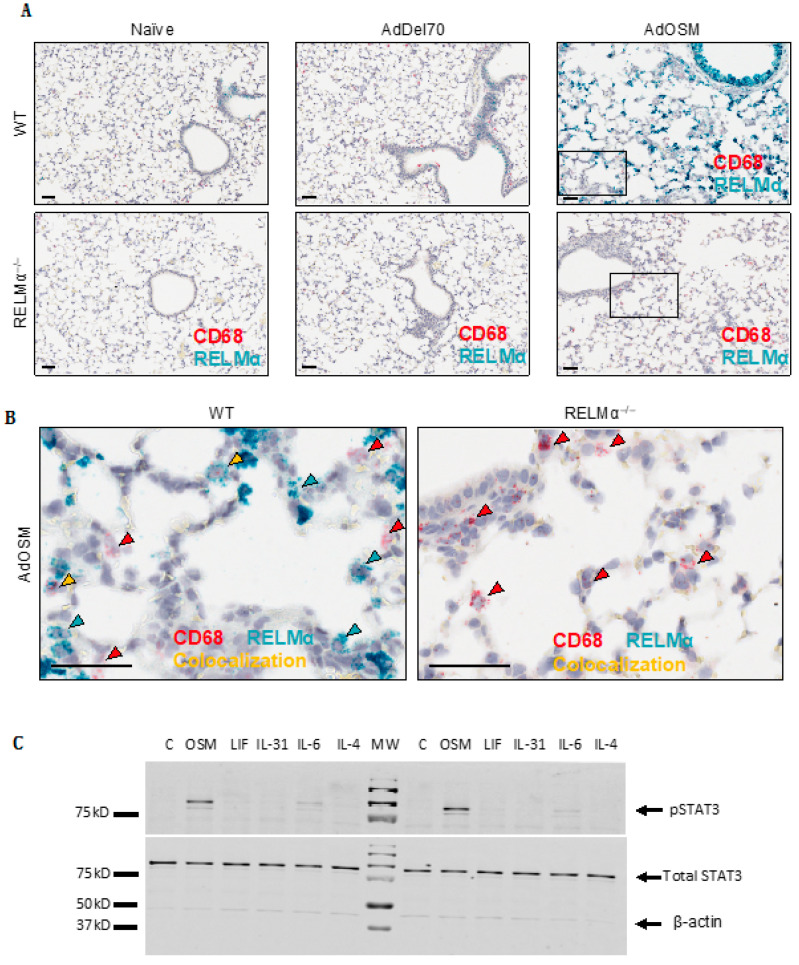
Figure 2.
RELMα mRNA is highly induced in columnar airway epithelial cells. (A) Representative images (n = 5 mice/group) are shown of CISH staining for RELMα (green-blue) and CD68 (red, a macrophage marker) in formalin-fixed paraffin-embedded lung tissue sections from naïve, AdDel70- or AdOSM-treated at day 7 as indicated in wild-type (upper panels) or RELMα–/– mice (lower panels). Scale bars, 50 µm. (B) High magnification images of indicated regions (boxed) from AdOSM-treated mouse lungs from right panels of (A) RELMα (green-blue) and CD68 (red) mRNA signals show as punctate dots unless at very high levels in which signals converge. Scale bars, 50 µm. Co-localization of both signals is indicated as yellow arrowheads in wild-type mice. (C) C57Bl/6-derived murine airway epithelial cells were stimulated in vitro for 1 h with 20ng/ml of OSM, leukemia inhibitory factor (LIF), IL-31, IL-6 or IL-4 and whole cell extracts probed for phospho-STAT3 (pSTAT3), total STAT3 and β-Actin by Western Blotting. Two separate cell culture experiments are shown.

