Abstract
One of the driving forces of carcinogenesis in humans is the aberrant activation of receptors; consequently, one of the most promising mechanisms for cancer treatment is receptor inhibition by chemotherapy. Although a variety of cancers are initially susceptible to chemotherapy, they eventually develop multi-drug resistance. Anti-tumor agents overcoming resistance and acting through two or more ways offer greater therapeutic benefits over single-mechanism entities. In this study, we report on a new family of bifunctional compounds that, offering the possibility of dual action (drug + radiotherapy combinations), may result in significant clinical benefits. This new family of compounds combines two fragments: the drug fragment is a lapatinib group, which inhibits the tyrosine kinase receptor activity, and an icosahedral boron cluster used as agents for neutron capture therapy (BNCT). The developed compounds were evaluated in vitro against different tyrosine kinase receptors (TKRs)-expressing tumoral cells, and in vitro–BNCT experiments were performed for two of the most promising hybrids, 19 and 22. We identified hybrid 19 with excellent selectivity to inhibit cell proliferation and ability to induce necrosis/apoptosis of glioblastoma U87 MG cell line. Furthermore, derivative 22, bearing a water-solubility-enhancing moiety, showed moderate inhibition of cell proliferation in both U87 MG and colorectal HT-29 cell lines. Additionally, the HT-29 cells accumulated adequate levels of boron after hybrids 19 and 22 incubations rendering, and after neutron irradiation, higher BNCT-effects than BPA. The attractive profile of developed hybrids makes them interesting agents for combined therapy.
Keywords: tyrosine kinase inhibitors; lapatinib; [1,2,3]triazolyl linker; boron clusters; in vitro BNCT effect
1. Introduction
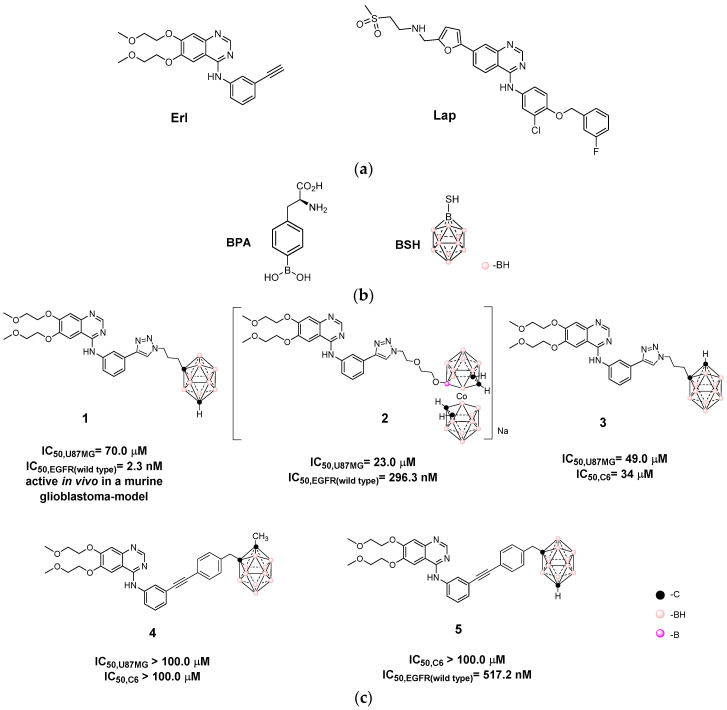
Tyrosine kinase receptors (TKRs) are transmembrane-type receptors with cytoplasmic tyrosine kinase domains, which transduce extracellular signals to a variety of intracellular signaling cascades involved in proliferation and differentiation of both normal and malignant cells [1]. The overexpression of some TKRs and their enhanced signaling contribute to the initiation, progression, and invasiveness of cancers [2]. Thus, TKRs are attractive targets for the development of therapeutic tools, for example, small inhibitors such as erlotinib (Erl)-targeting epidermal growth factor receptor (EGFR, ErbB1), and lapatinib (Lap) targeting EGFR and human epidermal growth factor receptor 2 (ErbB2, HER2) (Figure 1) [3,4,5]. Another anti-cancer strategy, boron neutron capture therapy (BNCT), has been recognized as a promising therapy for melanoma, locally malignant gliomas, and head and neck cancers [6,7,8,9,10]. BNCT is based on the nuclear capture and fission reactions of the 10B atom with low energy thermal/epithermal neutrons to yield high linear energy transfer α particles and recoiling 7Li nuclei. Since the path lengths of the particles are approximately 9−10 μm, similar to the dimensions of a single cell, 10B-containing cells are selectively destroyed by BNCT. Two compounds, p-borono-L-phenylalanine (BPA) and disodium mercapto-closo-undecahydrododecaborate (BSH), are clinically used in treatment of cancer with BNCT (Figure 1) [11,12]. BPA is selectively taken up into tumor cells, while BSH tumor selectivity is slightly low [13]. However, BSH and its derivatives are of increasing interest as boron carriers for BNCT, due to the ability to deliver large amounts of 10B atoms to tumor cells (12 times more B per BSH-molecule than BPA) [14,15,16]. The first boron drug Steboronine® to be utilized in BNCT was recently developed by Stella Pharma corporation on 25 March 2020 [17]. It not only provides another option to the oncologists but benefits locally unresectable recurrent or unresectable advanced head and neck cancer treatment.
Figure 1.
(a) Small tyrosine kinase receptors (TKRs) inhibitors erlotinib (Erl) and lapatinib (Lap); (b) Compounds BPA and BSH, clinically used in the treatment of cancer with boron neutron capture therapy (BNCT); (c) Closo-carboranyl and metallabis(dicarbollide) clusters as hybrid agents derived from erlotinib, which act as TKRs inhibitor-scaffold hybrids previously developed by our group [18,19,20,21]. IC50 were measured for U98MG cell line.
We have previously designed hybrid agents by combining substructures derived from Erl and icosahedral boron clusters with the aim to develop a new bimodal therapy of cancer. These bifunctional compounds, which combine the TKR-interaction/inhibition ability plus the selective and high boron-atoms-loading capacity in cells for the BNCT process, would act as anticancer bimodal agents (chemo- + radiotherapy) to result in significant clinical benefits such as reducing the doses to get the same therapeutic effect while diminishing the side effects suffered by the patient. We have demonstrated that the incorporation of the boron cluster has resulted in hybrids with enhanced and selective in vitro and in vivo anti-tumoral activities, i.e., compounds 1, 2, and 4 (Figure 1) [18,19,20,21]. From a structural point of view, relevant aspects were observed; the [1,2,3]triazolylalkyl-linker yielded hybrids with the most promising profiles, that is, being better than other links (see bio-profiles of 3 and 5, Figure 1). Herein, we describe the exploration of the EGFR and ErbB2 inhibitor Lap as scaffold to develop new hybrid molecules for bimodal therapy of cancer treatment. These newly synthesized hybrids were evaluated in vitro as cytotoxic agents on TKRs-overexpressing cells, and its selectivity against glioma-cells was also tested. The cellular death mechanism triggered by one of these hybrids was also studied on glioma cells and astrocytes. Additionally, for selected hybrids, the in vitro ability to inhibit active EGFR in a cell-free system was performed. Moreover, BNCT potentiality was evaluated analyzing cellular B-accumulations followed by neutron irradiation experiments.
2. Materials and Methods
2.1. Chemistry
Chemicals were reagent-grade and were used as received from commercial suppliers (Merck (Sigma-Aldrich), Darmstadt, Germany). 1,2-closo-C2B10H12, and B10H14 were obtained from Katchem (Prague, Czech Republic) and Lap from Baoji Guokang Bio-Technology Co (Baoji, China). 1-CH3-1,2-closo-C2B10H11 was synthesized from B10H14 as reported in the literature [22]; 1,7-closo-C2B10H12 and 1-CH3-1,7-closo-C2B10H11 were synthesized from 1,2-closo-C2B10H12 and 1-CH3-1,2-closo-C2B10H11, respectively, following the reported procedures [23]; and Cs[3,3’-Co(1,2-C2B9H11)2] (14) [24], [3,3’-Co(8-(CH2CH2O)2-1,2-C2B9H10)(1’,2’-C2B9H11)] (15) [25,26], and [3,3’-Co(8-N3-(CH2CH2O)2-1,2-C2B9H10)(1’,2’-C2B9H11)] (16) [27] were synthesized as reported,. Intermediates 6–13 were synthesized as reported in the literature [18,28]. Most reactions were performed under an atmosphere of nitrogen by employing standard Schlenk techniques. Analytical thin-layer chromatography (TLC) was carried out on pre-coated plates with silica gel 60 on aluminum foil F254 (Merck). Compounds were visualized by staining using UV light (254 nm) and/or by a 0.5% acidic solution of PdCl2 in HCl/methanol for boron-containing derivatives.
2.2. Instrumentation
Elemental analyses were performed using a Carlo Erba Model EA1108 elemental analyzer instrument (AB, Canada). All NMR spectra were recorded on Bruker ARX-300 or on Bruker DPX-400 spectrometers (Billerica, MA, USA) equipped with the appropriate decoupling accessories. The 1H (300.13 MHz or at 400.13 MHz), 11B{1H} (96.29 MHz), and 13C{1H} (75.47 MHz or at 100.77 MHz) NMR spectra (see Supplementary Material) were recorded in CDCl3, CD3COCD3, or (CD3)2SO at 298 K. Chemical shifts are reported in units of parts per million downfield from the reference, and all coupling constants are reported in Hertz (Hz). The 11B and 11B{1H} NMR shifts were referenced to external BF3·OEt2, while the 1H, 1H{11B}, and 13C{1H} NMR shifts were referenced to SiMe4. Multiplicity is abbreviated as follows: s is the singlet, d is the doublet, dd is the doublet of doublet, dt is doublet of triplets, dq is doublet of quartets, t is the triplet, m is the multiplet, and bs is the broad singlet. MS were performed at a Shimadzu QP-2010 spectrometer (Kyoto, Japan) at 70 eV ionizing voltage. Mass spectra are presented as m/z (% rel int.). MALDI-TOF mass spectra were recorded in the negative-ion mode using a Bruker Biflex MALDI-TOF (N2 laser; λexc = 337 nm; 0.5 ns pulses); voltage ion source 20.00 kV (Uis1) and 17.50 kV (Uis2)). UV measurements were performed on spectrofluorometer Varioskan flash, Thermo® (Waltham, MA, USA) at 298 K and using 1.0 cm cuvettes.
2.3. Synthesis of Lapatinib Derivative
Triethylamine (1 equiv., 0.1 mL, 0.69 mmol) was added drop by drop to a stirred suspension of Lap (1 equiv., 400 mg, 0.69 mmol) in CHCl3 (12 mL). The mixture was stirred for 1 h at room temperature. After that, 3-bromo-1-propyne solution (80% in toluene, 1.05 equiv., 0.075 mL, 0.72 mmol) was added over a period of 15 min. The mixture was stirred overnight at reflux, and then it was quenched with an aqueous saturated solution of NH4Cl (15 mL) and extracted with CHCl3 (3 × 20 mL). The organic layer was dried over MgSO4 and evaporated in vacuum to dryness. The orange residue was purified by SiO2 column chromatography (CH2Cl2:MeOH, 97:3) to give the desired compound as a yellow solid (398 mg, 74%). 1H-NMR (400 MHz, CDCl3) δ: 8.69 (s, 1H, pyrimidine-H), 8.40 (bs, 2H, -NH and Ar-H), 7.95 (dd, JH,H = 8.7; 1.5, 1H, Ar-H), 7.90 (d, JH,H = 2.5, 1H, Ar-H), 7.85 (d, JH,H = 8.7, 1H, Ar-H), 7.67 (dd, JH,H = 8.8; 1.5, 1H, Ar-H), 7.40–7.33 (m, 1H, Ar-H), 7.26–7.20 (m, 2H, Ar-H), 7.04 (dd, JH,H = 8.7; 2.1, 1H, Ar-H), 6.99 (d, JH,H = 8.7, 1H, Ar-H), 6.71 (d, JH,H = 3.3, 1H, furyl-H), 6.39 (d, JH,H = 3.3, 1H, furyl-H), 5.16 (s, 2H, -ArO-CH2-Ar), 3.87 (s, 2H, -N-CH2-furyl), 3.46 (d, JH,H = 2.3, 2H, -N-CH2-C≡CH), 3.37 (bs, 4H, -N-CH2-CH2-SO2), 2.97 (s, 3H, -SO2CH3), 2.33 (t, JH,H = 2.2, 1H, -N-CH2-C≡C). 13C{1H}-NMR (100.77 MHz, CDCl3) δ: 164.7, 161.4, 158.0, 154.9, 153.1, 150.9, 149.5, 144.8, 139.3, 132.8, 130.2, 129.0, 128.7, 125.3, 123.1, 122.7, 122.5, 115.9, 115.0, 114.7, 114.3, 113.9, 111.8, 106.9, 74.5, 70.50, 55.1, 52.4, 49.4, 48.3, 46.8, 42.1.
2.4. General Procedure for Hybrids 18–23 Preparation
Lapatinib derivative (1 equiv.) dissolved in THF:H2O (1:1, v/v) (6 mL for each 50 mg of Lapatinib derivative) was introduced into a 50-mL round-bottom flask equipped with a magnetic stirring bar. Then, sodium ascorbate (15 mol %), CuSO4·5H2O (10 mol %) and the corresponding azide (8, 9, 12, 13, 16, or 17) (1.1 equiv.) were added. The mixture was stirred overnight at room temperature. After that, the solvent was evaporated and the crude was dissolved in CH2Cl2 (15 mL for each 50 mg of lapatinib derivative) and washed with brine (3 × 5 mL for each 50 mg of lapatinib derivative). The organic layer was dried over MgSO4 and evaporated in vacuum to dryness. The product was purified by preparative SiO2-TLC or chromatographic column using CH2Cl2:MeOH (97:3) as eluent.
2.4.1. Hybrid 18
Yellow solid (123 mg, 75%). 1H{11B}-NMR (400 MHz, CDCl3) δ: 8.68 (s, 2H, pyrimidine-H and -NH), 8.51 (s, 1H, Ar-H), 7.92 (d, JH,H = 8.8, 1H, Ar-H), 7.85 (m, 2H, Ar-H), 7.72 (dd, JH,H = 8.7; 2.1, 1H, Ar-H), 7.54 (s, 1H, triazole-H), 7.52–7.42 (m, 1H, Ar-H), 7.24 (dd, JH,H = 15.3; 8.0, 2H, Ar-H), 7.04 (dd, JH,H = 8.5; 1.6, 1H, Ar-H), 6.98 (d, JH,H = 8.9, 1H, Ar-H), 6.68 (d, JH,H = 3.2, 1H, furyl-H), 6.36 (d, JH,H = 3.2, 1H, furyl-H), 5.14 (s, 2H, -ArO-CH2-Ar), 4.32 (t, JH,H = 6.2, 2H, triazole-CH2-CH2- CH2-Ccluster), 3.88 (s, 2H, -N-CH2-furyl), 3.79 (s, 2H, -N-CH2-triazole), 3.61 (s, 1H, Ccluster-H), 3.35 (t, JH,H = 6.4, 2H, CH2-CH2-SO2), 3.22 (t, JH,H = 6.5, 2H, -N-CH2-CH2-), 2.97 (s, 3H, -SO2CH3), 2.28–2.15 (bs, 4H, triazole-CH2-CH2-CH2-Ccluster). 13C{1H}-NMR (100.77 MHz, CDCl3) δ: 164.6, 161.4, 158.0, 154.8, 153.0, 150.9, 149.3, 144.9, 139.2, 132.7, 130.2, 130.1, 128.9, 128.8, 128.6, 125.3, 122.9, 122.5, 115.8, 115.6, 115.0, 114.8, 114.2, 113.8, 111.8, 107.0, 73.6, 70.5, 61.8, 52.3, 49.2, 49.1, 48.66, 46.5, 42.3, 34.8, 29.7. 11B{1H}-NMR (96.29 MHz, CDCl3) δ: −2.5 (1B), −5.9 (1B), −9.5 (2B), −12.2 (6B). Anal. calcd. for: C37H45B10ClFN7O4S: C, 52.50; H, 5.36; N, 11.58. Found: C, 52.39; H, 5.68, N, 11.95.
2.4.2. Hybrid 19
Yellow solid (191 mg, 84%). 1H{11B}-NMR (400 MHz, CDCl3) δ: 8.68 (s, 2H, pyrimidine-H and -NH), 8.52 (s, 1H, Ar-H), 7.93 (dd, JH,H= 8.8; 1.1, 1H, Ar-H), 7.90 (d, JH,H = 2.5, 1H, Ar-H), 7.84 (d, JH,H = 8.8, 1H, Ar-H), 7.73 (dd, JH,H = 8.9; 2.5, 1H, Ar-H), 7.57 (s, 1H, triazole-H), 7.41–7.33 (m, 1H, Ar-H), 7.27–7.18 (m, 2H, Ar-H), 7.04 (dd, JH,H = 8.4; 2.4, 1H, Ar-H), 6.99 (d, JH,H = 8.9, 1H, Ar-H), 6.69 (d, JH,H = 3.3, 1H, furyl-H), 6.37 (d, JH,H = 3.3, 1H, furyl-H), 5.15 (s, 2H, -ArO-CH2-Ar), 4.37 (t, JH,H = 5.6, 2H, -CH2-CH2-triazole), 3.91 (s, 2H, -N-CH2-furyl), 3.80 (s, 2H, -N-CH2-triazole), 3.40 (t, JH,H = 6.4, 2H, CH2-CH2-SO2), 3.26 (t, JH,H = 6.5, 2H, -N-CH2-CH2-), 2.98 (s, 3H, -SO2CH3), 2–23–2.11 (bs, 4H, triazole-CH2-CH2-CH2-Ccluster), 1.92 (s, 3H, Ccluster-CH3). 13C{1H}-NMR (100.77 MHz, CDCl3) δ: 164.5, 161.3, 157.9, 154.7, 152.9, 150.9, 150.8, 149.3, 144.6, 139.2, 132.7, 130.1, 128.8, 128.6, 128.4, 125.1, 123.0, 122.6, 122.4, 115.7, 114.9, 114.6, 114.2, 113.7, 111.7, 106.8, 74.8, 70.4, 52.3, 49.3, 49.0, 48.3, 46.7, 42.0, 33.4, 30.2, 24.3. 11B{1H}-NMR (96.29 MHz, CDCl3) δ: −4.4 (1B), −5.9 (1B), −10.6 (8B). Anal. calcd. for: C38H47B10ClFN7O4S: C, 53.04; H, 5.51; N, 11.40. Found: C, 53.22; H, 5.88, N, 11.02.
2.4.3. Hybrid 20
Yellow solid (188 mg, 79%). 1H{11B}-NMR (400 MHz, CDCl3) δ: 8.70 (s, 1H, pyrimidine-H), 8.66 (s, 1H, -NH) 8.53 (s, 1H, Ar-H), 7.94 (m, 2H, Ar-H), 7.85 (d, JH,H = 8.7, 1H, Ar-H), 7.74 (dd, JH,H = 8.8; 2.5, 1H, Ar-H), 7.51 (s, 1H, triazole-H), 7.41–7.34 (m, 1H, Ar-H), 7.27–7.18 (m, 2H, Ar-H), 7.05 (dd, JH,H = 8.6; 1.8, 1H, Ar-H), 7.00 (d, JH,H = 8.9, 1H, Ar-H), 6.70 (d, JH,H = 3.2, 1H, furyl-H), 6.37 (d, JH,H = 3.2, 1H, furyl-H), 5.16 (s, 2H, -ArO-CH2-Ar), 4.27 (t, JH,H = 5.6, 2H, -CH2-CH2-triazole) 3.91 (s, 2H, -N-CH2-furyl), 3.81 (s, 2H, -N-CH2-triazole), 3.43 (dd, JH,H = 19.5; 13.2, 2H, CH2-CH2-SO2), 3.27 (dt, JH,H = 17.9; 11.5, 2H, -N-CH2-CH2-), 2.99 (s, 3H, -SO2CH3), 2.92 (s, 1H, Ccluster-H ) 1.97 (bs, 4H, triazole-CH2-CH2-CH2-Ccluster). 13C{1H}-NMR (100.77 MHz, CDCl3) δ: 164.7, 161.4, 158.0, 154.9, 153.1, 150.9, 149.5, 144.8, 139.3, 132.8, 130.2, 130.1, 129.0, 128.7, 128.5, 125.3, 123.2, 122.5, 115.9, 115.7, 115.0, 114.7, 114.3, 113.9, 111.8, 106.9, 74.5, 70.5, 55.1, 52.4, 49.4, 49.2, 48.3, 46.8, 42.1, 33.6, 30.3. 11B{1H}-NMR (96.29 MHz, CDCl3) δ: −4.6 (1B), −11.1 (5B), −13.8 (2B), −15.7 (2B Anal. calcd. for: C37H45B10ClFN7O4S: C, 53.50; H, 5.36; N, 11.58. Found: C, 52.62; H, 5.71, N, 11.21.
2.4.4. Hybrid 21
Yellow solid (257 mg, 88%). 1H{11B}-NMR (400 MHz, CDCl3) δ: 8.69 (bs, 2H, pyrimidine-H and -NH), 8.54 (s, 1H, Ar-H), 7.94 (dd, JH,H= 6.2; 1.9, 2H, Ar-H), 7.85 (d, JH,H = 8.7, 1H, Ar-H), 7.73 (dd, JH,H = 8.8; 2.4, 1H, Ar-H), 7.51 (s, 1H, triazole-H), 7.41–7.33 (m, 1H, Ar-H), 7.26–7.21 (m, 2H, Ar-H), 7.04 (dd, JH,H = 8.7; 1.9, 1H, Ar-H), 6.99 (d, JH,H = 8.8, 1H, Ar-H), 6.69 (d, JH,H = 3.3, 1H, furyl-H), 6.37 (d, JH,H = 3.2, 1H, furyl-H), 5.16 (s, 2H, -ArO-CH2-Ar), 4.27 (t, JH,H = 5.6, 2H, -CH2-CH2-triazole), 3.91 (s, 2H, -N-CH2-furyl), 3.81 (s, 2H, -N-CH2-triazole), 3.41 (t, JH,H = 6.4, 2H, CH2-CH2-SO2), 3.33 (t, JH,H = 6.5 Hz, 2H, -N-CH2-CH2-), 2.98 (s, 3H, -SO2CH3), 1.95 (bs, 4H, triazole-CH2-CH2-CH2-Ccluster-, 1.67 (s, 3H, Ccluster-CH3). 13C{1H}-NMR (100.77 MHz, CDCl3) δ: 164.7, 161.4, 158.0, 154.8, 153.1, 151.0, 149.5, 144.7, 139.3, 132.8, 130.2, 130.1, 128.9, 128.7, 128.5, 125.2, 123.1, 122.5, 115.9, 115.7, 115.0, 114.7, 114.3, 113.9, 111.8, 106.9, 74.7, 70.8, 70.5, 52.4, 49.4, 49.1, 48.4, 46.8, 42.1, 33.5, 30.3, 24.4. 11B{1H}-NMR (96.29 MHz, CDCl3) δ: −4.8 (1B), −7.2 (1B), −9.2 (6B), −11.6 (2B). Anal. calcd. for C38H47B10ClFN7O4S: C, 53.04; H, 5.51; N, 11.40. Found: C, 53.35; H, 5.63, N, 11.77.
2.4.5. Hybrid 22
Orange solid (142 mg, 73%). 1H{11B}-NMR (400 MHz, CO(CD3)2) δ: 9.20 (bs, 1H, -NH), 8.69 (s, 1H, pyrimidine-H), 8.60 (bs, 1H, Ar-H), 8.18–8.15 (m, 2H, Ar-H), 8.06 (s, 1H, Ar-H), 7.84 (d, JH,H = 8.1, 1H, Ar-H), 7.88 (dd, JH,H = 8.9; 2.2, 1H, Ar-H), 7.53–7.43 (m, 1H, Ar-H), 7.40 (s, 1H, triazole-H), 7.38–7.31 (m, 1H, Ar-H), 7.23 (d, JH,H = 8.9, 1H, Ar-H), 7.15–7.09 (m, 1H, Ar-H), 6.95 (d, JH,H = 2.6, 1H, furyl-H), 6.55 (d, JH,H = 2.3, 1H, furyl-H), 5.29 (s, 2H, -ArO-CH2-Ar), 4.60 (t, JH,H = 5.6, 2H, -CH2-CH2-triazole), 4.22 (bs, 4H, Ccluster-H), 3.95 (s, 2H, -N-CH2-furyl), 3.93–3.87 (m, 2H, -CH2-CH2-triazole), 3.66–3.59 (m, 2H, -CH2-CH2-SO2), 3–58–3.52 (m, 2H, -N-CH2-CH2-), 3.45–3.35 (m, 6H, -CH2-CH2-Ccluster and -CH2-CH2-SO2) 3.09 (s, 3H, -SO2CH3). 13C{1H}-NMR (100.77 MHz, CO(CD3)2) δ: 164.5 161.3, 157.8, 152.5, 150.6, 144.6, 140.1, 140.0, 133.2, 130.4, 130.3, 129.1, 128.8, 128.5, 124.5, 123.1, 122.2, 122.1, 116.1, 114.6, 114.3, 114.3, 114.1, 113.8, 111.8, 107.7, 71.9 (2C), 69.9, 69.2 (2C), 68.5 (2C), 53.8 (2C), 52.3, 50.1, 48.1, 46.5, 41.6. 11B{1H}-NMR (96.29 MHz, CO(CD3)2) δ: 24.7 (1B), 6.0 (1B), 1.6 (1B), −1.3 (1B), −3.1 (2B), −6.1 (6B), −16.0 (2B), −19.0 (3B), −27.5 (1B). MALDI-TOF-MS: m/z calcd. for C40H57B18ClCoFN7O6S: 1074.48. Found: 1072.7446. Anal. calcd.: C: 44.82; H: 5.36; N: 9.15. Found: C: 44.61; H: 5.90; N: 9.27.
2.4.6. Bioisoster 23
Yellow solid (69 mg, 91%). 1H-NMR (400 MHz, CDCl3) δ: 8.74 (s, 1H, pyrimidine-H), 8.69 (s, 1H, Ar-H), 8.56 (bs, 1H, -NH), 7.95–7.91 (m, 1H, Ar-H), 7.90 (d, JH,H = 1.4, 1H, Ar-H), 7.84 (d, JH,H = 8.4, 1H, Ar-H), 7.72 (dd, JH,H = 8.9; 2.5, 1H, Ar-H), 7.51 (s, 1H, triazole-H), 7.41–7.29 (m, 2H, Ar-H), 7.27–7.18 (m, 4H, Ar-H), 7.14 (d, JH,H = 7.2, 2H, Ar-H), 7.03 (t, JH,H = 8.4, 1H, Ar-H), 6.96 (d, JH,H = 8.9, 1H, Ar-H), 6.68 (d, JH,H = 3.2, 1H, furyl-H), 6.36 (d, JH,H = 3.2, 1H, furyl-H), 5.12 (s, 2H, -ArO-CH2-Ar), 4.35 (t, JH,H = 7.1, 2H, -CH2-CH2-triazole), 3.91 (s, 2H, -N-CH2-furyl), 3.80 (s, 2H, -N-CH2-triazole), 3.40 (t, JH,H = 6.7, 2H, CH2-CH2-SO2), 3.27 (t, JH,H = 6.9, 2H, -N-CH2-CH2-), 2.98 (s, 3H, -SO2CH3), 2.64 (t, JH,H= 7.5, 2H, -CH2-CH2-Ar), 2.40–2.05 (m, 2H, -CH2-CH2-Ar). 13C{1H}-NMR (100.77 MHz, CDCl3) δ: 164.7, 164.6, 161.4, 158.1, 154.8, 152.9, 151.1, 150.9, 149.4, 144.3, 139.9, 139.2, 132.8, 130.2, 130.1, 128.8, 128.6, 128.5, 128.4, 126.4, 125.3, 123.1, 122.9, 122.5, 115.9, 115.7, 114.9, 114.7, 114.2, 113.8, 111.8, 106.9, 70.4, 52.4, 49.7, 49.1, 48.4, 46.8, 42.1, 32.5, 31.6. Anal. calcd. for C41H39ClFN7O4S: C, 63.11; H, 5.04; N, 12.57. Found: C, 63.51; H, 5.12, N, 12.21.
2.5. Biology
2.5.1. Tumor Cells
EGFR-overexpressing cell lines, glioma cell lines U87 MG (ATCC HTB-14) and C6 (ATCC CCL-107), and colorectal adenocarcinoma cell line HT-29 (ATCC® HTB-38) were obtained from ATCC culture collection (Virginia City, NV, USA). F98 rat glioma cells, histologically characterized as an anaplastic astrocytoma, were a kind gift from Dr. Rolf Barth (Dept. of Pathology, The Ohio State University, Columbus, OH, USA). Cells were cultured in Dulbecco modified Eagle’s medium high glucose (4.5 g/L) with stable glutamine (3.97 mM) (DMEM) supplemented with 10% heat-inactivated fetal bovine serum (i-FBS) and 1% of a solution containing penicillin-streptomycin (PS-B) at 37 °C in a humid atmosphere containing 5% CO2 concentration. Culturing materials were purchased from Capricorn Scientific. In order to obtain i-FBS, original FBS was heated at 60 °C for 30 min.
2.5.2. Glia Primary Cell Culture
A mixed glia primary cell culture was obtained from cerebral cortex of newborn mice as previously described with minor modifications [29]. Cerebral cortex of 3–4 newborn mice were dissected with the meninges carefully removed. Cerebral cortex were mechanically chopped and then enzymatically dissociated in 0.25% trypsin for 10 min at 37 °C. Fetal bovine serum (FBS) 10% in DMEM was then added to halt trypsin digestion. Repetitive pipetting thoroughly disaggregated the tissue, which was then strained through an 80 µm mesh and spun down. The pellet was re-suspended in glia culture medium and plated in T25 culture flasks. Glia culture medium composition is as follows: DMEM supplemented with 10% FBS, HEPES buffer (3.6 g/mL), penicillin (100 IU/mL), and streptomycin (100 µg/mL). Culture medium was replaced every 48 h until 90% confluence was reached.
2.6. Study Approval
All procedures using laboratory animals were performed in accordance with the national and international guidelines and were approved by the Institutional Animal Committee for animal experimentation. All experimental procedures were approved by the Ethical Committee for the use of Experimental Animals (CEUA) of the Institut Pasteur de Montevideo, Uruguay (CEUA Approved protocol: #003-17 to Dr. Emiliano Trias), and under the current ethical regulations of the Uruguayan Law 18.611 for animal experimentation that follows the Guide for the Care and Use of Laboratory Animals of the National Institutes of Health [30].
2.7. Cytotoxicity Assays
Cytotoxicity assays were performed as previously described with minor modifications [18]. EGFR-overexpressing cells or glia cells were seeded in 96-well plates (7 × 10³ – 1 × 10⁴ cells/well depending on the cell line for tumor cells and 2 × 10⁴ cells/well for glia cells) in 100 μL final volume of growing medium and were allowed to grow for 24 h. After that, 125 μL of fresh culture medium was added and the cells were allowed to grow for additional 24 h. Then, 25 μL of a solution 10× the desired final concentration of the tested compounds in culture medium was added to the culturing media. Cells were further incubated for 24 h. Afterwards, culture medium was removed and cells were washed twice with 200 μL of PBS. Cells were then fixed with 50 μL of ice-cold trichloroacetic acid for 1 h at 4 °C. Next, the plates were washed five times in distilled water and allowed to dry at room temperature. Sulphorhodamine B (SRB) solution (50 μL, 0.4 w/v in acetic acid 1% v/v) was added to each well of the dried 96-well plates [31]. Staining was performed at room temperature for 30 min. The SRB solution and unbound dye were removed by washing the plates quickly with aqueous solution of acetic acid (1%, v/v) at least five times (until excess dye was fully removed). The washed plates were allowed to dry at room temperature for at least 24 h. Finally, the bound SRB was solubilized by adding Tris Base buffer (pH 10, 10 mM, 100 μL) to each well, and the resulting solution was shaken for 5 min on a shaker platform. The optical density (OD) of each well solution was read in a 96-well plate reader at λ = 540 nm. The OD of SRB solution in each well is directly proportional to the cell number. Cell viability percentage was calculated according to the following equation: CV% = (A540 nm – B)/(C – B), where CV% stands for cell viability percentage, A540 nm corresponds to OD of a particular well, B is the OD of untreated wells with no cells seeded onto them, and C is the OD of control wells treated only with 1% of DMSO. CV% values were plotted against compound concentration, and the IC50 values were determined.
2.8. Kinase Enzymatic Assays
The enzymatic activity of hybrid 19 and Lap against EGFR was determined using ADP-Glo™ kinase Assay system from Promega Corporation (Catalog number V3831, Fitchburg, WI, USA). The experiments were conducted according to the manufacturer instructions. Briefly, an EGFR kinase reaction mixture of 10 μL was carried out by addition of 2 μL of the compound dilution, 3 μL of the enzyme dilution, and 5 μL of the ATP/substrate solution to get a final concentration of 50 μM ATP, 0.2 g/mL of Poly(Glu, Tyr) substrate and 5 mg/mL of enzyme. Each reaction was performed by triplicate on a 384-well plate and incubated for a duration of 1 h at room temperature. Final compound concentrations in the kinase reaction were 100, 10, 1, 0.1, 0.01, 0.001, and 0.0001 μM. After the reaction time, 5 μL of ADP-Glo™ was added and allowed to react during 40 min. Finally, 10 μL of Kinase Detection Reagent was added and incubated for additional 40 min. Afterwards, luminescence was detected on a BioTek® FLx800 Multi-Detection Microplate Reader (Integration time 0.5–1 s). Curve fitting and data presentations were performed using GraphPad Prism version 8.0.
2.9. Flow Cytometry Analysis
In order to study cell death mechanism (namely apoptosis or necrosis) triggered by exposure to hybrid 19 or Lap of glia and U87 MG cells, phosphatidylserine exposure was measured by flow cytometry using Annexin-V staining. Glia cells (6 × 10⁴ cells) were seeded into p60 dishes and were allowed to grow until 80% confluence. Next, 1 × 10⁵ U87 MG stained cells with PKH26 dye were seeded onto a p60 dish containing a growing glia primary cell culture. After a 48 h incubation in fresh growing medium, the p60 dishes were treated with IC50 dose of hybrid 19 or Lap at 10 µM or 100 µM. A p60 dish treated with 1% DMSO served as control. Treated cells were incubated for 24 h. Then, cells were harvested with trypsin (0.05%, supplemented with EDTA, 0.38 mg/mL) and centrifuged at 250 g speed. The resulting pellet was resuspended in an appropriate volume of Annexin binding buffer (0.01 M Hepes pH 7.4, 0.14 M NaCl, and 2.5 mM CaCl2) to get a cell suspension of 1 × 10⁶ cells per mL. After cell counts, samples were divided, and cells alone and isotype-matched control samples were generated to control for nonspecific binding of antibodies and for autofluorescence. An Annexin V-FITC antibody solution (catalog number: A13199) was used at a 1:20 concentration. After 30 min of incubation with the aforementioned antibody at 4 °C, samples were incubated with DAPI at a 1:5000 concentration and were immediately analyzed by flow cytometry. To perform the analysis, cells were first gated for PKH26 in order to distinguish U87 MG cells from glia cells. In each subpopulation of cells, the following markers were used to define four different populations: viable cells (Annexin-V−/DAPI−), necrotic cells (Annexin-V−/DAPI+), early apoptotic cells (Annexin-V+/DAPI−), and finally late apoptotic cells (Annexin-V+/DAPI+). Samples were acquired using FACSAria Fusion flow cytometer (Biosense, Irwindale, CA, USA). and BD FACSDiva™ software (Biosense). Three independent flow cytometry experiments were done, except for Lap, evaluated at 100 μM, which was included in only one of these experiments. Data obtained from these experiments were analyzed using FlowJo software (FlowJo LLC, Ashland, OR, USA).
2.10. Statistical Analysis
Statistical analysis was done using GraphPad Prism 8.0 software (GraphPad, LaJolla, CA, USA). For statistical analysis of flow cytometry data, unpaired T Test was performed to determine whether each population present on the dotplot DAPI vs. Annexin-V-FITC of each experimental condition was different from control populations. A p-value < 0.05 was considered significant.
2.11. Fluorescence Microscopy Analysis
Fluorescence microscopy studies were performed on in vitro co-cultures comprising U87 MG cells plated along with glia cells. Co-cultures were set by seeding glia cells (3 × 10⁴ cells) into p35 dishes. Glia cells were allowed to grow until 80% confluence. Next, 5 × 10⁴ U87 MG stained cells with PKH26 dye were seeded onto a p35 dish containing growing glia cells. After a 48 h-incubation in fresh growing medium, the p60 dishes were treated with IC50 dose of hybrid 19 or Lap at 10 µM or 100 µM. A p60 dish treated with 1% DMSO served as a negative control. Treated cells were incubated for further 24 h. After that, treated co-cultures were fixed for 20 min at 4 °C with PFA and washed 2 times with PBS. Then, samples were permeabilized for 10 min at room temperature with 0.1% Triton X-100 in PBS, passed through washing PBS, blocked with 5% BSA:PBS for 1 h at room temperature, and incubated overnight in a solution of 1% BSA:PBS containing the primary antibodies and DAPI at 4 °C. After washing, treated cells were incubated in 1:500-diluted secondary antibodies during 2 h at room temperature. The following antibodies were used for immunofluorescence staining: primary antibodies 1:400 mouse monoclonal anti-GFAP (Sigma-Aldrich, Darmstadt, Germany) and 1:50 rat monoclonal anti M2, and secondary antibodies conjugated to AlexaFluor 633 AlexaFluor 488 (Invitrogen, Carlsbad, CA, USA), respectively. DAPI was used at a 1:1000 dilution. Antibodies were detected by confocal microscopy using a confocal Zeiss LSM 800 microscope (Jena, Germany).
2.12. In Vitro BNCT Experiments
2.12.1. Determination of Intracellular Boron by Inductively Coupled Plasma Optical Emission Spectroscopy (ICP−OES).
Exponentially growing cells (F98 or HT-29) were seeded in plates of 24 wells and incubated with hybrid 19 (10 μM, dissolved in DMSO), 22 (10 μM, dissolved in DMSO) or 10B-BPA (0.925 mM, diluting with water the stock solution to a final volume of 500 μL). The stock solution of 10B-BPA was prepared at a concentration of 30 mg 10B-BPA-fructose (99% 10B enriched, L-isomer) (Glyconix Corp, Raleigh, NC, USA) per mL (0.14 M) as follows: 10B-BPA was combined in water with a 10% molar excess of fructose, adjusting the pH to 9.5–10.0 with aqueous solution of NaOH, and the resulted mixture was stirred until all solids were dissolved; finally, the pH was readjusted to 7.4 with aqueous solution of HCl [32]. After varying times of incubation (1, 2, and 4 h for F98 and 1, 2, 4, 6, 24, and 48 h for HT-29), the cells were washed twice with PBS 1× at 4 °C. The pellets were digested with formic acid (500 μL). An aliquot of 250 μL was diluted to 1.0 mL with an aqueous solution containing 1 mg/L of Y (0.75 mL) as internal standards. The boron uptake was measured by ICP–OES. Analytical and internal standard lines (in nm) were as follows: B: 249.677 and Y: 371.029. Matrix-matched standard solutions containing the internal standard elements and boron between 0.05 and 0.75 mg/mL were employed for daily calibration [33,34,35]. An internal measurement control was also used, with each certain number of samples adding a sample of known concentration (QC). The other aliquot of 250 μL was dried and re-suspended in aqueous solution of NaOH (0.3 N), and total proteins were determined by Lowry method. The boron amount was referred to the total proteins.
2.12.2. Neutron-Irradiation Procedures
Cells were irradiated at the thermal column of the RA-3 reactor, a 9 MW nuclear reactor located in Ezeiza (Argentina), where a highly thermalized and homogenous irradiation field is available. Thermal flux was near to (1.0 ± 0.1) × 1010 neutrons cm−2 s−1; the cadmium ratio was 4100 for gold foils, which allows neglecting fast neutron dose; and the gamma dose rate was approximately (6.0 ± 0.2) Gy h−1. Total dose was obtained by adding partial doses coming from photons, nitrogen capture (a 3.5% wt of nitrogen content is assumed), and boron capture. Before each irradiation, neutron flux at the irradiation position was checked using calibrated Rh-SPND detectors in a system that mimics the configuration that will be used (96-well plates with no cells inside), while, simultaneously, signal from a boron-coated ionization chamber was used as a monitor. Based on this measurement, irradiation times were calculated in order to deliver a dose of 1 and 2 Gy with an estimated uncertainty of 10% [36]. The dosimetry, for each treatment, the time, the fluence and each dose component of the total physical absorbed dose of the neutron beam without and with boron has been studied. The intracellular boron concentration at the time of irradiation was assumed uniformly distributed inside and outside the cells for the dosimetric calculations.
2.12.3. Irradiation of Hybrid 19-, 22- or BPA-Treated HT-29 Cells Assays
HT-29 cells were seeded at 2500 cells/well in 96-well plates. Eight different wells were seeded per treatment. The cells were then treated with 10.0 ppm of 10B of hybrids 19 or 22 (dissolved in DMSO) or 10B-BPA (dissolved in water) for 1 h after plating. Irradiated DMSO (5%)-treated cells and non-irradiated compound-treated cells served as controls. After this time, the cells were irradiated with thermal neutrons as described above.
2.12.4. Cell Surviving Assay
After irradiation, the medium was changed and the cells were cultured at 37 °C for 10 days. Afterwards, 20 μL of vital dye 3-(4,5-dimethylthiazol-2-yl)-2,5-diphenyltetrazolium bromide (MTT, Sigma 128, 0.5%, w/v in PBS) was added to the culture medium, and after 4 h of incubation at 37 °C, absorbance at 540 nm was observed. Results are expressed as percentage of untreated controls.
3. Results and Discussion
3.1. Design and Synthesis of Hybrids Carboranyl-Decorated Lapatinib-Scaffold
The following two structural features are responsible for effective Lap EGFR interaction [37]: i) the quinazoline ring, via its nitrogens that establish hydrogen bonds to Met769 and Thr830, and sandwiching between Ala719 and Leu820; and ii) the fluorobenzyloxyphenylamino moiety that makes hydrophobic interactions in the back of the ATP binding site. On the other hand, the methylsulfonylethylamino group is positioned at the solvent interface without significant interactions with the protein, establishing poor interaction to Asp776. For these reasons and considering the structural requirements, for the new designed hybrids we selected the solvent-exposed ethylamino-moiety to bind the high boron content cages using a polar linker, i.e., [1,2,3]triazolyl moiety [20] (Figure 1). Due to the Ccluster-H and B-H vertices, boron clusters could establish special hydrogen and dihydrogen bonds, such as C-H···X [38] and BH···H-X (X = N, C, O, and S), as well as BH···π, C-H···π hydrogen bonds [39,40], and C–H···Halogen interactions (Halogen = F, I [41,42]); three types of clusters were incorporated into the Lap scaffold, the neutral closo-carboranes—i.e., 1,2-dicarba-closo-dodecarborane and 1,7-dicarba-closo-dodecarborane—and the anionic cobaltabis(dicarbollide).
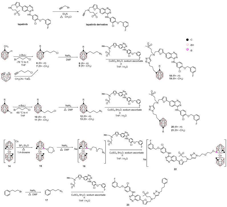
Consequently, our synthetic approach for the new hybrids starting from Lap is outlined in Scheme 1. Lap was selectively propargylated in the aliphatic amine in order to subsequently apply 1,3-dipolar cycloadditions with carboranyl-containing azides 8, 9, 12, 13, and 18. The desired hybrids, 18–22, connected by the polar [1,2,3]triazolyl moiety, were obtained in good to excellent yields. To confirm the relevance of carboranyl moiety in the displayed bioactivity (see below) we prepared the bioisoster [43,44] phenyl-analogue 23 (Scheme 1) of hybrids 18 and 20 using azide intermediate 17. The intermediates and final compounds were structurally characterized in terms of 1H-, 11B{1H}-, 13C{1H}-NMR, and UV-spectroscopies, mass spectrometry, and elemental microanalyses (C, H, N, and S).
Scheme 1.
Synthetic procedures used to prepare the hybrids carboranyl- and cobaltabis(dicarbollide)-lapatinib-scaffold (18–22) and the bioisoster 23.
3.2. Biological Studies
3.2.1. In Vitro Cytotoxicity Studies
To address the issue of human therapy, in vitro cytotoxicities of the hybrids 18–22 and the bioisoster 23 were determined on TKRs-overexpressing cells [18]: Homo sapiens colorectal adenocarcinoma HT-29 and brain glioblastoma U87 MG. For further animal in vivo experiments, Rattus norvegicus brain glioma C6 were also included in this study (Table 1). Compared to parent compound Lap, the hybrids resulted poorly active against HT-29 cells, being the most cytotoxic the Cobaltabis(dicarbollide) derivative 22 and the 1,2-dicarba-closo-dodecarborane 18. However, against glioma cells, a marked cytotoxic behavior was evidenced mainly for compound 19, resulting in 4.6- and over 10-times more active than the parent compound, Lap, against C6 and U87 MG cells, respectively. Additionally, hybrids 18, 21, and 22 were more active than Lap against U87 MG glioma cells. These results encouraged us to evaluate hybrid 19 against normal glial cells isolated from neonatal cortex to determine if this compound selectively killed tumor cells, while having no cytotoxic effects on normal glial cells (Table 1). The results showed that hybrid 19 displays tumor-cell selectivity with excellent selectivity indexes compared to the parent compound Lap. The benzyl-derivative 23 was less active than the corresponding bioisoster carborane hybrids 18 and 20 in all the studied cellular systems, confirming the relevance of the boron cages in the biological behaviors.
Table 1.
In vitro behavior of hybrids 18–22, bioisoster 23, and reference compounds, as IC50 (μM) or percentage of cells survival at 100 μM (%), on HT-29, C6, and U87 MG cells and astrocytes. Selectivity indexes (SI) for selected hybrid and reference compounds.
| Hybrid | IC50,HT-29 1 | IC50,U87 MG 1 | IC50,C6 1 | IC50,astrocytes | SI 2 | |
|---|---|---|---|---|---|---|
| U87 MG | C6 | |||||
| 18 | 80 ± 6 | >100 (65 ± 4) | >100 (100) | |||
| 19 | >100 (73 ± 6) | 10.0 ± 0.2 3 | 11.8 ± 0.4 4 | >100 | >10 | >8.5 |
| 20 | 100 ± 8 | >100 (85 ± 2) | >100 (100) | |||
| 21 | 100 ± 8 | >100 (77 ± 4) | >100 (100) | |||
| 22 | 50 ± 5 5 | >100 (66 ± 6) | >100 (100) | |||
| 23 | >100 (64 ± 4) | >100 (91 ± 8) | >100 (100) | |||
| Lap | 6.25 ± 0.05 | >100 (89 ± 5) | 54 ± 14 | 8 ± 3 | <0.08 | 0.15 |
1 Values in parenthesis correspond to the percentage of cell survival at 100 μM. 2 Selectivity indexes (SI) = IC50,astrocytes/IC50,indicated cells. 3 IC1 (dose necessary to inhibit 1% of growth cells) = 9.2 ± 0.8 μM, IC20 (dose necessary to inhibit 20% of growth cells) = 9.7 ± 0.1 μM, IC80 (dose necessary to inhibit 80% of growth cells) = 11.0 ± 0.1 μM. 4 IC1 = 1.0 ± 0.2 μM, IC20 = 9.5 ± 0.3 μM, IC80 = 12.3 ± 0.2 μM. 5 IC1 < 1.0 μM, IC20 = 1.0 ± 0.3 μM, IC80 = 90 ± 1 μM.
3.2.2. In Vitro Inhibition of EGRF by Hybrid 19
With EGFR being one of the bio-systems inhibited by Lap, we analyzed the inhibition of the most cellular-cytotoxic hybrid, 19, on this isolated system, comparing to Lap inhibition ability. The hybrid 19 was 10 times less potent than Lap against EGFR (IC50,19 ~3.0 μM, IC50,Lap ~0.3 μM, Figure S1, Supplementary Material). However, it should be noted that Lap, which is not a typical Type II inhibitor because of its unusual displacement of the α-C helix, resulted in in vitro high-throughput competition-binding assay (KINOMEscan, Figure S2, Supplementary Material), in an extremely selective inhibitor, against EGFR and Erb2. Thus, closo-carboranyl derivative 19 could be acting on glioblastoma cells, inhibiting other tyrosine kinase proteins, or also through different mechanisms of action.
3.2.3. Effect of Hybrid 19 on Simulated Tumor Environment
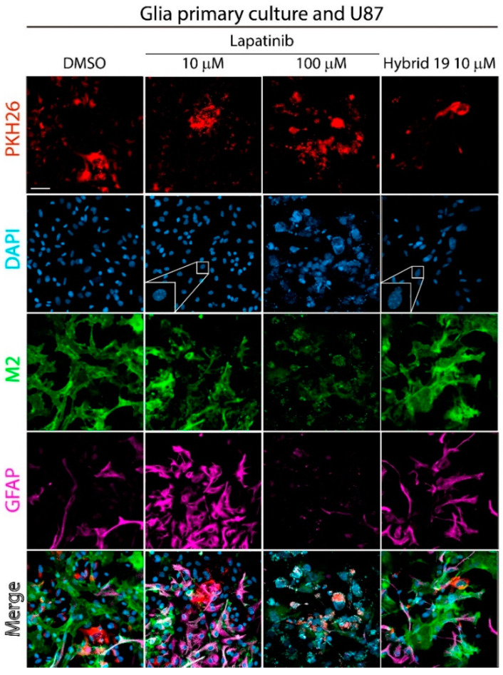
In order to study the effect of hybrid 19 more deeply, we simulated the cellular environment of a developing brain tumor co-culturing U87 MG cells, stained with PKH26 dye, together with neonatal murine astrocytes. Co-cultured cells were incubated for 24 h with hybrid 19 at U87 MG IC50 dose, Lap at 10 μM (near to IC50,astrocyte dose), and 100 μM (lower than IC50,U87 MG dose), or dimethylsulfoxide (DMSO, 1%) as negative control. Confocal microscopy revealed very different behaviors among the studied compounds (Figure 2, see merge images). As expected, PKH26 dye showed a decrease of U87 MG population upon 19-treatment compared to control vehicle-treated cells. However, according to astrocytic markers M2 for astrocytes membrane and GFAP for astrocytes activation [45,46], the population of tumor-associated astrocytes was observed mainly in the hybrid 19-incubated cells and in the control, while it was not present in the co-culture treated with Lap at the higher dose. Moreover, when comparing GFAP staining of lower dose Lap-treated cells and control cells, it was possible to observe that Lap made astrocytes become reactive, as indicated by increased GFAP expression, but hybrid 19 had a similar effect to that of the control (see merge images). Finally, DAPI nuclei staining showed on hybrid 19-treatment, like in Lap at the lower dose, some degree of chromatin condensation, which could be associated with the onset of apoptosis (Figure 2, zoomed-in inset).
Figure 2.
Effect of hybrid 19 or Lap on co-cultures of mixed glial cells with tumoral cells. Confocal images showing the co-culture of U87 MG cells (PKH26, red) with mouse neonatal cortical-derived astrocytes (M2, green and GFAP, magenta). The nuclei were stained with DAPI. Co-cultured cells were treated at IC50,U87 MG doses of hybrid 19 or at 10 and 100 μM for Lap and DMSO (1%) as control for 24 h.
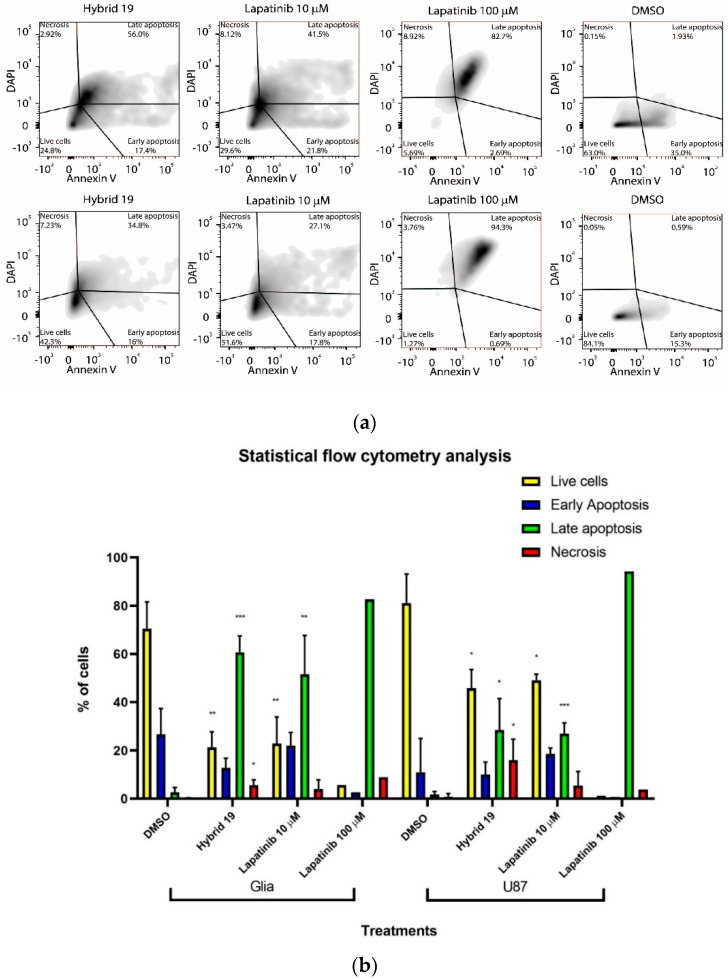
For this reason, the co-cultured systems were phosphatidylserine-exposure analyzed by flow cytometry. The flow cytometry analysis showed that hybrid 19 produced cellular death of U87 MG tumoral cells by apoptosis and necrosis. Compared to untreated control, hybrid 19 significantly increased the percentage of cells undergoing late apoptosis from ~0.6% to ~35% and necrosis from 0.05% to ~7% (Figure 3). Similarly, associated astrocytes mainly died by apoptosis as a result of hybrid 19 exposure (56% of late apoptosis), with nearly 3% of necrosis. Lap displayed very different death behavior according to the assayed dose. At 100 μM, a lower dose than its IC50 against U87 MG, Lap promoted on U87 MG mainly late apoptosis (~95%). However, at 10 μM (a dose near to the astrocytes-cytotoxic dose), Lap promoted late apoptosis, ~42% on associated astrocytes (Figure 3).
Figure 3.
(a) Representative examples of phosphatidylserine exposure results for primary glial cells (upper panels) and U87 MG (lower panels). Co-cultures were incubated with hybrid 19 or Lap for 24 h; (b) The graphs show the quantitative analysis flow cytometry (three independent experiments). (*) p < 0.05; (**) p < 0.01; (***) p < 0.001.
3.3. In Vitro BNCT Studies
For these studies, we selected two of the most relevant hybrids, i.e., 19 and 22. On the one hand, the closo-carboranyl derivative 19 displayed moderate cytotoxicity against HT-29 cell line, which displays a key factor in order to fulfill the requirements for successful BNCT. Additionally, 19 showed very interesting biological behaviors against glioblastoma cells; consequently, combined with neutron irradiation, it could be used in malignant gliomas. On the other hand, the derivative 22 displayed moderate activity against colorectal cells, optimum for BNCT treatments, and it bears a water-solubility-enhancing moiety, the metallacarboranyl group, that improves its drug-like properties. Besides, it is able to deliver larger amounts of 10B atoms to cells (18 times more B per 22-molecule than BPA, 1.8 times more B per 22-molecule than 19, 1.5 times more B per 22-molecule than BSH).
3.3.1. Boron Cellular Accumulation
First, we analyzed the presence of boron promoted by 19 and 22 into two different cellular systems, i.e., HT-29 and Rattus norvegicus brain glioblastoma F98 cells to address further in vivo animal BNCT studies. Among the different ways to calculate the boron cellular concentration (g of boron/g of tumor tissue, number of boron atoms/number cells [7,8,9] or g of boron/mg of protein [47,48]) reported nowadays, the latest one has been chosen in this article. Boron accumulation as a result of 19- and 22-incubations, at 10 μM doses, was detected in HT-29 cells even after 48 h of treatments (values near to 0.5 μg of boron/mg of protein content for both compounds, Figure 4a) with the highest accumulations within the first hour, i.e., 4.5 μg of boron/mg of protein content for 19 and 3.2 μg of boron/mg of protein content for 22. According to our previous results [33], HT-29 accumulates 0.5 μg of boron/mg of protein content when it was incubated for 24 h with BPA but with a dose 90 times higher than that indicated above for 19 and 22. In F98 19 and 22 incubation, for 4 h and at 10 μM doses, 10B-BPA was used at 0.925 mM dose as the reference compound. After 2 h of incubation the amount of boron produced by hybrids 19 and 22 was very important, decreasing with time for closo-carborane 19 and maintaining constantly a high value for metallacarborane 22 (Figure 4b). Additionally, F98 boron accumulation due to BPA was lower than 19 and 22 accumulations at 1 and 2 h of incubation, and unlike these compounds, it significantly decreased with time as a result of efflux by extracellular amino acids exchange (Figure 4b) [49].
Figure 4.
Amount of elemental boron incorporated into studied cells after compounds exposure, (a) in HT-29 cells treated with hybrids 19 or 22, at 10 μM for 48 h, and (b) in F98 astrocyte cells treated with hybrid 19 or 22, at 10 μM for 4 h, or BPA at 0.925 mM. (*) p < 0.05; (**) p < 0.01.
3.3.2. Neutron Irradiation Treatments
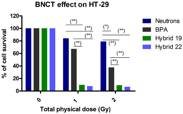
The previous information encouraged us to perform in vitro neutron irradiation experiments with both hybrids. In this sense, the HT-29 cells were incubated for 1 h with hybrid 19 or 22 using 10B-BPA as reference, and doses of 1 and 2 Gy were delivered. The irradiation times to deliver these doses, 1 and 2 Gy, were determined by dosimetric studies. Table 2 shows the dosimetry, for each treatment, the time, the fluence and each dose component of the total physical absorbed dose of the neutron beam without and with boron. All the studied compounds were incubated at doses equivalents to 10.0 ppm of 10B. The effect of in vitro BNCT displayed by hybrids 19 and 22 was very interesting, where a marked and statistically significant decrease was observed with respect to the irradiation with neutrons alone in the surviving cell fraction at 1 and 2 Gy-irradiated groups (Figure 5). Contrarily, at 1 and 2 Gy-irradiation and at 10.0 ppm of 10B, BPA produced a surviving cell fraction statistically significantly higher than hybrids 19 and 22 (Figure 5). The BPA effect was statistically significant, with respect to irradiation with neutrons alone, at 2-Gy of dose. Hybrid 19 was at least 7 times more effective than BPA, and hybrid 22 was at least 6 times more effective than BPA, in the in vitro model of BNCT.
Table 2.
Dosimetry for thermal neutron irradiation without and with boron (10 ppm 10B).
| Irradiation Time (min) | Fluence (×1012, n/cm2) | Dose γ (Gy) | Dose 14N (Gy) | Dose 10B (Gy) | Total Absorbed Dose (Gy) | Relative Error Dose |
|---|---|---|---|---|---|---|
| 5.55 | 1.94 | 0.46 | 0.54 | 0 | 1.0 | ±7% |
| 11.12 | 3.88 | 0.92 | 1.08 | 0 | 2.0 | ±7% |
| 2.07 | 0.722 | 0.17 | 0.20 | 0.63 | 1.0 | ±8% |
| 4.15 | 1.45 | 0.34 | 0.40 | 1.26 | 2.0 | ±8% |
Figure 5.
Effect on HT-29-cell survival without or with hybrids 19 and 22, or BPA treatment post-neutron-irradiation (1 and 2 Gy). Compounds were studied at doses equivalent to 10.0 ppm of 10B for 1 h of incubation. (*) p < 0.05; (**) p < 0.01.
4. Conclusions
A series of hybrid compounds using the tyrosine kinase receptors inhibitor lapatinib, as structural scaffold, and closo-carboranyl and metallacarboranyl moieties, as decorators, were developed as potential anti-tumor agents by dual mechanisms of action. The novel hybrids were evaluated in a panel of tumoral cells that overexpress tyrosine kinase proteins. Additionally, two hybrids were studied in a BNCT in vitro model and demonstrated promising behaviors.
The overall attractive profile of 19 and 22 makes them interesting compounds for further development. In this regard, further assessment will focus to extend insights into the mechanism of action through KINOMEscan’s in vitro competition binding assay, the preparation of hybrids 19 and 22 10B-enriched, and assessment of BNCT in vitro and in vivo efficacy.
Acknowledgments
We thank Arpita Saha for native English revision of this manuscript.
Supplementary Materials
The following are available online at https://www.mdpi.com/2073-4409/9/6/1408/s1: NMR- and MS-spectra of developed compounds. Figure S1: EGFR inhibition, and Figure S2: Profile of Lap against 468 protein kinases.
Author Contributions
Conceptualization, M.C., F.T., C.V., and H.C.; methodology, M.C., E.T., M.A.D., C.V., and H.C.; formal analysis, M.C., M.F.G., E.T., M.A.D., C.V., and H.C.; investigation, C.A., M.F.G., M.K., S.N., E.P., P.C., and S.T.; writing—original draft preparation, M.C., C.V., and H.C.; writing—review and editing, all the authors. All authors have read and agreed to the published version of the manuscript.
Funding
This research was funded by FCE-ANII (FCE_3_2018_1_148288), Institut Pasteur de Montevideo— FOCEM. M.C., M.F.G., E.T., and H.C. are Sistema Nacional de Investigadores- Agencia Nacional de Investigación e Innovación (ANII) researchers. M.C. thanks CSIC-Universidad de la República (UdelaR) (Grupo I + D, CSIC-421) for his scholarships. M.C. thanks funding from ANII for his doctoral-scholarship (POS_NAC_2015_1_110068). C.V. and F.T. thanks MINECO (CTQ2016-75150-R) for financial support.
Conflicts of Interest
The authors declare no conflict of interest.
References
- 1.Lemmon M.A., Schlessinger J. Cell signaling by receptor tyrosine kinases. Cell. 2010;141:1117–1134. doi: 10.1016/j.cell.2010.06.011. [DOI] [PMC free article] [PubMed] [Google Scholar]
- 2.Ritter C.A., Artega C.L. The epidermal growth factor receptor tyrosine kinase: A promising therapeutic target in solid tumors. Semin. Oncol. 2003;30:993–1011. doi: 10.1053/sonc.2003.50027. [DOI] [PubMed] [Google Scholar]
- 3.Steins M., Thomas M., Geißler M. Erlotinib. Recent results. Cancer Res. 2018;211:1–17. doi: 10.1007/978-3-319-91442-8_1. [DOI] [PubMed] [Google Scholar]
- 4.Medina P.J., Goodin S. Lapatinib: A dual inhibitor of human epidermal growth factor receptor tyrosine kinases. Clin. Ther. 2008;30:1426–1447. doi: 10.1016/j.clinthera.2008.08.008. [DOI] [PubMed] [Google Scholar]
- 5.Chow L.Q., Eckhardt S.G. Sunitinib: From rational design to clinical efficacy. J. Clin. Oncol. 2007;25:884–896. doi: 10.1200/JCO.2006.06.3602. [DOI] [PubMed] [Google Scholar]
- 6.Suzuki M. Boron neutron capture therapy (BNCT): A unique role in radiotherapy with a view to entering the accelerator-based BNCT era. Int. J. Clin. Oncol. 2020;25:43–50. doi: 10.1007/s10147-019-01480-4. [DOI] [PubMed] [Google Scholar]
- 7.Sauerwein W., Wittig A., Moss R., Nakagawa Y. Neutron Capture Therapy. Principles and Applications. Springer; Berlin/Heidelberg, Germany: 2012. [Google Scholar]
- 8.Hopewell J.W., Gorlia T., Pellettieri L., Giusti V., H-Stenstam B., Sköld K. Boron neutron capture therapy for newly diagnosed glioblastoma multiforme: An assessment of clinical potential. Appl. Radiat. Isot. 2011;12:1737–1740. doi: 10.1016/j.apradiso.2011.03.022. [DOI] [PubMed] [Google Scholar]
- 9.Schwint A.E., Monti Hughes A., Garabalino M.A., Pozzi E.C.C., Heber E.M., Trivillin V.A. Chapter 3.6. Optimizing the therapeutic efficacy of boron neutron capture therapy (BNCT) for different pathologies: Research in animal models employing different boron compounds and administration strategies. In: Hey-Hawkins E., Viñas C., editors. Boron-Based Compounds. Potential and Emerging applications in Medicine. John Wiley & Sons Ltd.; Chichester, UK: 2018. [Google Scholar]
- 10.Imamichi S., Masutani M. Investigation of biological effect of BNCT system in NCC. Cancer Sci. 2018;109:753. doi: 10.1111/cas.13499. [DOI] [Google Scholar]
- 11.Wang L.-W., Hsueh Liu Y.-W., Chou F.-I., Jiang S.-H. Clinical trials for treating recurrent head and neck cancer with boron neutron capture therapy using the Tsing-Hua open pool reactor. Cancer Commun. 2018;38:37. doi: 10.1186/s40880-018-0295-y. [DOI] [PMC free article] [PubMed] [Google Scholar]
- 12.Sander A., Wosniok W., Gabel D. Case numbers for a randomized clinical trial of boron neutron capture therapy for glioblastoma multiforme. Appl. Radiat. Isot. 2014;88:16–19. doi: 10.1016/j.apradiso.2013.11.092. [DOI] [PubMed] [Google Scholar]
- 13.Kageji T., Nakagawa Y., Kitamura K., Matsumoto K., Hatanaka H. Pharmacokinetics and boron uptake of BSH (Na2B12H11SH) in patients with intracranial tumors. J. Neuro-Oncol. 1997;33:117–130. doi: 10.1023/A:1005785718533. [DOI] [PubMed] [Google Scholar]
- 14.Isono A., Tsuji M., Sanada Y., Matsushita A., Masunaga S., Hirayama T., Nagasawa H. Design, synthesis, and evaluation of lipopeptide conjugates of mercaptoundecahydrododecaborate for Boron neutron capture therapy. ChemMedChem. 2019;14:823–832. doi: 10.1002/cmdc.201800793. [DOI] [PubMed] [Google Scholar]
- 15.Futamura G., Kawabata S., Nonoguchi N., Hiramatsu R., Toho T., Tanaka H., Masunaga S.-I., Hattori Y., Kirihata M., Ono K., et al. Evaluation of a novel sodium borocaptate containing unnatural amino acid as a Boron delivery agent for neutron capture therapy of the F98 rat glioma. Radiat. Oncol. 2017;12:26. doi: 10.1186/s13014-017-0765-4. [DOI] [PMC free article] [PubMed] [Google Scholar]
- 16.Masunaga S., Nagasawa H., Gotoh K., Uto Y., Hori H., Sakurai Y., Nagata K., Suzuki M., Maruhashi A., Kinashi Y., et al. Evaluation of hypoxia-specific cytotoxic bioreductive agent sodium borocaptate-10B conjugates, as 10B-carriers in Boron neutron capture therapy. Radiat. Med. 2006;24:98–107. doi: 10.1007/BF02493275. [DOI] [PubMed] [Google Scholar]
- 17.STELLA PHARMA. [(accessed on 28 April 2020)]; Available online: https://stella-pharma.co.jp/en/news/
- 18.Couto M., Mastandrea I., Cabrera M., Cabral P., Teixidor F., Cerecetto H., Viñas C. Small-molecule kinase-inhibitors-loaded boron cluster as hybrid agents for glioma-cell-targeting therapy. Chem. Eur. J. 2017;23:9233–9238. doi: 10.1002/chem.201701965. [DOI] [PubMed] [Google Scholar]
- 19.Couto M., García M.F., Alamón C., Cabrera M., Cabral P., Merlino A., Teixidor F., Cerecetto H., Viñas C. Discovery of potent EGFR inhibitors through the incorporation of a 3D-aromatic-boron-rich-cluster into the 4-anilinoquinazoline scaffold: Potential drugs for glioma treatment. Chem. Eur. J. 2018;24:3122–3126. doi: 10.1002/chem.201705181. [DOI] [PubMed] [Google Scholar]
- 20.Couto M., Alamón C., Sánchez C., Dávila B., Fernández M., Lecot N., Cabral P., Teixidor F., Viñas C., Cerecetto H. Carboranylanilinoquinazoline EGFR-inhibitors: Toward “lead-to-candidate” stage in the drug-development pipeline. Future Med. Chem. 2019;11:2273–2285. doi: 10.4155/fmc-2019-0060. [DOI] [PubMed] [Google Scholar]
- 21.Couto M. Ph. D. Thesis. Universidad de la República; Montevideo, Uruguay: 2019. Design, Synthesis and Biological Evaluation of New Organoboranes with Use in Anti-tumor Activity against Glioblastoma by 10B Neutron Capture Therapy. [Google Scholar]
- 22.Fein M.M., Grafstein D., Paustian J.E., Bobinski J., Lischtein B.M., Mayes N., Schwartz N.N., Cohen M.S. Carboranes. II. The preparation of 1- and 1,2-substituted carboranes. Inorg. Chem. 1963;2:1115–1119. doi: 10.1021/ic50010a008. [DOI] [Google Scholar]
- 23.Papetti S., Obenland C., Heying T.L. Vapor phase isomerization of o-carborane. Ind. Eng. Chem. Prod. Res. Dev. 1966;5:334–337. doi: 10.1021/i360020a008. [DOI] [Google Scholar]
- 24.Hawthorne M.F., Young D.C., Andrews T.D., Howe D.V., Pilling R.L., Pitts A.D., Reintjes M., Warren L.F., Wegner P.A. β-Dicarbollyl derivatives of the transition metals. Metallocene analogs. J. Am. Chem Soc. 1968;90:879–896. doi: 10.1021/ja01006a008. [DOI] [Google Scholar]
- 25.Teixidor F., Pedrajas J., Rojo I., Viñas C., Kivekäs R., Sillanpää R., Sivaev I., Bregadze V., Sjöberg S. Chameleonic capacity of [3,3‘-Co(1,2-C2B9H11)2]− in coordination. Generation of the highly uncommon S(thioether)-Na bond. Organometallics. 2003;22:3414–3423. doi: 10.1021/om030135o. [DOI] [Google Scholar]
- 26.Plešek J., Heřmánek S., Franken A., Císařová I., Nachtigan C. Dimethyl sulfate induced nucleophilic substitution of the [bis(1,2-dicarbollido)-3-cobalt(1-)]ate ion. Syntheses, properties and structures of its 8,8’-μ-sulfato, 8-phenyland 8-dioxane derivatives. Collect Czech Chem. Commun. 1997;62:47–56. doi: 10.1135/cccc19970047. [DOI] [Google Scholar]
- 27.Wojtczak B.A., Andrysiak A., Gruner B., Lesnikowski Z.J. “Chemical ligation”: A versatile method for nucleoside modification with boron clusters. Chem. Eur. J. 2008;14:10675–10682. doi: 10.1002/chem.200801053. [DOI] [PubMed] [Google Scholar]
- 28.Teixidor F., Gómez S., Lamrani M., Viñas C., Sillanpää R., Kivakäs R. Mixed cobaltacarboranes incorporating η5-pyrrolyl and dicarbollide ligands. Synthetic routes, structures, and mechanistic implications. Organometallics. 1997;16:1278–1283. doi: 10.1021/om960689+. [DOI] [Google Scholar]
- 29.Schildge S., Bohrer C., Beck K., Schachtrup C. Isolation and culture of mouse cortical astrocytes. J. Vis. Exp. 2013;71:e50079. doi: 10.3791/50079. [DOI] [PMC free article] [PubMed] [Google Scholar]
- 30.Guide for the Care and Use of Laboratory Animals . National Research Council (US) Committee for the Update of the Guide for the Care and Use of Laboratory Animals. 8th ed. National Academies Press; Washington, DC, USA: 2011. [PubMed] [Google Scholar]
- 31.Vichai V., Kirtikara K. Sulforhodamine B colorimetric assay for cytotoxicity screening. Nat. Protoc. 2006;1:1112–1116. doi: 10.1038/nprot.2006.179. [DOI] [PubMed] [Google Scholar]
- 32.Rossini A.E., Dagrosa M.A., Portu A., Saint Martin G., Thorp S., Casal M., Navarro A., Juvenal G.J., Pisarev M.A. Assessment of biological effectiveness of boron neutron capture therapy in primary and metastatic melanoma cell lines. Int. J. Radiat. Biol. 2015;91:81–89. doi: 10.3109/09553002.2014.942013. [DOI] [PubMed] [Google Scholar]
- 33.Dagrosa A., Carpano M., Perona M., Thomasz L., Nievas S., Cabrini R., Juvenal G., Pisarev M. Studies for the application of boron neutron capture therapy to the treatment of differentiated thyroid cancer. Appl. Radiat. Isotopes. 2011;69:1752–1755. doi: 10.1016/j.apradiso.2011.02.030. [DOI] [PubMed] [Google Scholar]
- 34.Dagrosa M.A., Viaggi M., Jimenez Rebagliati R., Castillo V.A., Batistoni D., Cabrini R.L., Castiglia S., Juvenal G.J., Pisarev M.A. Biodistribution of p-borophenylalanine (BPA) in dogs with spontaneous undifferentiated thyroid carcinoma (UTC) Appl. Radiat. Isotopes. 2004;61:911–915. doi: 10.1016/j.apradiso.2004.05.006. [DOI] [PubMed] [Google Scholar]
- 35.Dagrosa M.A., Viaggi M., Kreimann E., Farías S., Garavaglia R., Agote M., Cabrini R.L., Dadino J.L., Juvenal G.J., Pisarev M.A. Selective uptake of p-borophenylalanine by undifferentiated thyroid carcinoma for boron neutron capture therapy. Thyroid. 2002;12:7–12. doi: 10.1089/105072502753451904. [DOI] [PubMed] [Google Scholar]
- 36.Perona M., Rodríguez C., Carpano M., Thomasz L., Nievas S., Olivera M., Thorp S., Curotto P., Pozzi E., Kahl S., et al. Improvement of the boron neutron capture therapy (BNCT) by the previous administration of the histone deacetylase inhibitor sodium butyrate for the treatment of thyroid carcinoma. Radiat. Environ. Biophys. 2013;52:363–373. doi: 10.1007/s00411-013-0470-0. [DOI] [PubMed] [Google Scholar]
- 37.Wood E.R., Truesdale A.T., McDonald O.B., Yuan D., Hassell A., Dickerson S.H., Ellis B., Pennisi C., Horne E., Lackey K., et al. A unique structure for epidermal growth factor receptor bound to GW572016 (Lapatinib): Relationships among protein conformation, inhibitor off-rate, and receptor activity in tumor cells. Cancer Res. 2004;64:6652–6659. doi: 10.1158/0008-5472.CAN-04-1168. [DOI] [PubMed] [Google Scholar]
- 38.Donati A., Ristori S., Bonechi C., Panza L., Martini G., Rossi C. Evidences of Strong C-H···O Bond in an o-Carboranyl β-Lactoside in Solution. J. Am. Chem. Soc. 2002;124:8778–8779. doi: 10.1021/ja016697l. [DOI] [PubMed] [Google Scholar]
- 39.Zhang X., Dai H., Yan H., Zou W., Cremer D. B–H···π interaction: A new type of nonclassical hydrogen bonding. J. Am. Chem. Soc. 2016;138:4334–4337. doi: 10.1021/jacs.6b01249. [DOI] [PubMed] [Google Scholar]
- 40.Fox M.A., Hughes A.K. Cage C-H…X interactions in solid-state structures of icosahedral carboranes. Coord. Chem. Rev. 2004;248:457–476. doi: 10.1016/j.ccr.2003.10.002. [DOI] [Google Scholar]
- 41.Lee H., Knobler C.B., Hawthorne M.F. Supramolecular self-assembly directed by carborane C–H…F interactions. Chem Commun. 2000;24:2485–2486. doi: 10.1039/b007353i. [DOI] [Google Scholar]
- 42.Barberà G., Viñas C., Teixidor F., Rosair G.M., Welch A.J. Self-assembly of carborane molecules via C–H · I hydrogen bonding: The molecular and crystal structures of 3-I-1,2-closo-C2B10H11. J. Chem. Soc. Dalton Trans. 2002:3647–3648. doi: 10.1039/B206892N. [DOI] [Google Scholar]
- 43.Hawthorne M.F., Pushechnikov A. Polyhedral borane derivatives: Unique and versatile structural motifs. Pure Appl. Chem. 2012;84:2279–2288. doi: 10.1351/PAC-CON-12-02-11. [DOI] [Google Scholar]
- 44.Armstrong A.F., Valliant J.F. The bioinorganic and medicinal chemistry of carboranes: From new drug discovery to molecular imaging and therapy. Dalton Trans. 2007:4240–4251. doi: 10.1039/b709843j. [DOI] [PubMed] [Google Scholar]
- 45.Chen Z., Zhong D., Li G. The role of microglia in viral encephalitis: A review. J. Neuroinflamm. 2019;16 doi: 10.1186/s12974-019-1443-2. [DOI] [PMC free article] [PubMed] [Google Scholar]
- 46.Brandao M., Simon T., Critchley G., Giamas G. Astrocytes, the rising stars of the glioblastoma microenvironment. Glia. 2019;67:779–790. doi: 10.1002/glia.23520. [DOI] [PubMed] [Google Scholar]
- 47.Crossley E.L., Issa F., Scarf A.M., Kassiou M., Rendina L.M. Synthesis and cellular uptake of boron-rich pyrazolopyrimidines: Exploitation of the translocator protein for the efficient delivery of boron into human glioma cells. Chem. Commun. 2011;47:12179–12181. doi: 10.1039/c1cc14587h. [DOI] [PubMed] [Google Scholar]
- 48.Alberti D., Toppino A., Geninatti Crich S., Meraldi C., Prandi C., Protti N., Bortolussi S., Altieri S., Aime S., Deagostino A. Synthesis of a carborane-containing cholesterol derivative and evaluation as a potential dual agent for MRI/BNCT applications. Org. Biomol. Chem. 2014;12:2457–2467. doi: 10.1039/C3OB42414F. [DOI] [PubMed] [Google Scholar]
- 49.Nomoto T., Inoue Y., Yao Y., Suzuki M., Kanamori K., Takemoto H., Matsui M., Tomoda K., Nishiyama N. Poly(vinyl alcohol) boosting therapeutic potential of p-boronophenylalanine in neutron capture therapy by modulating metabolism. Sci. Adv. 2020;6:eaaz1722. doi: 10.1126/sciadv.aaz1722. [DOI] [PMC free article] [PubMed] [Google Scholar]
Associated Data
This section collects any data citations, data availability statements, or supplementary materials included in this article.


