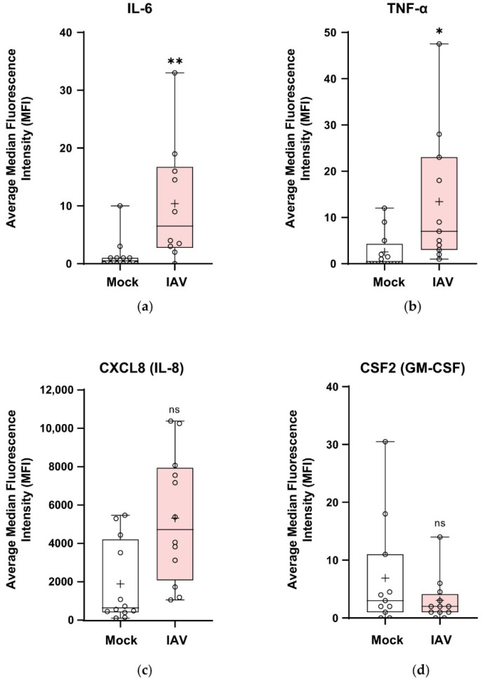
Figure 7.
The apical secretion of cytokine and chemokines by wdNHBE cells in response to infection with IAV H1N1pdm09. At 30 h postinfection, the proinflammatory cytokines IL-6, TNF-α, CSF2 and the CXCL8 chemokine secreted into the apical compartment by wdNHBE inoculated with H1N1pdm09 or mock-infected were assayed by multiplex immunoassays. (a) IL-6, (b) TNF-α, (c) CXCL8 and (d) CSF2. ALI media was added to the upper surface and the supernatant collected after a 15 min incubation. Data was pooled from three independent experiments and the average of the median fluorescence intensity (MFI) presented as box and whisker plots showing the mean (+), median, interquartile range and maximum and minimum values. The ends of each box represent the upper and lower quartiles, the horizontal line within the box shows the median value and error bars indicate the minimum and maximum values. Individual values are depicted by open circles. IAV apical cytokines: n = 12 for all treatments, with the exception of IAV, IL-6 (n = 10); IAV TNF-α (n = 11) and Mock CSF2 (n = 11); ns, not significant; *, p < 0.05; **, p < 0.01.

