

Abstract
The giant cell tumor of tendon sheath (GCTTS) or nodular tenosynovitis arises as discrete solitary nodule in the tendon sheath near joints of toes and fingers. Multifocal giant cell tumor of tendon sheath is a rare entity, of which the etiology is not yet fully understood and it is different from diffuse type of GCTTS. Diffuse type of GCTTS occurs around large joints having a main mass from which a small satellite nodule may arise. Multifocal GCTTS along a single tendon is a more rare entity. Herein, we describe a case of multifocal GCTTS along the tendon sheath of flexor digitorum profundus tendon of index finger. The patient was managed by surgical excision of the tumor swellings with no recurrence at two years follow up.
Keywords: Giant cell tumor of tendon sheath, Tendon, Tumor
1. Introduction
Giant cell tumor of tendon sheath (GCTTS) is the most common tumor of hand, only behind the ganglion cyst of hand.1 The occurrence of GCTTS is more commonly in women than in men, typically, it has a peak incidence between 3rd and 4th decade of life.2 Usually, the patient presents with a painless swelling in fingers, which is, located subcutaneously.3 Giant cell tumors of tendon sheath are usually solitary nodules. Multifocal giant cell tumor of tendon sheath is a rare entity.4 We report a patient with a multifocal giant cell tumor of tendon sheath arising along the flexor digitorum profundus tendon of the index finger.
2. Case report
A 44-year old, right-hand dominant, man presented with insidious onset, painless, lobulated swellings on the volar aspect of the index finger of the left hand. The swelling in the palm appeared first and the swelling around volar aspect of middle phalanx of the index finger appeared in the last, over period of six months. The patient was having difficulty in gripping objects due to swellings and occasional pain around the swellings. There was no history of trauma to the finger. Physical examination revealed four, subcutaneous, non-tender swellings over the volar aspect of index finger and palm (Fig. 1). The sizes of the swellings were 2 × 2 cm, 1 × 1 cm, 1 × 1cm and 3 × 2 cm from distal to proximal. There was no sensory impairment and the digital Allen’s test was negative for finger. The digital Allen’s test was done by doing gentle retrograde massage of the finger, till the finger become relatively pale, and then both radial digital and ulnar digital arteries were compressed with thumbs of both hands of the examiner separately. The pressure over the radial digital artery was released, it was observed that the finger became red again. The similar steps were repeated and the pressure over the ulnar digital vessel was released, the finger turned red again. The distal interphalangeal joint (DIP) range of motion was 0–70°. The range of motion of proximal interphalangeal joint (PIPJ) was 0–110°. There was terminal restriction of PIPJ, possibly due to swellings coming in close contact of each other at full flexion. The metacarpophalangeal joint (MCPJ) movement was 0–90°. The radiographs of the hand revealed soft tissue swellings over the volar aspect of hand. Magnetic resonance imaging (MRI) (Fig. 2) of the hand showed four separate swellings arising from the flexor tendons of index finger. These were iso-intense in T1 weighted images and hyper-intense in T2 weighted images. The MRI appearance suggested solid benign swellings arising from the flexor tendons. The patient underwent FNAC of the swelling which showed picture of giant cell tumor of tendon sheath.
Fig. 1.

The figure depicts the clinical photograph of the hand. The four swellings are marked with the asterixalong the course of flexor tendons of index finger of left hand.
Fig. 2.

The multiple GCTTS are seen arising from the flexor tendons in MRI of the hand. These were iso-intense in T1 weighted images (marked with white stars) and hyper-intense solid swellings in T2 weighted images (marked with black stars); in sagittal sections of MRI.
Total excision of the GCTTS was done. Intra-operatively, the lesions were separate from each other (Fig. 3). The A2, A3 pulleys were opened at the site of the tumors. The proximal most tumor was excised by doing tenosynovectomy. All four lesions were arising all along the flexor digitorum profundus (FDP). There was no involvement of any joint or the volar plates. The reconstruction of A2 pulley was not done, as it was excised partially only. Grossly, the tumor was yellowish brown in colour and firm in consistency (Fig. 4). The histopathology findings confirmed the diagnosis of GCTTS (Fig. 5).
Fig. 3.

Intraoperatively, the lesions were arising from the flexor tendons of index finger.
Fig. 4.
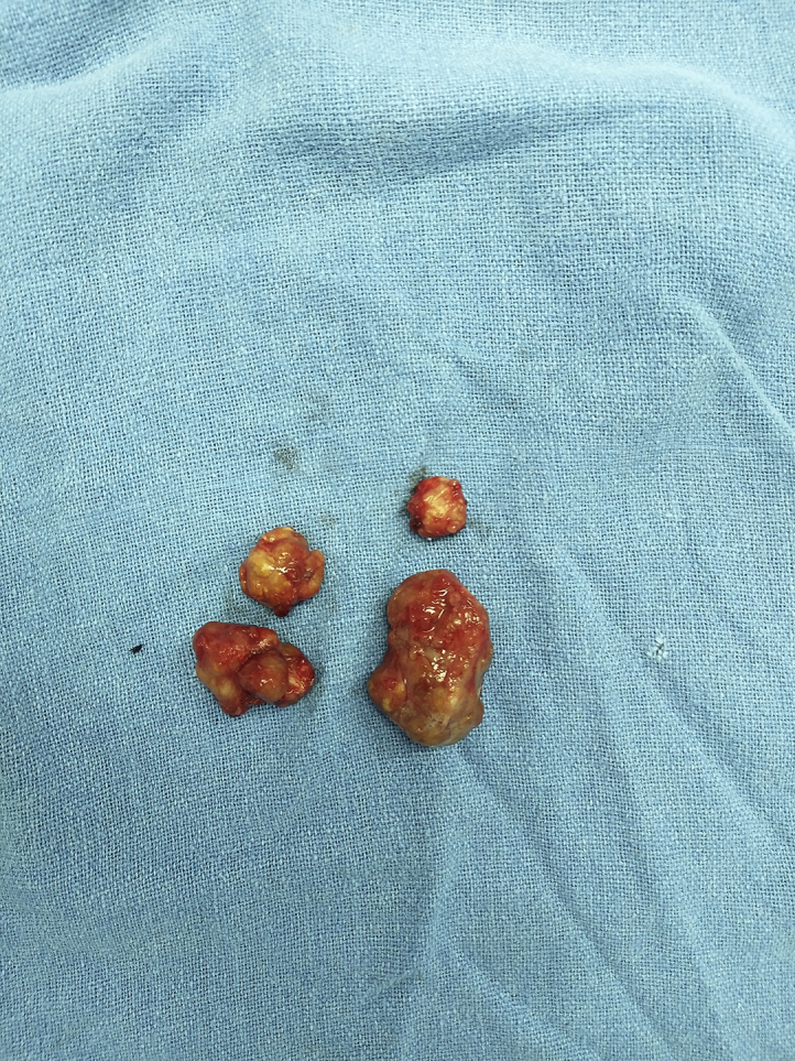
The measurement of the four nodules from distal to proximal was as:1 × 1 cm, 2 × 2cm, 3 × 2 cm and 3 × 3 cm.
Fig. 5.

Hematoxylin and eosin (HE) stained sections showed a nodular lesion with well-defined border (a, HE x40). It was comprised of mononuclear cells in diffuse arrangement admixed with scattered osteoclast-like giant cells (b, HE x100) and at places foam cells infiltration (c, HE x100). The mononuclear cells had histiocytic morphology with vesicular nuclei, small nucleolus, presence of nuclear grooves and vacuolated to pale eosinophilic cytoplasm (d, HE x400).
At two years follow up, the patient did not show any recurrence and he was able to do his normal day to day activities. The distal interphalangeal joint (DIP) range of motion was 0–70°. The range of motion of proximal interphalangeal joint (PIPJ) was 0–140°. The metacarpophalangeal joint (MCPJ) movement was 0–90°.
3. Discussion
It is rare to have multifocal GCTTS arising from same tendon sheath as it has been described in different case reports (Table 1). Ushijima et al. did a study of 207 patients of GCTTS, two tumors arising at different sites was noted; one in right ring finger and other in left great toe and it was observed that there was no lesion in same tendon.2 In retrospective analysis of 71 patients by Monaghan et al., there was no incidence of multifocal GCTTS.4 The first case of multifocal GCTTS along a single tendon was described in 2002 by Hitora et al.5 It involved the index finger and there was description of three nodules. Park et al. reported a case of 33 year old male who had 2 separate lesions in the tendon of FPL.3There was a gap of 7 months between the appearances of 2 lesions.3 Singh et al. reported multifocal GCTTS which had 5 lesions. Two lesions were noted before the operation on gross examination. Intra-operatively, five lesions were found.6Altaykan et al. reported a case of multifocal GCTTS in little finger with two lesions along the same tendon (FDS)9. Although, there was no recurrence in any of the reported case, the follow up of these patients has been less than two years. There is a chance that in case of multifocal GCTTS, the swelling may come in different time frame and it may be labelled as recurrence but in fact these were multifocal GCTTS along the same tendon. Zenistra et al. reported a case as a recurrence of GCTTS at the level of DIP joint and proximal to the wrist where the primary tumor was excised from the flexor tendons of the same finger at the level of the MCPJ. .8
Table 1.
The review of literature has shown very few case reports where the multifocal GCTTS has been reported along a single tendon. The reports are mentioned below in a tabulated form.
| Serial No | Study done by | Place and Year of study | Patient Age/Sex | Tendon of origin and digit | Number of GCTTS | Follow up and recurrence |
|---|---|---|---|---|---|---|
| 1. | Hitora et al.5 | JAPAN; 2002 | 76yr/female | Flexor tendon – indices, no specification available | 3 | 6 months/No recurrence |
| 2. | Park et al.3 | Korea; 2006 | 33yr/Male | Flexor pollicis longus | 2 | 1 year/No recurrence |
| 3. | Singh et al.6 | UK; 2009 | 37yr/Male | Flexor digitorum superficialis tendon of the little finger | 5 | 2 year/No recurrence |
| 4. | Altaykan et al.7 | Turkey; 2009 | 26 yr/male | Flexor digitorum superficialis tendon of the little finger | 2 | 6 months/no recurrence |
| 5. | Zeinstra et al.8 | Netherlands; 2011 | 58 yr/male | Flexor digitorum superficialis tendon of the third finger | 3 | Follow up not mentioned |
The description of the GCT along the tendon length as separate swellings suggests that there may be travelling tumor cells along the synovial fluid of the tendon sheath. These tumor cells would have stucked along the narrowest point. The presence of the tumors more commonly in border digits (little finger) and near the joint further supports this hypothesis, as these are the narrowest points along the tendon sheath. In our case, the proximal most swelling appeared first and the distal most swelling appeared in the last. It appears that the tumor cells would have travelled from this swelling to distally in finger, giving rise to appearance of tumor to other sites. Multifocal GCTTS along the single tendon sheath should be considered to have high recurrence because the tumor may be of true multicentric origin in different time frames.
In conclusion, multifocal GCTTS along a single tendon is a rare entity. We suggest that MRI of whole hand should be done in every case of GCTTS, so that multifocal tumors are not missed. Complete excision of all tumor swellings is essential.
Funding statement
The authors received no financial support for research, authorship, and/or publication of this article.
Declaration of competing interest
The authors declared no potential conflicts of interest with respect to the research, authorship, and/or publication of this article.
Acknowledgements
Nil.
References
- 1.Sapra S., Prokopetz R., Murray A.H. Giant cell tumor of tendon sheath. Int J Dermatol. 1989;28(9):587–590. doi: 10.1111/j.1365-4362.1989.tb02533.x. [DOI] [PubMed] [Google Scholar]
- 2.Ushijima M., Hashimoto H., Tsuneyoshi M., Enjoji M. Giant cell tumor of the tendon sheath (nodular tenosynovitis). A study of 207 cases to compare the large joint group with the common digit group. Cancer. 1986;57(4):875–884. doi: 10.1002/1097-0142(19860215)57:4<875::aid-cncr2820570432>3.0.co;2-y. [DOI] [PubMed] [Google Scholar]
- 3.Park J.W. Multiple separated giant cell tumors of the tendon sheath in a thumb. J Am Acad Dermatol. 2006;54(3):540–542. doi: 10.1016/j.jaad.2005.06.024. [DOI] [PubMed] [Google Scholar]
- 4.Monaghan H., Salter D.M., Al-Nafussi A. Giant cell tumor of tendon sheath (localised nodular tenosynovitis): clinicopathological features of 71 cases. J Clin Pathol. 2001;54(5):404–407. doi: 10.1136/jcp.54.5.404. [DOI] [PMC free article] [PubMed] [Google Scholar]
- 5.Hitora T., Yamamoto T., Akisue T. Multicentric localized giant cell tumor of the tendon sheath: two separate lesions at different sites in a finger. Br J Dermatol. 2002;147(2) doi: 10.1046/j.1365-2133.2002.484115.x. 403–5. [DOI] [PubMed] [Google Scholar]
- 6.Singh T., Noor S., Simons A.W. Multiple localized giant cell tumor of the tendon sheath (GCTTS) affecting a single tendon: a very rare case report and review of recent cases. Hand Surg. 2011;16:367–369. doi: 10.1142/S0218810411005710. [DOI] [PubMed] [Google Scholar]
- 7.Altaykan A., Yildiz K., Hapa O., Cukur S. Multifocal giant cell tumor of the tendon sheath occurring at different localizations of the same tendon of a finger: a case report and review of the literature. Eklem Hastalik Cerrahisi. 2009;20(2):119–123. [PubMed] [Google Scholar]
- 8.Zeinstra J.S.F., Kwee R.M., Kavanagh E.C., van Hemert W.L.W., Adriaensen M.E. Multifocal giant cell tumor of the tendon sheath: case report and literature review. Skeletal Radiol. 2013;42(3):447–450. doi: 10.1007/s00256-012-1552-9. [DOI] [PubMed] [Google Scholar]