Abstract
After decades of pioneering and improvement, kidney transplantation is now the renal replacement therapy of choice for most patients with end-stage kidney disease (ESKD). Where focus has traditionally been on surgical techniques and immunosuppressive treatment with prevention of rejection and infection in relation to short-term outcomes, nowadays, so many people are long-living with a transplanted kidney that lifestyle, including diet and exposure to toxic contaminants, also becomes of importance for the kidney transplantation field. Beyond hazards of immunological nature, a systematic assessment of potentially modifiable—yet rather overlooked—risk factors for late graft failure and excess cardiovascular risk may reveal novel targets for clinical intervention to optimize long-term health and downturn current rates of premature death of kidney transplant recipients (KTR). It should also be realized that while kidney transplantation aims to restore kidney function, it incompletely mitigates mechanisms of disease such as chronic low-grade inflammation with persistent redox imbalance and deregulated mineral and bone metabolism. While the vicious circle between inflammation and oxidative stress as common final pathway of a multitude of insults plays an established pathological role in native chronic kidney disease, its characterization post-kidney transplant remains less than satisfactory. Next to chronic inflammatory status, markedly accelerated vascular calcification persists after kidney transplantation and is likewise suggested a major independent mechanism, whose mitigation may counterbalance the excess risk of cardiovascular disease post-kidney transplant. Hereby, we first discuss modifiable dietary elements and toxic environmental contaminants that may explain increased risk of cardiovascular mortality and late graft failure in KTR. Next, we specify laboratory and clinical readouts, with a postulated role within persisting mechanisms of disease post-kidney transplantation (i.e., inflammation and redox imbalance and vascular calcification), as potential non-traditional risk factors for adverse long-term outcomes in KTR. Reflection on these current research opportunities is warranted among the research and clinical kidney transplantation community.
Keywords: nephrology, kidney transplant, kidney transplant recipients, long-term outcomes, graft failure, cardiovascular mortality, lifestyle, inflammation, vascular calcification, bone mineral density, dual-energy X-ray absorptiometry
1. Introduction
Chronic kidney disease (CKD) is a major public health problem, with a current worldwide prevalence of approximately 843 million individuals [1]. Global mean prevalence was recently reported at 13.4% for all CKD stages together (1–5) and at 10.6% if only the more severe CKD stages (3–5) are considered [2]. Whereas the prevalence of all stages of CKD rises with age, older patients with similar levels of eGFR are less likely than their younger counterparts to progress to the need of renal replacement therapy, which has raised the question of whether all older patients who meet criteria for CKD actually have CKD [3].
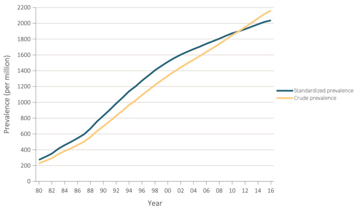
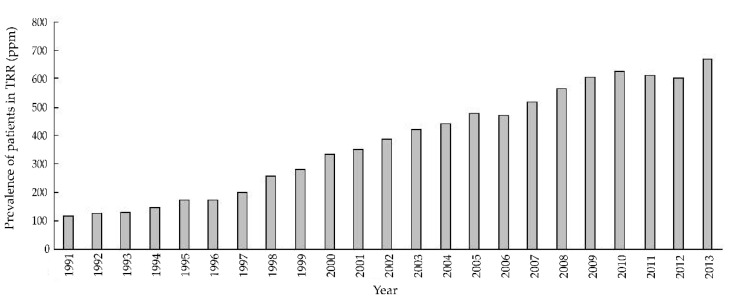
The prevalence of CKD, its detection, treatment, and impact on health have been mainly studied in economically developed countries [1]. Nevertheless, even in these circumstances, it usually remains a silent, smoldering health threat, with, e.g., rates of awareness of being afflicted with kidney disease of approximately 10% among patients with CKD in an economically developed country like the United States [4]. Along the same line, in 2016, approximately 35% of patients diagnosed with incident end-stage kidney disease (ESKD) received little or no nephrology care prior to actually being diagnosed with ESKD [4]. Regrettably, prevalence of ESKD and prevalence of renal replacement therapy continue to increase (Figure 1 and Figure 2) [4].
Figure 1.
Prevalence of end-stage kidney disease (ESKD) in the United States (US) population, 1980–2016. This figure shows a steady increase in ESKD prevalence over recent ~35 years in the US. Standardized for age, sex, and race. Data Source: USRDS 2018 Annual Data Report [4].
Figure 2.
Prevalence of renal replacement therapy in Latin America, 1991−2013. This figure shows a steady increase in prevalence of renal replacement therapy over recent ~25 years in Latin America. Reprinted from “Latin American Dialysis and Transplant Registry: Experience and contributions to end-stage kidney disease epidemiology” [5].
Compared to chronic dialysis treatment, kidney transplantation is considered the renal replacement therapy of choice and the gold-standard treatment for most ESKD patients because it offers superior cost-effectiveness, quality of life, and life expectancy [6,7,8,9,10]. However, the latter has largely been due to significant improvements of short-term outcomes [11]. Advances in immunosuppression, tissue typing, treatment of infections, and surgical techniques led rates of 1-year graft survival at a pinnacle, whereas improvement of long-term outcomes post-transplant remains a major challenge in the kidney transplantation field [11].
On the one hand, the life-saving benefit of a kidney transplant remains largely hampered by cumulative injury of a multitude of hazards through immune and non-immune mechanisms of kidney damage. Over time, these mechanisms lead to chronic interstitial fibrosis and tubular atrophy as histopathological consequence and end-stage kidney allograft failure as functional repercussion, eventually requiring restart of dialysis or re-transplantation as final adverse clinical event (i.e., graft failure) [11,12,13,14,15].
On the other hand, kidney transplant recipients (KTR) are at particularly high risk of premature death, depicting overall mortality rates considerably higher than that of age-matched controls in the general population [16,17].
Indeed, approximately half of all kidney allograft losses are due to premature death with a functioning graft, a long-standing pattern that has remained largely unchanged over recent years [17,18].
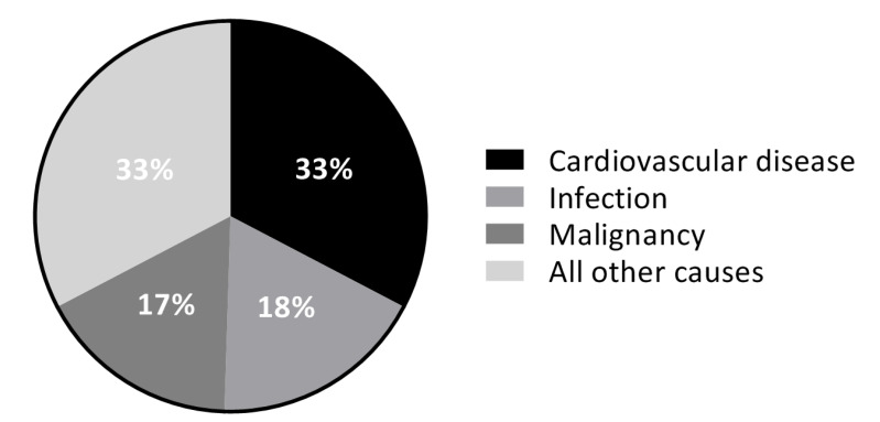
Next, under the general understanding that cardiovascular disease is the leading cause of premature death post-kidney transplant (Figure 3) and thereby importantly challenging the improvement of longevity of KTR, great efforts have focused on the improvement of long-term cardiovascular outcomes [19,20,21].
Figure 3.
Mortality by causes of death with graft function in US KTR in 2015. This figure shows that cardiovascular disease was the leading cause of mortality among US KTR in 2015. Cardiovascular disease included acute myocardial infarction, atherosclerotic heart disease, congestive heart failure, cerebrovascular accident, and arrhythmia/cardiac arrest. Adapted from USRDS 2018 Annual Data Report [4].
In the clinical setting of KTR after the first-year post-transplant, beyond hazards of immunological nature, there is a pressing need to systematically study and characterize the clinical impact of potentially modifiable risk factors, such as lifestyle, diet, and exposure to toxic contaminants, which are underexplored areas in the kidney transplantation field [22,23,24,25,26]. This evidence is needed to guide decision making by clinicians and policy-makers in post-transplantation care. Furthermore, because kidney transplantation aims to restore kidney function but it incompletely mitigates collateral mechanisms of disease, such as chronic low-grade inflammation with persistent redox imbalance and deregulated mineral and bone metabolism, further research investigating specific clinical and laboratory readouts with a proposed involvement in such pathological pathways may point towards non-traditional risk factors and reveal novel targets for clinical intervention [27,28,29,30,31,32].
In the kidney transplantation field, future advances are expected from amelioration of adverse long-term outcomes by increasing recognition and developing novel, early, and cost-effective risk-management strategies focused on the non-immune aspects of post-kidney transplantation care and thus optimize long-term health and downturn current rates of premature death in stable KTR [11].
2. Lifestyle: Healthy Diet and Toxic Contaminants
One area with great potential for improvement is lifestyle, in particular diet and exposure to toxic contaminants. Systematic investigation of traditional and potentially modifiable risks factors in the post-kidney transplant setting may point towards otherwise overlooked early risk-management opportunities and thus provide the basis for the development of cost-effective interventional approaches to increase the lifespan of KTR. Healthy diet is a cornerstone element of cardio-metabolic health in the general population [33,34,35,36,37,38]. In general, a healthy diet is recommended as essential for cardiovascular disease prevention in all individuals. Surprisingly, however, little is known about the potential impact of a healthy diet on cardiovascular health and survival benefit in kidney patients across the continuum of CKD stages, in patients undergoing kidney replacement therapy, and remarkably limited evidence is available in the post-kidney transplantation clinical setting [39,40,41,42]. Moreover, native CKD and pre-transplant ESKD patients are generally advised to follow seemingly conflicting and challenging dietary recommendations with the aim of restricting individual nutrients such as potassium, salt, phosphorus, and protein [43]. It should be realized that there is scant evidence to support such restrictive dietary recommendations [44,45,46]. Finally, there is a notorious lack of studies aimed to aid on the development of evidence-based recommendations to appropriately adjust any pre-transplant dietary advice to the patient after kidney transplantation has been performed [26,43,44,47,48]. Below, we provide several examples of where opportunities may lie (Box 1).
Box 1. Characteristics of a healthy diet [49].
≥200 g of fruit per day (2–3 servings).
≥200 g of vegetables per day (2–3 servings).
Fish 1–2 times per week, one of which to be oily fish.
Saturated fatty acids to account for <10% of total energy intake through replacement by polyunsaturated fatty acids.
Trans unsaturated fatty acids: as little as possible, preferably no intake from processed food and <1% of total energy intake from natural origin.
30 g unsalted nuts per day.
<5 g of salt per day.
Consumption of alcoholic beverages should be limited to 2 glasses per day (20 g/d of alcohol) for men and 1 glass per day (10 g/d of alcohol) for women.
Sugar-sweetened soft drinks and alcoholic beverages consumption must be discouraged.
2.1. Fruit and Vegetable Consumption Post-Kidney Transplantation
With the aim of limiting potassium intake, for example, pre-transplant ESKD patients have largely been discouraged from a high consumption of fruits and vegetables, which are, however, well-known essential components of a healthy diet [50,51,52,53,54]. Beyond being rich in potassium, fruits and vegetables are rich in fibers, polyunsaturated and monounsaturated fatty acids, magnesium, iron, and generate less acid and contain smaller amounts of saturated fatty acids, protein, and absorbable phosphorus in comparison to meat [39,55]. At least four servings of fruit and vegetables per day are widely recommended for the prevention of major chronic diseases in the general population [49]. Indeed, increased consumption of fruits and vegetables has consistently shown to confer superior cardiovascular prognosis in the general population [52,53,54,56].
Recent studies show that KTR consume less fruits and vegetables than the general population, which has been associated with higher risk of cardiovascular mortality and posttransplant diabetes [57,58]. At present, however, post-kidney transplant, there is no clear incentive by transplant healthcare providers to prescribe restoration of the consumption of these basic items of a healthy diet. This attitude may respond to the fact that it remains relatively unexplored whether an increase of fruits and vegetables consumption post-kidney transplantation positively impacts outcomes of KTR, which would be hypothetically expected mainly by decreasing the excess cardiovascular burden and premature cardiovascular death. Epidemiological studies aimed to estimate a theoretical benefit of a relative increase of these specific food items are warranted as first step to, thereafter, investigate potential interventional strategies promoting novel, cost-effective, and patient-centered approaches to the nutritional management of KTR, adequately informing clinical practice and policy.
2.2. Fish Intake Post-Kidney Transplantation and Mercury Exposure
Similarly, fish are rich in the omega-3 polyunsaturated fatty acids (n-3 PUFA) EPA (eicosapentaenoic acid) and DHA (docosahexaenoic acid), which are suggested to yield several beneficial effects for cardiovascular health [59,60,61,62]. Circulating levels of EPA and DHA have been associated with reduced cardiovascular risk in both healthy populations and in patients with pre-existing cardiovascular disease [59,60,61,62]. Proposed beneficial health effects of marine-derived n-3 PUFA are wide-ranging, favorably impacting inflammation, fibrosis, lipid modulation, plaque stabilization, blood pressure, artery calcification processes, and endothelial function [63,64,65,66]. These properties render EPA and DHA as of encompassing therapeutic potential in the management of cardiovascular risk of KTR. Indeed, in this particular setting, recent observational studies showed that plasma levels of marine-derived n-3 PUFA are inversely associated with cardiovascular mortality risk [67,68].
It should be realized, however, that the results of randomized control trials using supplementation of these individual nutrients are not yet sufficiently powered to draw definitive conclusions and recommendations for KTR [69,70]. Moreover, no study has been devoted to evaluating the potential beneficial effect of a relatively high dietary fish intake, as mostly shown in the general population [71,72,73,74,75]. Indeed, fish is the main dietary source of n-3 PUFA, and its inclusion in diet seems reasonable because it is a good source of protein without potentially adverse effects of accompanying intake of high saturated fat as present in fatty meat products. Not exempt of drawbacks, however, fish is also the major source of human exposure to organic mercury (with the exception of industrial accidents or particular occupational exposures) [76,77,78]. Therefore, alongside the study of the potential health benefits of marine-derived n-3 PUFA, weighted investigation of a relatively higher fish intake has been performed as necessary step towards developing cautious evidence-based dietary guidelines for clinical uptake [79], suggesting that beneficial effects of a higher dietary intake of n-3 PUFA by increasing fish consumption post-kidney transplantation may not be mitigated by postulated increased cardiovascular risk due to concomitant exposure to mercury [79].
2.3. Cadmium Exposure and Nephrotoxicity in the Post-Kidney Transplant Setting
Cadmium is another heavy metal of environmental and lifestyle-related concern, with tobacco and diet as primary sources of exposure. Previous studies have demonstrated that cadmium may induce hypertension, which in turn is associated with accelerated kidney function decline and particularly demonstrated in KTR, by shortened allograft survival [80,81,82,83,84]. Most importantly, a strong body of evidence shows that the kidney is the most sensitive target organ of cadmium-induced body burden, through postulated direct mechanisms of cadmium-induced injury in this organ, wherein it accumulates with a half-life of up to 45 years [85,86,87,88,89]. It is important to note that, particularly in settings of long-term oxidative stress such as that of KTR, cadmium-induced nephrotoxicity may be associated with impaired kidney function at concentrations that are otherwise considered non-toxic [90,91,92]. Taking also into account that the most effective way to reduce cardiovascular disease in KTR may indeed be preservation of graft function, the aforementioned constellation of factors turn the investigation of cadmium-associated risk of encompassing relevance within the study of long-term outcomes of kidney allograft function [21,84]. Furthermore, bodily cadmium is susceptible to therapeutic interventions [93]. Thus, cadmium-targeted interventional strategies may offer novel opportunities to decrease the long-standing high burden of late kidney graft failure; however, whether the nephrotoxic exposure to cadmium represents an overlooked hazard for preserved graft functioning remains unknown.
3. Inflammation and Oxidative Stress and Vascular Calcification
Another area of great opportunities for further improvement may lie in a better evaluation of disease mechanisms long-term after transplantation. Traditional risk factors such as diabetes mellitus, smoking, and hypertension, among others, do not suffice to account for the excess burden of premature cardiovascular death of, otherwise, stable KTR [94,95,96,97]. Indeed, cardiovascular disease has an atypical nature in KTR when compared with the general population [20,21]. Unexplained cardiovascular risk subsidizes current efforts to provide cutting-edge evidence on the potential independent hazard of novel (non-traditional) cardiovascular risk factors post-kidney transplantation [98,99,100,101,102].
It should be taken into account that while kidney transplantation aims to restore kidney function, it incompletely abrogates mechanisms of disease. Moreover, an aggregate of factors specific to the transplant milieu such as a chronic low-grade immunologic response to the kidney allograft, long-term toxicity of maintenance immunosuppressive, as well as various degrees of progressive uremia, contribute to perpetuate chronic inflammation, redox imbalance, and deregulated mineral and bone metabolism, which have to be proposed as major independent and evolving pathophysiological mechanisms, whose mitigation may counterbalance—at least to a considerable extent—the excess risk of cardiovascular disease and graft failure post-kidney transplantation [30,32,101,103,104]. Below, we provide several examples of where opportunities may lie.
3.1. Inflammation and Oxidative Stress Post-Kidney Transplantation
Indeed, while the vicious circle between inflammation and oxidative stress as final common pathway of a multitude of insults plays an established pathological role in native chronic kidney disease (CKD), its characterization post-kidney transplant has been less than satisfactory [105,106,107,108,109]. This is relevant because, at a physiological level, the cornerstone role of the complex interplay between inflammation and oxidative stress (Box 2) provides a theoretical and conceptual framework upon which upcoming research may deepen the understanding of the pathophysiological status of KTR once they reach a seemingly stable clinical stage [105].
Box 2. Oxidative stress.
Oxidative stress is defined as an imbalance between the generation and removal of oxidant species. The most representative biological oxidant agents are reactive oxygen species (ROS) and reactive nitrogen species (RNS). The former group includes hydrogen peroxide, superoxide anion, and hydroxyl radical, whereas within the latter group relevant species are peroxynitrite anion, nitric oxide, and nitrogen dioxide radicals. Oxidative stress occurs when ROS and/or RNS production overwhelms the endogenous antioxidant defense system, either by excess production and/or inadequate removal. The antioxidant defense system is constituted by enzymatic antioxidant agents, including catalase, glutathione peroxidase, and superoxide dismutase. Non-enzymatic antioxidant components include a diversity of biological molecules, such as ascorbic acid (vitamin C), α-tocopherol (vitamin E), reduced glutathione, carotenoids, flavonoids, polyphenols, and several other exogenous antioxidants [110].
3.1.1. Vitamin C as Anti-Inflammatory and Antioxidant Agent and Its Depletion Post-Kidney Transplant
Inflammation, specifically the established inflammatory biomarker high-sensitivity C-reactive protein (hs-CRP)—which is also an indirect marker of increased oxidant production—has been previously shown to be independently associated with increased mortality risk in KTR [98,100]. Supported by data consistently showing an inverse correlation with hs-CRP in different settings, vitamin C is well-known by its anti-inflammatory effects [111,112,113,114]. Moreover, vitamin C is a physiological antioxidant agent, with radical-scavenger and reducing activities, of paramount importance for protection against diseases and degenerative processes caused by oxidant stress [115]. This particular composite of biochemical properties renders vitamin C as compelling research candidate to broaden the understanding of the interaction of inflammation and oxidative stress in the mechanisms leading to excess risk of premature death post-kidney transplantation. It should be realized, moreover, that pre-transplant ESKD patients often have an imbalance of several critical trace elements and vitamins [39]. Vitamin C, particularly, has been shown to be removed by conventional hemodialysis membranes, leading to drastic vitamin C depletion and oxidative stress [116,117,118]. Through an inverse mediating effect on inflammatory signaling biomarkers, sub-physiological levels of vitamin C (depletion) may be hypothesized to be implicated in mechanisms that associate with increased risk of adverse long-term outcomes [119,120,121,122,123]. To date, however, relatively little is known regarding the prevalence of abnormal vitamin C status post-kidney transplantation, yet recent studies have shown that low plasma vitamin C contributes to excess risk for premature death post-kidney transplantation [124,125].
3.1.2. Advanced Glycation End products as Amplifiers of Oxidative Stress and Inflammatory Responses
Inflammation is referred to as a redox-sensitive mechanism on the basis that reactive oxygen species may activate transcription factors such as nuclear factor kappa B (NF-kB), which regulates inflammatory mediator genes expression [126]. In this regard, advanced glycation end products (AGE) are particularly interesting oxidative stress biomarkers because it has been demonstrated that, upon binding to AGE-specific receptors, AGE activate intracellular pathways that amplify inflammatory and oxidative stress responses and regulate the transcription of adhesion molecules through NF-kB activation [127]. In agreement, data derived from clinical studies in pre-transplant ESKD patients support the implication of AGE in the complex feedback loop between oxidative stress and inflammation leading to endothelial dysfunction and adverse cardiovascular effects [128,129,130].
Several studies have observed accumulation of AGE in native and transplant CKD patients, and a strong body of evidence on the general theory of AGE pathophysiology supports its pivotal role in the initiation and progression of mechanisms underlying cardiovascular disease. However, few attempts have been made to investigate the association of AGE with cardiovascular risk post-kidney transplantation [99,131]. Through a mediating effect on up-regulation of inflammatory, oxidative stress and endothelial dysfunction biomarkers, a relative increase of AGE may be hypothesized to actively contribute to the intracellular signaling pathways that ultimately yield excess risk of premature cardiovascular death in KTR. It remains unknown whether a hypothetical association with risk of cardiovascular mortality is independent of estimates of kidney function and traditional cardiovascular risk factors such as body mass index, diabetes, blood pressure, and smoking status.
3.1.3. Inflammation, Galectin-3, and Fibrosis
Inflammation is also referred to as a unifying mechanism of injury because—through a cornerstone signaling link with interstitial fibrosis and tubular atrophy—it may hold observations that connect hazards of several natures with structural damage and detrimental function of the kidney [12,13,14,15,132,133]. Of note, the concept that chronic rejection is responsible for all progressive long-term kidney graft failure has long ago been reformulated to a hypothesis of cumulative damage [12,13,14,15]. Thus, repeated insults of both immune and non-immune nature damage the graft by leading to interstitial fibrosis and tubular atrophy, which represents a final common pathway of injury with adverse functional consequences [13]. Galectin-3 is a β-galactoside-binding lectin with a postulated key mediating role on kidney tissue fibrosis [134,135,136,137,138]. In different models, it has been shown that whether a variety of insults incur on irreversible kidney fibrosis or not depends on the expression and secretion of galectin-3 [135,136,137,138]. In the general population, moreover, an increasing body of prospective evidence has related plasma galectin-3 with incident CKD [139,140,141]. Because galectin-3 is both a biomarker of systemic inflammation and kidney fibrosis, it may broaden our understanding and provide data to further support a unifying link between repeated inflammatory and pro-oxidant insults and increased risk of graft failure beyond the first-year post-kidney transplantation. Finally, it should be realized that the dependent role of galectin-3 on kidney fibrosis has been specifically shown in the particular post-kidney transplant setting in a murine model [138]. Within the clinical kidney transplantation field, however, a number of crucial questions remain unanswered. Especially with galectin-3, targeted pharmacological therapies are increasingly becoming available, and evidence of a hypothetical association between galectin-3 levels and risk of long-term graft survival may point towards novel interventional avenues to potentially decrease the long-standing burden of late graft failure.
3.2. Bone Disease and Vascular Calcification
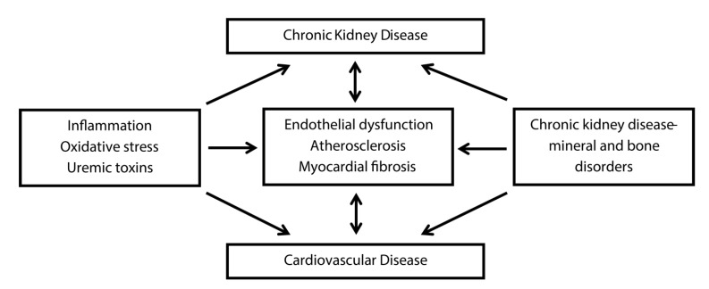
Chronic kidney disease-mineral and bone disorders (CKD-MBD) is the clinical entity or syndrome that KDIGO (Kidney Disease: Improving Global Outcomes) more than a decade ago has coined to embody the disruption of the complex systems biology enclosed by the kidney, skeleton, and cardiovascular system [142]. In line with previous evidence, the results of a recent elegant study by Yilmaz et al. support the hypothesis that decline in cardiovascular risk post-kidney transplantation depends on partial resolution of inflammation but also on resolution of the CKD-MBD [143,144]. The findings of the aforementioned research group support the notion that beyond restoration of organ function post-kidney transplant, amelioration of inflammation and correction of CKD-MBD may attenuate excess cardiovascular disease through separate biological pathways. In agreement, Cozzolino et al. recently depicted inflammation and oxidative stress, on one hand, and CKD-MBD, on the other hand, as major mechanisms underlying a feedback loop that exacerbates cardiovascular disease in CKD patients (Figure 4) [145].
Figure 4.
Cardiovascular disease in chronic kidney disease. This figure shows inflammation, oxidative stress, and uremic toxins on one side and chronic kidney disease-mineral and bone disorders on the other side of independent mechanisms linking chronic kidney and cardiovascular disease. Adapted from: “Cardiovascular disease in dialysis patients” by M. Cozzolino et al., 2019, Nephrol Dial Transplant, 33: iii28–34 [145].
Within the context of CKD-MBD, vascular calcification—a currently established cardiovascular risk factor in KTR, as shown by previous studies of our group and others [146,147,148,149,150,151]—is linked with bone disease through inter-related pathophysiological mechanisms that comprise the bone-vascular axis hypothesis, which contributes to the exceedingly high cardiovascular risk in native CKD [152,153,154,155,156]. Post-kidney transplant bone disease is certainly a topic of epidemiological relevance due to its high prevalence and its association with fragility fractures and reduced mobility [157,158,159,160,161,162]. Previous studies remarked that existing research had failed to explore a hypothetical contributing role of post-kidney transplant bone disease to increased risk of vascular calcification in KTR [154,158,163]. Recent evidence, however, has come to support the existence of a bone-vascular axis post-kidney transplantation, providing data to evaluate its epidemiological relevance post-kidney transplant and pointing towards an otherwise overlooked therapeutic opportunity to at least partially decrease the markedly high cardiovascular burden post-kidney transplant [164].
It has also been proposed that mediators of inflammation (e.g., interleukin 6 and tumor necrosis factor) contribute to fibroblast growth factor (FGF)-23 elevation and that, in turn, FGF-23 increases cytokine production, thus linking systemic inflammation with dysregulated phosphate metabolism in a vicious cycle [165,166]. It has been proposed that inflammatory mediators function as drug targets to decrease the burden of FGF23-associated injury in various tissues, thus offering a novel therapeutic opportunity to decrease the burden of cardiovascular diseases including vascular calcification in kidney disease patients [165,167]. Nevertheless, even in CKD patients within normal range of serum phosphate levels, vascular calcification is often observed. Calciprotein particles are calcium-phosphate nanoparticles that increase with CKD progression, which have been associated with inflammatory responses, endothelial damage, vascular stiffness, and calcification [168]. Calciprotein particles may play a pathophysiological role in the link between chronic inflammation and vascular calcification. Further research is warranted to evaluate its contribution to overall cardiovascular burden in KTR and to develop novel pharmacological strategies targeting calciprotein particles to encourage protection against the risk of vascular calcification post-kidney transplantation [169].
3.3. Immunosuppressive Therapy and Traditional Risk Factors of Vascular Calcification
The contribution of several traditional risk factors of vascular calcification may be particularly relevant in the post-kidney transplantation setting due to the effect of maintenance immunosuppressive therapy on diabetes, dyslipidemia, and vitamin D metabolism [170]. Previous studies have shown that low vitamin D along with low vitamin K may synergistically associate with higher risk of hypertension [171] and thereby contribute to higher risk of vascular calcification [172]. In KTR, particularly, we have recently shown that combined vitamin D and K deficiency is highly prevalent and is associated with increased mortality and graft failure [173]. Further research is needed to investigate both the direct and indirect role of immunosuppressive drugs in the progression of vascular calcification. There may, however, be opposing effects, because it has been described that steroids and calcineurin inhibitors inhibit inducible nitric oxide and may thereby lead to progression of vascular calcification through endothelial dysfunction [170], while mycophenolate mofetil inhibits vascular smooth muscle cell proliferation and may be protective against vascular calcification [174,175]. Similarly, we recently reported that use of cyclosporine rather than tacrolimus correlated with prevalence of osteopenia, while osteopenia was associated with higher risk of vascular calcification after kidney transplantation [164]. Future studies are warranted to assess the association between immunosuppressive agents and risk of vascular calcification, which may provide new cardiovascular risk management opportunities post-kidney transplantation.
4. Conclusions
Further research on lifestyle-related factors including diet and exposure to toxic contaminants, as well as persisting mechanisms of disease post-kidney transplantation (i.e., inflammation and redox imbalance and vascular calcification) is needed as it may bring about powerful opportunities to improve long-term outcomes post-kidney transplantation. Reflection on these current research opportunities is warranted among the research and clinical kidney transplantation community. Forthcoming analyses of the data to be generated by the long-lasting Transplant Lines Prospective Cohort Study and Biobank of Solid Organ Transplant Recipients [176] may shed light on these questions.
Author Contributions
Conceptualization, C.G.S. and S.J.L.B.; methodology, C.G.S., M.H.d.B., G.J.N., and S.J.L.B.; investigation, C.G.S. and S.J.L.B.; resources, C.G.S., M.H.d.B., and S.J.L.B.; writing—original draft preparation, C.G.S.; writing—review and editing, C.G.S., M.H.d.B., C.A.t.V.-K., G.J.N., and S.J.L.B.; supervision, M.H.d.B., G.J.N., and S.J.L.B.; project administration, C.G.S. and S.J.L.B.; funding acquisition, C.G.S. and S.J.L.B. All authors have read and agreed to the published version of the manuscript.
Funding
Camilo G. Sotomayor is supported by a personal grant from CONICYT (F 72190118).
Conflicts of Interest
The authors declare no conflict of interest. The funders had no role in the design of the study; in the collection, analyses, or interpretation of data; in the writing of the manuscript, or in the decision to publish the results.
Disclaimer
The data reported in Figure 1 have been supplied by the United States Renal Data System. The interpretation and reporting of these data are the responsibility of the author(s) of this manuscript and in no way should be seen as an official policy or interpretation of the U.S. government.
References
- 1.Jager K.J., Kovesdy C., Langham R., Rosenberg M., Jha V., Zoccali C. A single number for advocacy and communication-worldwide more than 850 million individuals have kidney diseases. Nephrol. Dial. Transpl. 2019;34:1803–1805. doi: 10.1093/ndt/gfz174. [DOI] [PubMed] [Google Scholar]
- 2.Hill N.R., Fatoba S.T., Oke J.L., Hirst J.A., O’callaghan C.A., Lasserson D.S., Hobbs F.D.R. Global Prevalence of Chronic Kidney Disease-A Systematic Review and Meta-Analysis. PLoS ONE. 2016;11:e0158765. doi: 10.1371/journal.pone.0158765. [DOI] [PMC free article] [PubMed] [Google Scholar]
- 3.Eriksen B.O., Ingebretsen O.C. The progression of chronic kidney disease: A 10-year population-based study of the effects of gender and age. Kidney Int. 2006;69:375–382. doi: 10.1038/sj.ki.5000058. [DOI] [PubMed] [Google Scholar]
- 4.United States Renal Data System . In: USRDS 2018 Annual Data Report: Atlas of Chronic Kidney Disease and End-Stage Renal Disease in the United States. Bethesda M., editor. National Institutes of Health; National Institute of Diabetes and Digestive and Kidney Diseases; Bethesda, MD, USA: 2018. [(accessed on 8 September 2019)]. Available online: https://www.usrds.org/Default.aspx. [Google Scholar]
- 5.Cusumano A.M., Rosa-Diez G.J., Gonzalez-Bedat M.C. Latin American Dialysis and Transplant Registry: Experience and contributions to end-stage renal disease epidemiology. World J. Nephrol. 2016;5:389. doi: 10.5527/wjn.v5.i5.389. [DOI] [PMC free article] [PubMed] [Google Scholar]
- 6.Mohnen S.M., Van Oosten M.J.M., Los J., Leegte M.J.H., Jager K.J., Hemmelder M.H., Logtenberg S.J.J., Stel V.S., Hakkaart-Van Roijen L., De Wit G.A. Healthcare costs of patients on different renal replacement modalities – Analysis of Dutch health insurance claims data. PLoS ONE. 2019;14:e0220800. doi: 10.1371/journal.pone.0220800. [DOI] [PMC free article] [PubMed] [Google Scholar]
- 7.Suthanthiran M., Strom T.B. Renal Transplantation. N. Engl. J. Med. 1994;331:365–376. doi: 10.1056/NEJM199408113310606. [DOI] [PubMed] [Google Scholar]
- 8.Wolfe R.A., Ashby V.B., Milford E.L., Ojo A.O., Ettenger R.E., Agodoa L.Y.C., Held P.J., Port F.K. Comparison of Mortality in All Patients on Dialysis, Patients on Dialysis Awaiting Transplantation, and Recipients of a First Cadaveric Transplant. N. Engl. J. Med. 1999;341:1725–1730. doi: 10.1056/NEJM199912023412303. [DOI] [PubMed] [Google Scholar]
- 9.Oniscu G.C., Brown H., Forsythe J.L.R. Impact of Cadaveric Renal Transplantation on Survival in Patients Listed for Transplantation. J. Am. Soc. Nephrol. 2005;16:1859–1865. doi: 10.1681/ASN.2004121092. [DOI] [PubMed] [Google Scholar]
- 10.Tonelli M., Wiebe N., Knoll G., Bello A., Browne S., Jadhav D., Klarenbach S., Gill J. Systematic Review: Kidney Transplantation Compared With Dialysis in Clinically Relevant Outcomes. Am. J. Transpl. 2011;11:2093–2109. doi: 10.1111/j.1600-6143.2011.03686.x. [DOI] [PubMed] [Google Scholar]
- 11.Lamb K.E., Lodhi S., Meier-Kriesche H.U. Long-term renal allograft survival in the United States: A critical reappraisal. Am. J. Transpl. 2011;11:450–462. doi: 10.1111/j.1600-6143.2010.03283.x. [DOI] [PubMed] [Google Scholar]
- 12.Nankivell B.J., Borrows R.J., Fung C.L.-S., O’Connell P.J., Allen R.D.M., Chapman J.R. The natural history of chronic allograft nephropathy. N. Engl. J. Med. 2003;349:2326–2333. doi: 10.1056/NEJMoa020009. [DOI] [PubMed] [Google Scholar]
- 13.Nankivell B.J., Chapman J.R. Chronic allograft nephropathy: Current concepts and future directions. Transplantation. 2006;81:643–654. doi: 10.1097/01.tp.0000190423.82154.01. [DOI] [PubMed] [Google Scholar]
- 14.Nankivell B.J., Kuypers D.R.J. Diagnosis and prevention of chronic kidney allograft loss. Lancet. 2011;378:1428–1437. doi: 10.1016/S0140-6736(11)60699-5. [DOI] [PubMed] [Google Scholar]
- 15.Nankivell B.J., Borrows R.J., Fung C.L.-S., O’Connell P.J., Allen R.D.M., Chapman J.R. Natural history, risk factors, and impact of subclinical rejection in kidney transplantation. Transplantation. 2004;78:242–249. doi: 10.1097/01.TP.0000128167.60172.CC. [DOI] [PubMed] [Google Scholar]
- 16.Arend S.M., Mallat M.J.K., Westendorp R.J.W., Van Der Woude F.J., Van Es L.A. Patient survival after renal transplantation; more than 25 years follow-up. Nephrol. Dial. Transpl. 1997;12:1672–1679. doi: 10.1093/ndt/12.8.1672. [DOI] [PubMed] [Google Scholar]
- 17.Oterdoom L.H., de Vries A.P.J., van Ree R.M., Gansevoort R.T., van Son W.J., van der Heide J.J.H., Navis G., de Jong P.E., Gans R.O.B., Bakker S.J.L. N-Terminal Pro-B-Type Natriuretic Peptide and Mortality in Renal Transplant Recipients Versus the General Population. Transplantation. 2009;87:1562–1570. doi: 10.1097/TP.0b013e3181a4bb80. [DOI] [PubMed] [Google Scholar]
- 18.U.S. Renal Data System . USRDS 2010 Annual Data Report: Atlas of Chronic Kidney Disease and End-Stage Renal Disease in the United States, National Institutes of Health. National Institute of Diabetes and Digestive and Kidney Diseases; Bethesda, MD, USA: 2010. [Google Scholar]
- 19.Ojo A.O., Hanson J.A., Wolfe R.A., Leichtman A.B., Agodoa L.Y., Port F.K. Long-term survival in renal transplant recipients with graft function. Kidney Int. 2000;57:307–313. doi: 10.1046/j.1523-1755.2000.00816.x. [DOI] [PubMed] [Google Scholar]
- 20.Shirali A.C., Bia M.J. Management of cardiovascular disease in renal transplant recipients. Clin. J. Am. Soc. Nephrol. 2008;3:491–504. doi: 10.2215/CJN.05081107. [DOI] [PMC free article] [PubMed] [Google Scholar]
- 21.Jardine A.G., Gaston R.S., Fellstrom B.C., Holdaas H. Prevention of cardiovascular disease in adult recipients of kidney transplants. Lancet. 2011;378:1419–1427. doi: 10.1016/S0140-6736(11)61334-2. [DOI] [PubMed] [Google Scholar]
- 22.Fry K., Patwardhan A., Ryan C., Trevillian P., Chadban S., Westgarth F., Chan M. Development of Evidence-Based Guidelines for the Nutritional Management of Adult Kidney Transplant Recipients. J. Ren. Nutr. 2009;19:101–104. doi: 10.1053/j.jrn.2008.10.010. [DOI] [PubMed] [Google Scholar]
- 23.Zelle D.M., Kok T., Dontje M.L., Danchell E.I., Navis G., van Son W.J., Bakker S.J.L., Corpeleijn E. The role of diet and physical activity in post-transplant weight gain after renal transplantation. Clin. Transpl. 2013;27:E484–E490. doi: 10.1111/ctr.12149. [DOI] [PubMed] [Google Scholar]
- 24.Nolte Fong J.V., Moore L.W. Nutrition Trends in Kidney Transplant Recipients: The Importance of Dietary Monitoring and Need for Evidence-Based Recommendations. Front. Med. 2018;5:302. doi: 10.3389/fmed.2018.00302. [DOI] [PMC free article] [PubMed] [Google Scholar]
- 25.Klaassen G., Zelle D.M., Navis G.J., Dijkema D., Bemelman F.J., Bakker S.J.L., Corpeleijn E. Lifestyle intervention to improve quality of life and prevent weight gain after renal transplantation: Design of the Active Care after Transplantation (ACT) randomized controlled trial. BMC Nephrol. 2017;18:296. doi: 10.1186/s12882-017-0709-0. [DOI] [PMC free article] [PubMed] [Google Scholar]
- 26.Sabbatini M., Ferreri L., Pisani A., Capuano I., Morgillo M., Memoli A., Riccio E., Guida B. Nutritional management in renal transplant recipients: A transplant team opportunity to improve graft survival. Nutr. Metab. Cardiovasc. Dis. 2019;29:319–324. doi: 10.1016/j.numecd.2019.01.002. [DOI] [PubMed] [Google Scholar]
- 27.Monier-Faugere M.-C., Mawad H., Qi Q., Friedler R.M., Malluche H.H. High Prevalence of Low Bone Turnover and Occurrence of Osteomalacia after Kidney Transplantation. J. Am. Soc. Nephrol. 2000;11:1093–1099. doi: 10.1681/ASN.V1161093. [DOI] [PubMed] [Google Scholar]
- 28.Sprague S.M., Belozeroff V., Danese M.D., Martin L.P., Olgaard K. Abnormal bone and mineral metabolism in kidney transplant patients—A review. Am. J. Nephrol. 2008;28:246–253. doi: 10.1159/000110875. [DOI] [PubMed] [Google Scholar]
- 29.Evenepoel P., Lerut E., Naesens M., Bammens B., Claes K., Kuypers D., Vermeersch P., Meijers B., Van Damme B., Vanrenterghem Y. Localization, etiology and impact of calcium phosphate deposits in renal allografts. Am. J. Transpl. 2009;9:2470–2478. doi: 10.1111/j.1600-6143.2009.02792.x. [DOI] [PubMed] [Google Scholar]
- 30.Kalantar-Zadeh K., Molnar M.Z., Kovesdy C.P., Mucsi I., Bunnapradist S. Management of mineral and bone disorder after kidney transplantation. Curr. Opin. Nephrol. Hypertens. 2012;21:389–403. doi: 10.1097/MNH.0b013e3283546ee0. [DOI] [PMC free article] [PubMed] [Google Scholar]
- 31.Lou I., Foley D., Odorico S.K., Leverson G., Schneider D.F., Sippel R., Chen H. How Well Does Renal Transplantation Cure Hyperparathyroidism? Ann. Surg. 2015;262:653–659. doi: 10.1097/SLA.0000000000001431. [DOI] [PMC free article] [PubMed] [Google Scholar]
- 32.Wolf M., Weir M.R., Kopyt N., Mannon R.B., Von Visger J., Deng H., Yue S., Vincenti F. A Prospective Cohort Study of Mineral Metabolism After Kidney Transplantation. Transplantation. 2016;100:184–193. doi: 10.1097/TP.0000000000000823. [DOI] [PMC free article] [PubMed] [Google Scholar]
- 33.Willett W. Diet and health: What should we eat? Science. 1994;264:532–537. doi: 10.1126/science.8160011. [DOI] [PubMed] [Google Scholar]
- 34.Stampfer M.J., Hu F.B., Manson J.E., Rimm E.B., Willett W.C. Primary Prevention of Coronary Heart Disease in Women through Diet and Lifestyle. N. Engl. J. Med. 2000;343:16–22. doi: 10.1056/NEJM200007063430103. [DOI] [PubMed] [Google Scholar]
- 35.Hu F.B., Manson J.E., Stampfer M.J., Colditz G., Liu S., Solomon C.G., Willett W.C. Diet, lifestyle, and the risk of type 2 diabetes mellitus in women. N. Engl. J. Med. 2001;345:790–797. doi: 10.1056/NEJMoa010492. [DOI] [PubMed] [Google Scholar]
- 36.Chiuve S.E., McCullough M.L., Sacks F.M., Rimm E.B. Healthy Lifestyle Factors in the Primary Prevention of Coronary Heart Disease Among Men. Circulation. 2006;114:160–167. doi: 10.1161/CIRCULATIONAHA.106.621417. [DOI] [PubMed] [Google Scholar]
- 37.Chiuve S.E., Rexrode K.M., Spiegelman D., Logroscino G., Manson J.E., Rimm E.B. Primary prevention of stroke by healthy lifestyle. Circulation. 2008;118:947–954. doi: 10.1161/CIRCULATIONAHA.108.781062. [DOI] [PMC free article] [PubMed] [Google Scholar]
- 38.Chiuve S.E., Fung T.T., Rexrode K.M., Spiegelman D., Manson J.E., Stampfer M.J., Albert C.M. Adherence to a low-risk, healthy lifestyle and risk of sudden cardiac death among women. JAMA. 2011;306:62–69. doi: 10.1001/jama.2011.907. [DOI] [PMC free article] [PubMed] [Google Scholar]
- 39.Kalantar-Zadeh K., Fouque D. Nutritional Management of Chronic Kidney Disease. N. Engl. J. Med. 2017;377:1765–1776. doi: 10.1056/NEJMra1700312. [DOI] [PubMed] [Google Scholar]
- 40.Kelly J.T., Palmer S.C., Wai S.N., Ruospo M., Carrero J.-J., Campbell K.L., Strippoli G.F.M. Healthy Dietary Patterns and Risk of Mortality and ESRD in CKD: A Meta-Analysis of Cohort Studies. Clin. J. Am. Soc. Nephrol. 2017;12:272–279. doi: 10.2215/CJN.06190616. [DOI] [PMC free article] [PubMed] [Google Scholar]
- 41.Chauveau P. Nutrition in chronic kidney disease: Nephrology dialysis transplantation notable advances in 2018. Nephrol. Dial. Transpl. 2019;34:893–896. doi: 10.1093/ndt/gfz077. [DOI] [PubMed] [Google Scholar]
- 42.Saglimbene V.M., Wong G., Ruospo M., Palmer S.C., Garcia-Larsen V., Natale P., Teixeira-Pinto A., Campbell K.L., Carrero J.-J., Stenvinkel P., et al. Article Fruit and Vegetable Intake and Mortality in Adults undergoing Maintenance Hemodialysis. Clin. J. Am. Soc. Nephrol. 2019;14:250–260. doi: 10.2215/CJN.08580718. [DOI] [PMC free article] [PubMed] [Google Scholar]
- 43.Levin A., Stevens P.E., Bilous R.W., Coresh J., De Francisco A.L.M., De Jong P.E., Griffith K.E., Hemmelgarn B.R., Iseki K., Lamb E.J., et al. Notice. Kidney Int. Suppl. 2013;3:1. doi: 10.1007/BF02805891. [DOI] [Google Scholar]
- 44.Palmer S.C., Hanson C.S., Craig J.C., Strippoli G.F.M., Ruospo M., Campbell K., Johnson D.W., Tong A. Dietary and fluid restrictions in CKD: A thematic synthesis of patient views from qualitative studies. Am. J. Kidney Dis. 2015;65:559–573. doi: 10.1053/j.ajkd.2014.09.012. [DOI] [PubMed] [Google Scholar]
- 45.Mcmahon E.J., Campbell K.L., Bauer J.D., Mudge D.W. Altered dietary salt intake for people with chronic kidney disease. Cochrane Database Syst. Rev. 2015;18:CD010070. doi: 10.1002/14651858.CD010070.pub2. [DOI] [PubMed] [Google Scholar]
- 46.Liu Z., Su G., Guo X., Wu Y., Liu X., Zou C., Zhang L., Yang Q., Xu Y., Ma W. Dietary interventions for mineral and bone disorder in people with chronic kidney disease. Cochrane Database Syst. Rev. 2015;16:CD010350. doi: 10.1002/14651858.CD010350.pub2. [DOI] [PMC free article] [PubMed] [Google Scholar]
- 47.Gordon E.J., Prohaska T.R., Gallant M., Siminoff L.A. Self-care strategies and barriers among kidney transplant recipients: A qualitative study. Chronic Illn. 2009;5:75–91. doi: 10.1177/1742395309103558. [DOI] [PMC free article] [PubMed] [Google Scholar]
- 48.Stanfill A., Bloodworth R., Cashion A. Lessons learned: Experiences of gaining weight by kidney transplant recipients. Prog. Transpl. 2012;22:71–78. doi: 10.7182/pit2012986. [DOI] [PubMed] [Google Scholar]
- 49.Piepoli M.F., Hoes A.W., Agewall S., Albus C., Brotons C., Catapano A.L., Cooney M.-T., Corrà U., Cosyns B., Deaton C., et al. European Guidelines on cardiovascular disease prevention in clinical practice. Eur. Heart J. 2016;37:2315–2381. doi: 10.1093/eurheartj/ehw106. [DOI] [PMC free article] [PubMed] [Google Scholar]
- 50.Lloyd-Jones D.M., Hong Y., Labarthe D., Mozaffarian D., Appel L.J., Van Horn L., Greenlund K., Daniels S., Nichol G., Tomaselli G.F., et al. Defining and setting national goals for cardiovascular health promotion and disease reduction: The american heart association’s strategic impact goal through 2020 and beyond. Circulation. 2010;121:586–613. doi: 10.1161/CIRCULATIONAHA.109.192703. [DOI] [PubMed] [Google Scholar]
- 51.Crowe F.L., Roddam A.W., Key T.J., Appleby P.N., Overvad K., Jakobsen M.U., Tjonneland A., Hansen L., Boeing H., Weikert C., et al. Fruit and vegetable intake and mortality from ischaemic heart disease: Results from the European Prospective Investigation into Cancer and Nutrition (EPIC)-Heart study. Eur. Heart J. 2011;32:1235–1243. doi: 10.1093/eurheartj/ehq465. [DOI] [PMC free article] [PubMed] [Google Scholar]
- 52.Lim S.S., Vos T., Flaxman A.D., Danaei G., Shibuya K., Adair-Rohani H., AlMazroa M.A., Amann M., Anderson H.R., Andrews K.G., et al. A comparative risk assessment of burden of disease and injury attributable to 67 risk factors and risk factor clusters in 21 regions, 1990–2010: A systematic analysis for the Global Burden of Disease Study 2010. Lancet. 2012;380:2224–2260. doi: 10.1016/S0140-6736(12)61766-8. [DOI] [PMC free article] [PubMed] [Google Scholar]
- 53.Larsson S.C., Virtamo J., Wolk A. Total and specific fruit and vegetable consumption and risk of stroke: A prospective study. Atherosclerosis. 2013;227:147–152. doi: 10.1016/j.atherosclerosis.2012.12.022. [DOI] [PubMed] [Google Scholar]
- 54.Zhang X., Shu X.-O., Xiang Y.-B., Yang G., Li H., Gao J., Cai H., Gao Y.-T., Zheng W. Cruciferous vegetable consumption is associated with a reduced risk of total and cardiovascular disease mortality. Am. J. Clin. Nutr. 2011;94:240–246. doi: 10.3945/ajcn.110.009340. [DOI] [PMC free article] [PubMed] [Google Scholar]
- 55.Cases A., Cigarrán-Guldrís S., Mas S., Gonzalez-Parra E. Vegetable-Based Diets for Chronic Kidney Disease? It Is Time to Reconsider. Nutrients. 2019;11:1263. doi: 10.3390/nu11061263. [DOI] [PMC free article] [PubMed] [Google Scholar]
- 56.Forouzanfar M.H., Alexander L., Anderson H.R., Bachman V.F., Biryukov S., Brauer M., Burnett R., Casey D., Coates M.M., Cohen A., et al. Global, regional, and national comparative risk assessment of 79 behavioural, environmental and occupational, and metabolic risks or clusters of risks in 188 countries, 1990–2013: A systematic analysis for the Global Burden of Disease Study 2013. Lancet. 2015;386:2287–2323. doi: 10.1016/S0140-6736(15)00128-2. [DOI] [PMC free article] [PubMed] [Google Scholar]
- 57.Sotomayor C.G., Gomes-Neto A.W., Eisenga M.F., Nolte I.M., Anderson J.L., de Borst M.H., Osté M.C., Rodrigo R., Gans R.O., Berger S.P., et al. Consumption of fruits and vegetables and cardiovascular mortality in renal transplant recipients: A prospective cohort study. Nephrol. Dial. Transpl. 2020;35:357–365. doi: 10.1093/ndt/gfy248. [DOI] [PubMed] [Google Scholar]
- 58.Gomes-Neto A.W., Osté M.C., Sotomayor C., Berg E.V.D., Geleijnse J.M., Gans R.O., Navis G.J., Bakker S.J. Fruit and Vegetable Intake and Risk of Posttransplantation Diabetes in Renal Transplant Recipients. Diabetes Care. 2019;42:1644–1652. doi: 10.2337/dc19-0224. [DOI] [PubMed] [Google Scholar]
- 59.Mozaffarian D., Gottdiener J.S., Siscovick D.S. Intake of tuna or other broiled or baked fish versus fried fish and cardiac structure, function, and hemodynamics. Am. J. Cardiol. 2006;97:216–222. doi: 10.1016/j.amjcard.2005.08.025. [DOI] [PubMed] [Google Scholar]
- 60.Chin J.P., Gust A.P., Nestel P.J., Dart A.M. Marine oils dose-dependently inhibit vasoconstriction of forearm resistance vessels in humans. Hypertension. 1993;21:22–28. doi: 10.1161/01.HYP.21.1.22. [DOI] [PubMed] [Google Scholar]
- 61.Nestel P.J. Fish oil and cardiovascular disease: Lipids and arterial function. Am. J. Clin. Nutr. 2000;71:228S–231S. doi: 10.1093/ajcn/71.1.228S. [DOI] [PubMed] [Google Scholar]
- 62.Kristensen S.D., Bach Iversen A.M., Schmidt E.B. n−3 polyunsaturated fatty acids and coronary thrombosis. Lipids. 2001;36:S79–S82. doi: 10.1007/s11745-001-0686-8. [DOI] [PubMed] [Google Scholar]
- 63.Bar-Or A., Bashinskaya V.V., Kulakova O.G., Boyko A.N., Favorov A.V., Favorova O.O., Medicina F.D.E., Bos S.D., Berge T., Celius E.G., et al. The genetics of multiple sclerosis: SNPs to pathways to pathogenesis. PLoS ONE. 2012;7:457. doi: 10.1038/nrg2395. [DOI] [Google Scholar]
- 64.Christensen J.H. Omega-3 polyunsaturated Fatty acids and heart rate variability. Front. Med. 2011;2:84. doi: 10.3389/fphys.2011.00084. [DOI] [PMC free article] [PubMed] [Google Scholar]
- 65.Masson S., Marchioli R., Mozaffarian D., Bernasconi R., Milani V., Dragani L., Tacconi M., Marfisi R.M., Borgese L., Cirrincione V., et al. Plasma n-3 polyunsaturated fatty acids in chronic heart failure in the GISSI-heart failure trial: Relation with fish intake, circulating biomarkers, and mortality. Am. Heart J. 2013;165:208–215. doi: 10.1016/j.ahj.2012.10.021. [DOI] [PubMed] [Google Scholar]
- 66.Calder P.C. Omega-3 fatty acids and inflammatory processes. Nutrients. 2010;2:355–374. doi: 10.3390/nu2030355. [DOI] [PMC free article] [PubMed] [Google Scholar]
- 67.Eide I.A., Jenssen T., Hartmann A., Diep L.M., Dahle D.O., Reisæter A.V., Bjerve K.S., Christensen J.H., Schmidt E.B., Svensson M. The association between marine n-3 polyunsaturated fatty acid levels and survival after renal transplantation. Clin. J. Am. Soc. Nephrol. 2015;10:1246–1256. doi: 10.2215/CJN.11931214. [DOI] [PMC free article] [PubMed] [Google Scholar]
- 68.Gomes-Neto A.W., Sotomayor C.G., Pranger I., van den Berg E., Gans R.O., Soedamah-Muthu S., Navis G.J., Bakker S.J. Intake of Marine-Derived Omega-3 Polyunsaturated Fatty Acids and Mortality in Renal Transplant Recipients. Nutrients. 2017;9:363. doi: 10.3390/nu9040363. [DOI] [PMC free article] [PubMed] [Google Scholar]
- 69.Tatsioni A., Chung M., Sun Y., Kupelnick B., Lichtenstein A.H., Perrone R., Chew P., Lau J., Bonis P.A. Effects of fish oil supplementation on kidney transplantation: A systematic review and meta-analysis of randomized, controlled trials. J. Am. Soc. Nephrol. 2005;16:2462–2670. doi: 10.1681/ASN.2005020176. [DOI] [PubMed] [Google Scholar]
- 70.Lim A.K., Manley K.J., Roberts M.A., Fraenkel M.B. Fish oil for kidney transplant recipients. Cochrane Database Syst. Rev. 2016;8:CD005282. doi: 10.1002/14651858.CD005282.pub3. [DOI] [PMC free article] [PubMed] [Google Scholar]
- 71.Kromhout D., Feskens E.J., Bowles C.H. The protective effect of a small amount of fish on coronary heart disease mortality in an elderly population. Int. J. Epidemiol. 1995;24:340–345. doi: 10.1093/ije/24.2.340. [DOI] [PubMed] [Google Scholar]
- 72.Stone N.J. Fish consumption, fish oil, lipids, and coronary heart disease. Circulation. 1996;94:2337–2340. doi: 10.1161/01.CIR.94.9.2337. [DOI] [PubMed] [Google Scholar]
- 73.Krauss R.M., Eckel R.H., Howard B., Appel L.J., Daniels S.R., Deckelbaum R.J., Erdman J.W., Kris-Etherton P., Goldberg I.J., Kotchen T.A., et al. AHA Dietary Guidelines: revision 2000: A statement for healthcare professionals from the Nutrition Committee of the American Heart Association. Circulation. 2000;102:2284–2299. doi: 10.1161/01.CIR.102.18.2284. [DOI] [PubMed] [Google Scholar]
- 74.Psota T.L., Gebauer S.K., Kris-Etherton P. Dietary Omega-3 Fatty Acid Intake and Cardiovascular Risk. Am. J. Cardiol. 2006;98:3–18. doi: 10.1016/j.amjcard.2005.12.022. [DOI] [PubMed] [Google Scholar]
- 75.Wang C., Harris W.S., Chung M., Lichtenstein A.H., Balk E.M., Kupelnick B., Jordan H.S., Lau J. n-3 Fatty acids from fish or fish-oil supplements, but not α-linolenic acid, benefit cardiovascular disease outcomes in primary- and secondary-prevention studies: A systematic review. Am. J. Clin. Nutr. 2006;84:5–17. doi: 10.1093/ajcn/84.1.5. [DOI] [PubMed] [Google Scholar]
- 76.World Health Organization . Preventing Disease through Healthy Environment—Exposure to Mercury: A Major Public Health Concern. WHO; Geneva, Switzerland: 2010. [Google Scholar]
- 77.Hightower J.M., Moore D. Mercury levels in high-end consumers of fish. Environ. Health Perspect. 2003;111:604–608. doi: 10.1289/ehp.5837. [DOI] [PMC free article] [PubMed] [Google Scholar]
- 78.Joshi A., Douglass C.W., Kim H.D., Joshipura K.J., Park M.C., Rimm E.B., Carino M.J., Garcia R.I., Morris J.S., Willett W.C. The relationship between amalgam restorations and mercury levels in male dentists and nondental health professionals. J. Public Health Dent. 2003;63:52–60. doi: 10.1111/j.1752-7325.2003.tb03474.x. [DOI] [PubMed] [Google Scholar]
- 79.Sotomayor C.G., Gomes-Neto A.W., Gans R.O., de Borst M., Berger S.P., Rodrigo R., Navis G.J., Touw D.J., Bakker S.J., Sotomayor C.G., et al. Fish Intake, Circulating Mercury and Mortality in Renal Transplant Recipients. Nutrients. 2018;10:1419. doi: 10.3390/nu10101419. [DOI] [PMC free article] [PubMed] [Google Scholar]
- 80.Schroeder J.C., DiNatale B.C., Murray I.A., Flaveny C.A., Liu Q., Laurenzana E.M., Lin J.M., Strom S.C., Omiecinski C.J., Amin S., et al. The uremic toxin 3-indoxyl sulfate is a potent endogenous agonist for the human aryl hydrocarbon receptor. Biochemistry. 2010;49:393–400. doi: 10.1021/bi901786x. [DOI] [PMC free article] [PubMed] [Google Scholar]
- 81.Tellez-Plaza M., Navas-Acien A., Crainiceanu C.M., Guallar E. Cadmium exposure and hypertension in the 1999–2004 National Health and Nutrition Examination Survey (NHANES) Environ. Health Perspect. 2008;116:51–56. doi: 10.1289/ehp.10764. [DOI] [PMC free article] [PubMed] [Google Scholar]
- 82.Mange K.C., Cizman B., Joffe M., Feldman H.I. Arterial hypertension and renal allograft survival. JAMA. 2000;283:633–638. doi: 10.1001/jama.283.5.633. [DOI] [PubMed] [Google Scholar]
- 83.Mange K.C., Feldman H.I., Joffe M.M., Fa K., Bloom R.D. Blood Pressure and the Survival of Renal Allografts from Living Donors. J. Am. Soc. Nephrol. 2004;15:187–193. doi: 10.1097/01.ASN.0000104574.04006.08. [DOI] [PubMed] [Google Scholar]
- 84.Weir M.R., Burgess E.D., Cooper J.E., Fenves A.Z., Goldsmith D., McKay D., Mehrotra A., Mitsnefes M.M., Sica D.A., Taler S.J. Assessment and management of hypertension in transplant patients. J. Am. Soc. Nephrol. 2015;26:1248–1260. doi: 10.1681/ASN.2014080834. [DOI] [PMC free article] [PubMed] [Google Scholar]
- 85.Akesson A., Lundh T., Vahter M., Bjellerup P., Lidfeldt J., Nerbrand C., Samsioe G., Strömberg U., Skerfving S. Tubular and glomerular kidney effects in Swedish women with low environmental cadmium exposure. Environ. Health Perspect. 2005;113:1627–1631. doi: 10.1289/ehp.8033. [DOI] [PMC free article] [PubMed] [Google Scholar]
- 86.Buser M.C., Ingber S.Z., Raines N., Fowler D.A., Scinicariello F. Urinary and blood cadmium and lead and kidney function: NHANES 2007–2012. Int. J. Hyg. Environ. Health. 2016;219:261–267. doi: 10.1016/j.ijheh.2016.01.005. [DOI] [PMC free article] [PubMed] [Google Scholar]
- 87.Huang M., Choi S.J., Kim D.W., Kim N.Y., Park C.H., Do Yu S., Kim D.S., Park K.S., Song J.S., Kim H., et al. Risk assessment of low-level cadmium and arsenic on the kidney. J. Toxicol. Environ. Health A. 2009;72:1493–1498. doi: 10.1080/15287390903213095. [DOI] [PubMed] [Google Scholar]
- 88.Geeth Gunawardana C., Martinez R.E., Xiao W., Templeton D.M., Geeth C. Cadmium inhibits both intrinsic and extrinsic apoptotic pathways in renal mesangial cells. Am. J. Physiol. Ren. Physiol. 2006;290:1074–1082. doi: 10.1152/ajprenal.00067.2005. [DOI] [PubMed] [Google Scholar]
- 89.Prozialeck W.C., Edwards J.R. Mechanisms of cadmium-induced proximal tubule injury: New insights with implications for biomonitoring and therapeutic interventions. J. Pharm. Exp. 2012;343:2–12. doi: 10.1124/jpet.110.166769. [DOI] [PMC free article] [PubMed] [Google Scholar]
- 90.Shaikh Z.A., Vu T.T., Zaman K. Oxidative stress as a mechanism of chronic cadmium-induced hepatotoxicity and renal toxicity and protection by antioxidants. Toxicol. Appl. Pharm. 1999;154:256–263. doi: 10.1006/taap.1998.8586. [DOI] [PubMed] [Google Scholar]
- 91.Johri N., Jacquillet G., Unwin R. Heavy metal poisoning: The effects of cadmium on the kidney. BioMetals. 2010;23:783–792. doi: 10.1007/s10534-010-9328-y. [DOI] [PubMed] [Google Scholar]
- 92.Liu J., Qu W., Kadiiska M.B. Role of oxidative stress in cadmium toxicity and carcinogenesis. Toxicol. Appl. Pharm. 2009;238:209–214. doi: 10.1016/j.taap.2009.01.029. [DOI] [PMC free article] [PubMed] [Google Scholar]
- 93.Andersen O. Chelation of cadmium. Environ. Health Perspect. 1984;54:249–266. doi: 10.1289/ehp.8454249. [DOI] [PMC free article] [PubMed] [Google Scholar]
- 94.Kasiske B.L., Guijarro C., Massy Z.A., Wiederkehr M.R., Ma J.Z. Cardiovascular disease after renal transplantation. J. Am. Coll. Cardiol. 1996;7:158–165. doi: 10.1681/ASN.V71158. [DOI] [PubMed] [Google Scholar]
- 95.Kasiske B.L., Vazquez M.A., Harmon W.E., Brown R.S., Danovitch G.M., Gaston R.S., Roth D., Scandling J.D., Singer G.G., The American Society of Transplantation Recommendations for the outpatient surveillance of renal transplant recipients. American Society of Transplantation. J. Am. Soc. Nephrol. 2000;11:S1–S86. [PubMed] [Google Scholar]
- 96.Ducloux D., Kazory A., Chalopin J.M. Predicting coronary heart disease in renal transplant recipients: A prospective study. Kidney Int. 2004;66:441–447. doi: 10.1111/j.1523-1755.2004.00751.x. [DOI] [PubMed] [Google Scholar]
- 97.Kiberd B., Panek R. Cardiovascular outcomes in the outpatient kidney transplant clinic: The Framingham risk score revisited. Clin. J. Am. Soc. Nephrol. 2008;3:822–828. doi: 10.2215/CJN.00030108. [DOI] [PMC free article] [PubMed] [Google Scholar]
- 98.Winkelmayer W.C., Lorenz M., Kramar R., Födinger M., Hörl W.H., Sunder-Plassmann G. C-reactive protein and body mass index independently predict mortality in kidney transplant recipients. Am. J. Transpl. 2004;4:1148–1154. doi: 10.1111/j.1600-6143.2004.00477.x. [DOI] [PubMed] [Google Scholar]
- 99.Hartog J.W.L. Risk factors for chronic transplant dysfunction and cardiovascular disease are related to accumulation of advanced glycation end-products in renal transplant recipients. Nephrol. Dial. Transpl. 2006;21:2263–2269. doi: 10.1093/ndt/gfl132. [DOI] [PubMed] [Google Scholar]
- 100.Abedini S., Holme I., März W., Weihrauch G., Fellström B., Jardine A., Cole E., Maes B., Neumayer H.H., Grønhagen-Riska C., et al. Inflammation in renal transplantation. Clin. J. Am. Soc. Nephrol. 2009;4:1246–1254. doi: 10.2215/CJN.00930209. [DOI] [PMC free article] [PubMed] [Google Scholar]
- 101.Turkmen K., Tonbul H.Z., Toker A., Gaipov A., Erdur F.M., Cicekler H., Anil M., Ozbek O., Selcuk N.Y., Yeksan M., et al. The relationship between oxidative stress, inflammation, and atherosclerosis in renal transplant and end-stage renal disease patients. Ren. Fail. 2012;34:1229–1237. doi: 10.3109/0886022X.2012.723580. [DOI] [PubMed] [Google Scholar]
- 102.Ocak N., Dirican M., Ersoy A., Sarandol E. Adiponectin, leptin, nitric oxide, and C-reactive protein levels in kidney transplant recipients: Comparison with the hemodialysis and chronic renal failure. Ren. Fail. 2016;38:1639–1646. doi: 10.1080/0886022X.2016.1229965. [DOI] [PubMed] [Google Scholar]
- 103.van Gennip A.C.E., Broers N.J.H., ter Meulen K.J., Canaud B., Christiaans M.H.L., Cornelis T., Gelens M.A.C.J., Hermans M.M.H., Konings C.J.A.M., van der Net J.B., et al. Endothelial dysfunction and low-grade inflammation in the transition to renal replacement therapy. PLoS ONE. 2019;14:e0222547. doi: 10.1371/journal.pone.0222547. [DOI] [PMC free article] [PubMed] [Google Scholar]
- 104.Mazzaferro S., Pasquali M., Taggi F., Baldinelli M., Conte C., Muci M.L., Pirozzi N., Carbone I., Francone M., Pugliese F. Progression of coronary artery calcification in renal transplantation and the role of secondary hyperparathyroidism and inflammation. Clin. J. Am. Soc. Nephrol. 2009;4:685–690. doi: 10.2215/CJN.03930808. [DOI] [PMC free article] [PubMed] [Google Scholar]
- 105.Himmelfarb J., Stenvinkel P., Ikizler T.A., Hakim R.M. The elephant in uremia: Oxidant stress as a unifying concept of cardiovascular disease in uremia. Kidney Int. 2002;62:1524–1538. doi: 10.1046/j.1523-1755.2002.00600.x. [DOI] [PubMed] [Google Scholar]
- 106.Cachofeiro V., Goicochea M., de Vinuesa S.G., Oubiña P., Lahera V., Luño J. Oxidative stress and inflammation, a link between chronic kidney disease and cardiovascular disease. Kidney Int. 2008;74:S4–S9. doi: 10.1038/ki.2008.516. [DOI] [PubMed] [Google Scholar]
- 107.Liu J., Tian J., Chaudhry M., Maxwell K., Yan Y., Wang X., Shah P.T., Khawaja A.A., Martin R., Robinette T.J., et al. Attenuation of Na/K-ATPase Mediated Oxidant Amplification with pNaKtide Ameliorates Experimental Uremic Cardiomyopathy. Sci. Rep. 2016;6:34592. doi: 10.1038/srep34592. [DOI] [PMC free article] [PubMed] [Google Scholar]
- 108.Jerotic D., Matic M., Suvakov S., Vucicevic K., Damjanovic T., Savic-Radojevic A., Pljesa-Ercegovac M., Coric V., Stefanovic A., Ivanisevic J., et al. Association of Nrf2, SOD2 and GPX1 Polymorphisms with Biomarkers of Oxidative Distress and Survival in End-Stage Renal Disease Patients. Toxins. 2019;11:431. doi: 10.3390/toxins11070431. [DOI] [PMC free article] [PubMed] [Google Scholar]
- 109.La Russa D., Pellegrino D., Montesanto A., Gigliotti P., Perri A., La Russa A., Bonofiglio R. Oxidative Balance and Inflammation in Hemodialysis Patients: Biomarkers of Cardiovascular Risk? Oxid. Med. Cell Longev. 2019;2019:1–7. doi: 10.1155/2019/8567275. [DOI] [PMC free article] [PubMed] [Google Scholar]
- 110.Sies H., editor. Oxidative Stress II: Oxidants and Antioxidants. Academic Press; New York, NY, USA: 1991. [Google Scholar]
- 111.Langlois M., Duprez D., Delanghe J., De Buyzere M., Clement D.L. Serum vitamin C concentration is low in peripheral arterial disease and is associated with inflammation and severity of atherosclerosis. Circulation. 2001;103:1863–1868. doi: 10.1161/01.CIR.103.14.1863. [DOI] [PubMed] [Google Scholar]
- 112.Korantzopoulos P., Kolettis T.M., Kountouris E., Dimitroula V., Karanikis P., Pappa E., Siogas K., Goudevenos J.A. Oral vitamin C administration reduces early recurrence rates after electrical cardioversion of persistent atrial fibrillation and attenuates associated inflammation. Int. J. Cardiol. 2005;102:321–326. doi: 10.1016/j.ijcard.2004.12.041. [DOI] [PubMed] [Google Scholar]
- 113.Mikirova N., Casciari J., Rogers A., Taylor P. Effect of high-dose intravenous vitamin C on inflammation in cancer patients. J. Transl. Med. 2012;10 doi: 10.1186/1479-5876-10-189. [DOI] [PMC free article] [PubMed] [Google Scholar]
- 114.Mikirova N., Casciari J., Riordan N., Hunninghake R. Clinical experience with intravenous administration of ascorbic acid: Achievable levels in blood for different states of inflammation and disease in cancer patients. J. Transl. Med. 2013;11:191. doi: 10.1186/1479-5876-11-191. [DOI] [PMC free article] [PubMed] [Google Scholar]
- 115.Frei B., England L., Ames B.N. Ascorbate is an outstanding antioxidant in human blood plasma. Proc. Natl. Acad. Sci. USA. 1989;86:6377–6381. doi: 10.1073/pnas.86.16.6377. [DOI] [PMC free article] [PubMed] [Google Scholar]
- 116.Wang S., Eide T.C., Sogn E.M., Berg K.J., Sund R.B. Plasma ascorbic acid in patients undergoing chronic haemodialysis. Eur. J. Clin. Pharm. 1999;55:527–532. doi: 10.1007/s002280050668. [DOI] [PubMed] [Google Scholar]
- 117.Morena M., Cristol J.P., Bosc J.Y., Tetta C., Forret G., Leger C.L., Delcourt C., Papoz L., Descomps B., Canaud B. Convective and diffusive losses of vitamin C during haemodiafiltration session: A contributive factor to oxidative stress in haemodialysis patients. Nephrol. Dial. Transpl. 2002;17:422–427. doi: 10.1093/ndt/17.3.422. [DOI] [PubMed] [Google Scholar]
- 118.Sullivan J.F., Eisenstein A.B. Ascorbic acid depletion during hemodialysis. JAMA. 1972;220:1697–1699. doi: 10.1001/jama.1972.03200130029006. [DOI] [PubMed] [Google Scholar]
- 119.Jacob R.A. Assessment of Human Vitamin C Status. J. Nutr. 1990;120:1480–1485. doi: 10.1093/jn/120.suppl_11.1480. [DOI] [PubMed] [Google Scholar]
- 120.Johnston C.S., Solomon R.E., Corte C. Vitamin C depletion is associated with alterations in blood histamine and plasma free carnitine in adults. J. Am. Coll. Nutr. 1996;15:586–591. doi: 10.1080/07315724.1996.10718634. [DOI] [PubMed] [Google Scholar]
- 121.Zhang K., Li Y., Cheng X., Liu L., Bai W., Guo W., Wu L., Zuo L. Cross-over study of influence of oral vitamin C supplementation on inflammatory status in maintenance hemodialysis patients. BMC Nephrol. 2013;14:252. doi: 10.1186/1471-2369-14-252. [DOI] [PMC free article] [PubMed] [Google Scholar]
- 122.Attallah N., Osman-Malik Y., Frinak S., Besarab A. Effect of intravenous ascorbic acid in hemodialysis patients with EPO-hyporesponsive anemia and hyperferritinemia. Am. J. Kidney Dis. 2006;47:644–654. doi: 10.1053/j.ajkd.2005.12.025. [DOI] [PubMed] [Google Scholar]
- 123.May J.M., Harrison F.E. Role of vitamin C in the function of the vascular endothelium. Antioxid. Redox Signal. 2013;19:2068–2083. doi: 10.1089/ars.2013.5205. [DOI] [PMC free article] [PubMed] [Google Scholar]
- 124.Sotomayor C.G., Eisenga M.F., Gomes Neto A.W., Ozyilmaz A., Gans R.O.B., de Jong W.H.A., Zelle D.M., Berger S.P., Gaillard C.A.J.M., Navis G.J., et al. Vitamin C Depletion and All-Cause Mortality in Renal Transplant Recipients. Nutrients. 2017;9:568. doi: 10.3390/nu9060568. [DOI] [PMC free article] [PubMed] [Google Scholar]
- 125.Gacitúa T.A., Sotomayor C.G., Groothof D., Eisenga M.F., Pol R.A., de Borst M.H., Gans R.O.B., Berger S.P., Rodrigo R., Navis G.J., et al. Plasma Vitamin C and Cancer Mortality in Kidney Transplant Recipients. J. Clin. Med. 2019;8:2064. doi: 10.3390/jcm8122064. [DOI] [PMC free article] [PubMed] [Google Scholar]
- 126.Schreck R., Rieber P., Baeuerle P.A. Reactive oxygen intermediates as apparently widely used messengers in the activation of the NF-kappa B transcription factor and HIV-1. EMBO J. 1991;10:2247–2258. doi: 10.1002/j.1460-2075.1991.tb07761.x. [DOI] [PMC free article] [PubMed] [Google Scholar]
- 127.Yan S.D., Schmidt A.M., Anderson G.M., Zhang J., Brett J., Zou Y.S., Pinsky D., Stern D. Enhanced cellular oxidant stress by the interaction of advanced glycation end products with their receptors/binding proteins. J. Biol. Chem. 1994;269:9889–9897. [PubMed] [Google Scholar]
- 128.Linden E., Cai W., He J.C., Xue C., Li Z., Winston J., Vlassara H., Uribarri J. Endothelial dysfunction in patients with chronic kidney disease results from advanced glycation end products (AGE)-mediated inhibition of endothelial nitric oxide synthase through RAGE activation. Clin. J. Am. Soc. Nephrol. 2008;3:691–698. doi: 10.2215/CJN.04291007. [DOI] [PMC free article] [PubMed] [Google Scholar]
- 129.Stinghen A.E.M., Massy Z.A., Vlassara H., Striker G.E., Boullier A. Uremic Toxicity of Advanced Glycation End Products in CKD. J. Am. Soc. Nephrol. 2016;27:354–370. doi: 10.1681/ASN.2014101047. [DOI] [PMC free article] [PubMed] [Google Scholar]
- 130.Stam F., van Guldener C., Becker A., Dekker J.M., Heine R.J., Bouter L.M., Stehouwer C. DA Endothelial Dysfunction Contributes to Renal Function-Associated Cardiovascular Mortality in a Population with Mild Renal Insufficiency: The Hoorn Study. J. Am. Soc. Nephrol. 2006;17:537–545. doi: 10.1681/ASN.2005080834. [DOI] [PubMed] [Google Scholar]
- 131.Calviño J., Cigarran S., Gonzalez-Tabares L., Menendez N., Latorre J., Cillero S., Millan B., Cobelo C., Sanjurjo-Amado A., Quispe J., et al. Advanced glycation end products (AGEs) estimated by skin autofluorescence are related with cardiovascular risk in renal transplant. PLoS ONE. 2018;13:e0201118. doi: 10.1371/journal.pone.0201118. [DOI] [PMC free article] [PubMed] [Google Scholar]
- 132.Choi B.S., Shin M.J., Shin S.J., Kim Y.S., Choi Y.J., Kim Y.-S., Moon I.S., Kim S.Y., Koh Y.B., Bang B.K., et al. Clinical significance of an early protocol biopsy in living-donor renal transplantation: Ten-year experience at a single center. Am. J. Transpl. 2005;5:1354–1360. doi: 10.1111/j.1600-6143.2005.00830.x. [DOI] [PubMed] [Google Scholar]
- 133.Heilman R.L., Devarapalli Y., Chakkera H.A., Mekeel K.L., Moss A.A., Mulligan D.C., Mazur M.J., Hamawi K., Williams J.W., Reddy K.S. Impact of Subclinical Inflammation on the Development of Interstitial Fibrosis and Tubular Atrophy in Kidney Transplant Recipients. Am. J. Transpl. 2010;10:563–570. doi: 10.1111/j.1600-6143.2009.02966.x. [DOI] [PubMed] [Google Scholar]
- 134.Henderson N.C., Mackinnon A.C., Farnworth S.L., Kipari T., Haslett C., Iredale J.P., Liu F.T., Hughes J., Sethi T. Galectin-3 expression and secretion links macrophages to the promotion of renal fibrosis. Am. J. Pathol. 2008;172:288–298. doi: 10.2353/ajpath.2008.070726. [DOI] [PMC free article] [PubMed] [Google Scholar]
- 135.Kolatsi-Joannou M., Price K.L., Winyard P.J., Long D.A. Modified citrus pectin reduces galectin-3 expression and disease severity in experimental acute kidney injury. PLoS ONE. 2011;6:18683. doi: 10.1371/journal.pone.0018683. [DOI] [PMC free article] [PubMed] [Google Scholar]
- 136.Frenay A.-R.S., Yu L., van der Velde A.R., Vreeswijk-Baudoin I., López-Andrés N., van Goor H., Silljé H.H., Ruifrok W.P., de Boer R.A. Pharmacological inhibition of galectin-3 protects against hypertensive nephropathy. Am. J. Physiol. Ren. Physiol. 2015;308:F500–F509. doi: 10.1152/ajprenal.00461.2014. [DOI] [PubMed] [Google Scholar]
- 137.Martinez-Martinez E., Ibarrola J., Calvier L., Fernandez-Celis A., Leroy C., Cachofeiro V., Rossignol P., Lopez-Andres N. Galectin-3 blockade reduces renal fibrosis in two normotensive experimental models of renal damage. PLoS ONE. 2016;11:e0166272. doi: 10.1371/journal.pone.0166272. [DOI] [PMC free article] [PubMed] [Google Scholar]
- 138.Dang Z., Mackinnon A., Marson L.P., Sethi T. Tubular Atrophy and Interstitial Fibrosis After Renal Transplantation Is Dependent on Galectin-3. Transplantation. 2012;93:477–484. doi: 10.1097/TP.0b013e318242f40a. [DOI] [PubMed] [Google Scholar]
- 139.O’Seaghdha C., Hwang S., Ho J., Vasan R., Levy D., Fox C. Elevated Galectin-3 Precedes the Development of CKD. J. Am. Soc. Nephrol. 2013;24:1470–1477. doi: 10.1681/ASN.2012090909. [DOI] [PMC free article] [PubMed] [Google Scholar]
- 140.Rebholz C.M., Selvin E., Liang M., Ballantyne C.M., Hoogeveen R.C., Aguilar D., McEvoy J.W., Grams M.E., Coresh J. Plasma galectin-3 levels are associated with the risk of incident chronic kidney disease. Kidney Int. 2018;93:252–259. doi: 10.1016/j.kint.2017.06.028. [DOI] [PMC free article] [PubMed] [Google Scholar]
- 141.Alam M.L., Katz R., Bellovich K.A., Bhat Z.Y., Brosius F.C., de Boer I.H., Gadegbeku C.A., Gipson D.S., Hawkins J.J., Himmelfarb J., et al. Soluble ST2 and Galectin-3 and Progression of CKD. Kidney Int. Rep. 2018;4:103–111. doi: 10.1016/j.ekir.2018.09.013. [DOI] [PMC free article] [PubMed] [Google Scholar]
- 142.Moe S.M., Drüeke T., Lameire N., Eknoyan G. Chronic Kidney Disease-Mineral-Bone Disorder: A New Paradigm. Adv. Chronic. Kidney Dis. 2007;14:3–12. doi: 10.1053/j.ackd.2006.10.005. [DOI] [PubMed] [Google Scholar]
- 143.Simmons E.M., Langone A., Sezer M.T., Vella J.P., Recupero P., Morrow J.D., Ikizler T.A., Himmelfarb J. Effect of renal transplantation on biomarkers of inflammation and oxidative stress in end-stage renal disease patients. Transplantation. 2005;79:914–919. doi: 10.1097/01.TP.0000157773.96534.29. [DOI] [PubMed] [Google Scholar]
- 144.Yilmaz M.I., Sonmez A., Saglam M., Cayci T., Kilic S., Unal H.U., Karaman M., Cetinkaya H., Eyileten T., Gok M., et al. A longitudinal study of inflammation, CKD-mineral bone disorder, and carotid atherosclerosis after renal transplantation. Clin. J. Am. Soc. Nephrol. 2015;10:471–479. doi: 10.2215/CJN.07860814. [DOI] [PMC free article] [PubMed] [Google Scholar]
- 145.Cozzolino M., Mangano M., Stucchi A., Ciceri P., Conte F., Galassi A. Cardiovascular disease in dialysis patients. Nephrol. Dial. Transpl. 2018;33:iii28–iii34. doi: 10.1093/ndt/gfy174. [DOI] [PMC free article] [PubMed] [Google Scholar]
- 146.DeLoach S.S., Joffe M.M., Mai X., Goral S., Rosas S.E. Aortic calcification predicts cardiovascular events and all-cause mortality in renal transplantation. Nephrol. Dial. Transpl. 2009;24:1314–1319. doi: 10.1093/ndt/gfn753. [DOI] [PMC free article] [PubMed] [Google Scholar]
- 147.Nguyen P.T., Henrard S., Coche E., Goffin E., Devuyst O., Jadoul M. Coronary artery calcification: A strong predictor of cardiovascular events in renal transplant recipients. Nephrol. Dial. Transpl. 2010;25:3773–3778. doi: 10.1093/ndt/gfq268. [DOI] [PubMed] [Google Scholar]
- 148.Roe P., Wolfe M., Joffe M., Rosas S.E. Inflammation, coronary artery calcification and cardiovascular events in incident renal transplant recipients. Atherosclerosis. 2010;212:589–594. doi: 10.1016/j.atherosclerosis.2010.05.016. [DOI] [PMC free article] [PubMed] [Google Scholar]
- 149.Claes K.J., Heye S., Bammens B., Kuypers D.R., Meijers B., Naesens M., Vanrenterghem Y., Evenepoel P. Aortic calcifications and arterial stiffness as predictors of cardiovascular events in incident renal transplant recipients. Transpl. Int. 2013;26:973–981. doi: 10.1111/tri.12151. [DOI] [PubMed] [Google Scholar]
- 150.Davis B., Marin D., Hurwitz L.M., Ronald J., Ellis M.J., Ravindra K.V., Collins B.H., Kim C.Y. Application of a Novel CT-Based Iliac Artery Calcification Scoring System for Predicting Renal Transplant Outcomes. Am. J. Roent. 2016;206:436–441. doi: 10.2214/AJR.15.14794. [DOI] [PubMed] [Google Scholar]
- 151.Benjamens S., Pol R.A., Glaudemans A.W.J.M., Wieringa I., Berger S.P., Bakker S.J.L., Slart R.H.J.A. A high abdominal aortic calcification score by dual X-ray absorptiometry is associated with cardiovascular events after kidney transplantation. Nephrol. Dial. Transpl. 2018;33:2253–2259. doi: 10.1093/ndt/gfy158. [DOI] [PubMed] [Google Scholar]
- 152.Braun J., Oldendorf M., Moshage W., Heidler R., Zeitler E., Luft F.C. Electron beam computed tomography in the evaluation of cardiac calcifications in chronic dialysis patients. Am. J. Kidney Dis. 1996;27:394–401. doi: 10.1016/S0272-6386(96)90363-7. [DOI] [PubMed] [Google Scholar]
- 153.London G., Marty C., Marchais S.J., Guerin A.P., Metivier F., de Vernejoul M. Arterial Calcifications and Bone Histomorphometry in End-Stage Renal Disease. J. Am. Soc. Nephrol. 2004;15:1943–1951. doi: 10.1097/01.ASN.0000129337.50739.48. [DOI] [PubMed] [Google Scholar]
- 154.Seifert M.E., Hruska K.A. The Kidney-Vascular-Bone Axis in the Chronic Kidney Disease-Mineral Bone Disorder. Transplantation. 2016;100:497–505. doi: 10.1097/TP.0000000000000903. [DOI] [PMC free article] [PubMed] [Google Scholar]
- 155.Adragao T., Herberth J., Monier-Faugere M.-C., Branscum A.J., Ferreira A., Frazao J.M., Dias Curto J., Malluche H.H. Low Bone Volume-A Risk Factor for Coronary Calcifications in Hemodialysis Patients. Clin. J. Am. Soc. Nephrol. 2009;4:450–455. doi: 10.2215/CJN.01870408. [DOI] [PMC free article] [PubMed] [Google Scholar]
- 156.Asci G., Ok E., Savas R., Ozkahya M., Duman S., Toz H., Kayikcioglu M., Branscum A.J., Monier-Faugere M.C., Herberth J., et al. The link between bone and coronary calcifications in CKD-5 patients on haemodialysis. Nephrol. Dial. Transpl. 2011;26:1010–1015. doi: 10.1093/ndt/gfq491. [DOI] [PMC free article] [PubMed] [Google Scholar]
- 157.Bouquegneau A., Salam S., Delanaye P., Eastell R., Khwaja A. Mini-Review Bone Disease after Kidney Transplantation. Clin. J. Am. Soc. Nephrol. 2016;11:1282–1296. doi: 10.2215/CJN.11371015. [DOI] [PMC free article] [PubMed] [Google Scholar]
- 158.Malluche H.H., Monier-Faugere M.-C., Herberth J. Bone disease after renal transplantation. Nat. Rev. Neprhol. 2010;6:32–40. doi: 10.1038/nrneph.2009.192. [DOI] [PMC free article] [PubMed] [Google Scholar]
- 159.Drüeke T.B., Evenepoel P. The Bone after Kidney Transplantation. Clin. J. Am. Soc. Nephrol. 2019;14:795–797. doi: 10.2215/CJN.04940419. [DOI] [PMC free article] [PubMed] [Google Scholar]
- 160.Neves C.L., Dos Reis L.M., Batista D.G., Custodio M.R., Graciolli F.G., Martin R.D.C.T., Neves K.R., Dominguez W.V., Moyses R.M., Jorgetti V. Persistence of bone and mineral disorders 2 years after successful kidney transplantation. Transplantation. 2013;96:290–296. doi: 10.1097/TP.0b013e3182985468. [DOI] [PubMed] [Google Scholar]
- 161.Iyer S.P., Nikkel L.E., Nishiyama K.K., Dworakowski E., Cremers S., Zhang C., Mcmahon D.J., Boutroy S., Liu X.S., Ratner L.E., et al. Kidney Transplantation with Early Corticosteroid Withdrawal: Paradoxical Effects at the Central and Peripheral Skeleton. J. Am. Soc. Nephrol. 2014;25:1331–1341. doi: 10.1681/ASN.2013080851. [DOI] [PMC free article] [PubMed] [Google Scholar]
- 162.Keronen S., Martola L., Finne P., Burton I.S., Kröger H., Honkanen E. Changes in Bone Histomorphometry after Kidney Transplantation. Clin. J. Am. Soc. Nephrol. 2019;14:894–903. doi: 10.2215/CJN.09950818. [DOI] [PMC free article] [PubMed] [Google Scholar]
- 163.Moe S.M. Vascular calcification and renal osteodystrophy relationship in chronic kidney disease. Eur. J. Clin. Investig. 2006;36:51–62. doi: 10.1111/j.1365-2362.2006.01665.x. [DOI] [PubMed] [Google Scholar]
- 164.Sotomayor C.G., Benjamens S., Gomes-Neto A.W., Pol R.A., Groothof D., te Velde-Keyzer C.A., Chong G., Glaudemans A.W.J.M., Berger S.P., Bakker S.J.L., et al. Bone Mineral Density and Aortic Calcification. Transplantation. 2020 doi: 10.1097/TP.0000000000003226. [DOI] [PubMed] [Google Scholar]
- 165.Czaya B., Faul C. FGF23 and inflammation—a vicious coalition in CKD. Kidney Int. 2019;96:813–815. doi: 10.1016/j.kint.2019.05.018. [DOI] [PubMed] [Google Scholar]
- 166.Singh S., Grabner A., Yanucil C., Schramm K., Czaya B., Krick S., Czaja M.J., Bartz R., Abraham R., Di Marco G.S., et al. Fibroblast growth factor 23 directly targets hepatocytes to promote inflammation in chronic kidney disease. Kidney Int. 2016;90:985–996. doi: 10.1016/j.kint.2016.05.019. [DOI] [PMC free article] [PubMed] [Google Scholar]
- 167.Egli-Spichtig D., Imenez Silva P.H., Glaudemans B., Gehring N., Bettoni C., Zhang M.Y.H., Pastor-Arroyo E.M., Schönenberger D., Rajski M., Hoogewijs D., et al. Tumor necrosis factor stimulates fibroblast growth factor 23 levels in chronic kidney disease and non-renal inflammation. Kidney Int. 2019;96:890–905. doi: 10.1016/j.kint.2019.04.009. [DOI] [PubMed] [Google Scholar]
- 168.Hamano T., Matsui I., Mikami S., Tomida K., Fujii N., Imai E., Rakugi H., Isaka Y. Fetuin-mineral complex reflects extraosseous calcification stress in CKD. J. Am. Soc. Nephrol. 2010;21:1998–2007. doi: 10.1681/ASN.2009090944. [DOI] [PMC free article] [PubMed] [Google Scholar]
- 169.Kuro-O M. Klotho and endocrine fibroblast growth factors: markers of chronic kidney disease progression and cardiovascular complications? Nephrol. Dial. Transpl. 2019;34:15–21. doi: 10.1093/ndt/gfy126. [DOI] [PubMed] [Google Scholar]
- 170.Cianciolo G., Capelli I., Angelini M.L., Valentini C., Baraldi O., Scolari M.P., Stefoni S. Importance of vascular calcification in kidney transplant recipients. Am. J. Nephrol. 2014;39:418–426. doi: 10.1159/000362492. [DOI] [PubMed] [Google Scholar]
- 171.Van Ballegooijen A.J., Cepelis A., Visser M., Brouwer I.A., Van Schoor N.M., Beulens J.W. Joint Association of Low Vitamin D and Vitamin K Status with Blood Pressure and Hypertension. Hypertension. 2017;69:1165–1172. doi: 10.1161/HYPERTENSIONAHA.116.08869. [DOI] [PubMed] [Google Scholar]
- 172.Hou Y.-C., Lu C.-L., Zheng C.-M., Chen R.-M., Lin Y.-F., Liu W.-C., Yen T.-H., Chen R., Lu K.-C. Correction: Hou et al. Emerging Role of Vitamins D and K in Modulating Uremic Vascular Calcification: The Aspect of Passive Calcification. Nutrients. 2019;11:152. doi: 10.3390/nu11010152. [DOI] [PMC free article] [PubMed] [Google Scholar]
- 173.van Ballegooijen A.J., Beulens J.W.J., Keyzer C.A., Navis G.J., Berger S.P., de Borst M.H., Vervloet M.G., Bakker S.J.L. Joint association of vitamins D and K status with long-term outcomes in stable kidney transplant recipients. Nephrol. Dial. Transpl. 2020;35:706–714. doi: 10.1093/ndt/gfy399. [DOI] [PubMed] [Google Scholar]
- 174.Nguyen P.T.H., Coche E., Goffin E., Beguin C., Vlassenbroek A., Devuyst O., Robert A., Jadoul M. Prevalence and determinants of coronary and aortic calcifications assessed by chest CT in renal transplant recipients. Am. J. Nephrol. 2007;27:329–335. doi: 10.1159/000102978. [DOI] [PubMed] [Google Scholar]
- 175.Shimizu H., Takahashi M., Takeda S.I., Inoue S., Fujishiro J., Hakamata Y., Kaneko T., Murakami T., Takeuchi K., Takeyoshi I., et al. Mycophenolate mofetil prevents transplant arteriosclerosis by direct inhibition of vascular smooth muscle cell proliferation. Transplantation. 2004;77:1661–1667. doi: 10.1097/01.TP.0000127592.13707.B6. [DOI] [PubMed] [Google Scholar]
- 176.Eisenga M.F., Gomes-Neto A.W., Van Londen M., Ziengs A.L., Douwes R.M., Stam S.P., Osté M.C.J., Knobbe T.J., Hessels N.R., Buunk A.M., et al. Rationale and design of TransplantLines: A prospective cohort study and biobank of solid organ transplant recipients. BMJ Open. 2018;8:24502. doi: 10.1136/bmjopen-2018-024502. [DOI] [PMC free article] [PubMed] [Google Scholar]


