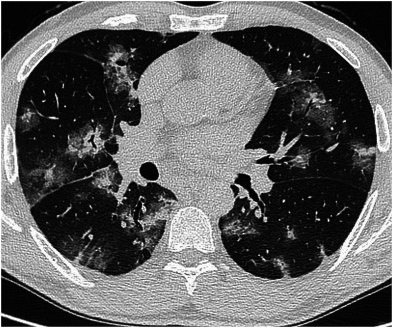
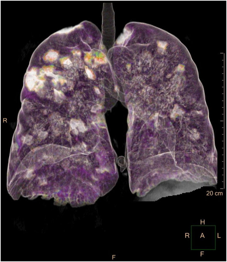
A 54-year-old man presented with a 5-day fever, cough, dyspnea, and chest pain. Physical examination revealed bilateral crackles on lung auscultation; temperature was 39°C. C-reactive protein was 27.2 mg/L (normal range: 0–5 mg/L) and serum lactate level was 274 U/L (normal range: 135–225 U/L); other blood tests showed normal results. COVID-19 was detected in two oropharyngeal swab samples by RT-PCR on consecutive days; Legionella and Streptococcus pneumoniae urinary antigens and PCR for other respiratory viruses on nasopharyngeal swabs were all negative. A bronchoalveolar lavage excluded pulmonary aspergillosis. On day 3, the patient developed severe dyspnea with decreased oxygen saturation (90%). Unenhanced chest computed tomography (CT) imaging showed diffuse bilateral peribronchial consolidations surrounded by ground-glass opacities (Figures 1 and 2).
Figure 1.
Chest computed tomography showing bilateral nodules and consolidations, surrounded by ground-glass opacities, and resulting in a halo sign, with prevalent peribronchovascular distribution. Air bronchograms are bilaterally recognizable.
Figure 2.
Volume-rendered three-dimensional reconstruction chest CT image shows the presence of bilateral opacities, with prevalent peribronchovascular distribution and sparing of the peripheral lung zones.
The typical CT pattern of COVID-19 pneumonia consists of ground-glass opacities (GGO) with bilateral and peripheral distribution.1 However, less common imaging findings have also been reported. A pattern of peribronchial infiltrate with surrounding GGO is not well described in COVID-19, let alone to this extent.2 Radiologists should be aware of the wide spectrum of CT manifestation of this infection.
REFERENCES
- 1.Chung M, et al. 2020. CT imaging features of 2019 novel coronavirus (2019-nCoV). Radiology 295: 202–207. [DOI] [PMC free article] [PubMed] [Google Scholar]
- 2.Zhou Z, Guo D, Li C, Fang Z, Chen L, Yang R, Li X, Zeng W, 2020. Coronavirus disease 2019: initial chest CT findings. Eur Radiol 2020: 1–9. [DOI] [PMC free article] [PubMed] [Google Scholar]


