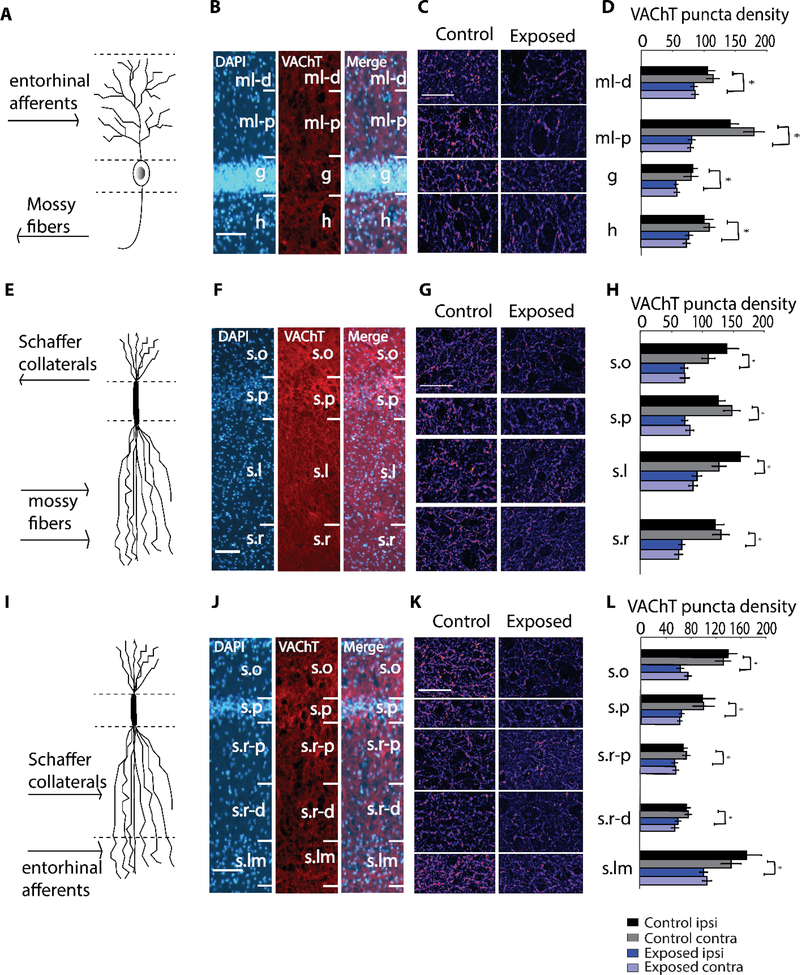
Figure 2. Robust decreases in VAChT puncta density in dentate gyrus (DG), area CA3, and area CA1 two weeks following noise exposure.
(A) Schematic granule cell, depicting organization of inputs corresponding to the layers in (B), which are images of VAChT labeling in DG at 100X magnification. (C) Representative images at 400X magnification in DG with layers corresponding to (D), which depicts mean (±SEM) VAChT puncta density (per 104 μm2) in the indicated layers.
(E) Schematic pyramidal neuron, depicting organization of inputs corresponding to the layers in (F), which are images of VAChT labeling in area CA3 at 100X magnification. (G) Representative images at 400X magnification in area CA3 with layers corresponding to (H), which depicts mean (±SEM) VAChT puncta density (per 104 μm2) in the indicated layers.
(I) Schematic pyramidal neuron, depicting organization of inputs corresponding to the layers in (J), which are images of VAChT labeling in area CA1 at 100X magnification. (K) Representative images at 400X magnification in area CA1 with layers corresponding to (L), which depicts mean (±SEM) VAChT puncta density (per 104 μm2) in the indicated layers.
In (B), (F), (J), scale bar is 100 μm. In (C), (G), (K), scale bar is 50 μm.
Abbreviations: ml-d, distal region of molecular layer; ml-p, proximal region of molecular layer; g, granule cell layer; h, hilus; s.o, stratum oriens; s.p, stratum pyramidale; s.l, stratum lucidum; s.r, stratum radiatum; s.r-p, proximal region of stratum radiatum; s.r-d, distal region of stratum radiatum; s.lm, stratum lacunosum-moleculare. *p < 0.05

