Abstract
ATP-dependent chromatin-remodeling complexes epigenetically modulate transcription of target genes to impact a variety of developmental processes. Our lab previously demonstrated that CHD4—a central ATPase and catalytic enzyme of the NuRD chromatin-remodeling complex—plays an important role in murine embryonic endothelial cells by transcriptionally regulating vascular integrity at midgestation. Since NuRD complexes can incorporate the ATPase CHD3 as an alternative to CHD4, we questioned whether the CHD3 enzyme likewise modulates vascular development or integrity. We generated a floxed allele of Chd3 but saw no evidence of lethality or vascular anomalies when we deleted it in embryonic endothelial cells in vivo (Chd3ECKO). Furthermore, double-deletion of Chd3 and Chd4 in embryonic endothelial cells (Chd3/4ECKO) did not dramatically alter the timing and severity of embryonic phenotypes seen in Chd4ECKO mutants, indicating that CHD3 does not play a cooperative role with CHD4 in early vascular development. However, excision of Chd3 at the epiblast stage of development with a Sox2-Cre line allowed us to generate global heterozygous Chd3 mice (Chd3Δ/+), which were subsequently intercrossed and revealed partial lethality of Chd3Δ/Δ mutants prior to weaning. Tissues from surviving Chd3Δ/Δ mutants helped us confirm that CHD3 was efficiently deleted in these animals and that CHD3 is highly expressed in the gonads and brains of adult wildtype mice. Therefore, Chd3-flox mice will be beneficial for future studies about roles for this chromatin-remodeling enzyme in viable embryonic development and in gonadal and brain physiology.
Introduction
The mammalian Nucleosome Remodeling and Histone Deacetylase (NuRD/Mi-2) complex is an ATP-dependent chromatin-remodeling complex that can transiently displace nucleosomes within gene regulatory regions and thereby impact transcription [1]. NuRD complexes are composed of multiple proteins, many of which have varying isoforms that can contribute to differential NuRD assembly in specific cell types and tissues [2, 3]. Among these proteins, the Chromodomain-Helicase-DNA-Binding ATPases CHD3 and CHD4 (also called Mi-2α and Mi-2β) are critical for NuRD activity because they provide the energy and helicase function required for chromatin remodeling [4]. Recent evidence suggests that CHD3 and CHD4 are mutually exclusive within NuRD complexes, despite the fact that they are co-expressed in various cell types [5, 6].
NuRD complexes play various roles in developmental and cellular differentiation processes, such as suppression of embryonic stem cell pluripotency, maintenance and lineage commitment of hematopoietic stem cells, maintenance of nephron progenitors in the kidney, peripheral nerve myelination, and cortical development in the brain [6–10]. Many of these and additional discoveries resulted from studies focused on genetic deletion of a single NuRD component or a NuRD-interacting cofactor [11]. A Chd4-flox allele generated by Katia Georgopoulos’ lab [12] has been particularly useful for defining the function of CHD4-NuRD complexes in specific cell types in vivo.
Our lab has a long-standing interest in the role of chromatin-remodeling enzymes in vascular development. We genetically delete chromatin-remodeling enzymes like CHD4 in murine embryonic endothelial cells in order to determine the impact on developing blood vessels and to identify target genes that require tight transcriptional regulation during vascular development [13]. We previously reported that CHD4 is required for maintenance of vascular integrity at midgestation and during liver development [14–16]. We also described how CHD4 counteracts the activity of the chromatin-remodeling enzyme BRG1 to regulate the Wnt signaling pathway in developing blood vessels [17]. Together these studies demonstrate that CHD4 plays critical roles in embryonic vascular development and maintenance, presumably through its participation in NuRD-related transcriptional regulation events in endothelial cells.
Because of the interesting and varied vascular phenotypes we saw associated with Chd4 deletion in developing vasculature, we wondered what role the alternative NuRD ATPase CHD3 might play in embryonic blood vessels. No knockout or conditional allele of Chd3 currently exists for genetic deletion studies in mice. Instead, one report describes electroporating shRNAs against CHD3 into embryonic mouse brains in order to inhibit CHD3 in vivo [6]. In this study we sought to analyze CHD3 during vascular development by generating a Chd3-flox allele and deleting the gene in developing endothelial cells. Although we found no obvious roles for CHD3 in early vascular development, global deletion of the gene resulted in partial embryonic lethality. Our genetic crosses and expression data from adult mouse tissues—along with recent reports about CHD3 roles in neural development—suggest that CHD3 may contribute to viable embryonic brain development.
Materials and methods
Mice
All animal use protocols were approved by the Oklahoma Medical Research Foundation Institutional Animal Care and Use Committee (protocol #20–15), and all efforts were made to minimize animal suffering. Animals were originally described and obtained from the following sources: Chd3-flox, generated by Cyagen Biosciences as described in this paper; Chd4-flox [12], gift of Dr. Katia Georgopoulos (Harvard Medical School); Tie2-Cre [18], The Jackson Laboratory #008863; Sox2-Cre [19], The Jackson Laboratory #008454; and FLPe [20], The Jackson Laboratory #005703. Timed matings were assessed by checking daily for a copulation plug; noon on the day of plug detection was designated as embryonic day 0.5 (E0.5). Embryos and yolk sacs were dissected from maternal tissue, and yolk sacs were digested for genotyping assays. Gross embryonic images were obtained with a Nikon SMZ800 stereomicroscope and Nikon DS-Fi1 camera and monitor. Genotyping primers and conditions are described in Table 1.
Table 1. Genotyping primers and conditions.
| Allele | Genotyping Primers | Annealing Temperature | Amplicon Size |
|---|---|---|---|
| Chd3-flox | Forward (F): 5’-GGGTGGAGGTGGAAAGTGTA-3’ | 55°C | Wildtype: 152 bp |
| Reverse (R1): 5’-AGAGGACAGGTCACAGGACAA-3’ | Floxed: 213 bp | ||
| Chd3Δ | Forward (F): 5’-GGGTGGAGGTGGAAAGTGTA-3’ | 59°C | ~230 bp |
| Reverse (R2): 5’-GGCACAGCTTCTCTGAGACC-3’ | |||
| Chd4-flox | Forward: 5’-TCCAGAAGAAGACGGCAGAT-3’ | 55°C | Wildtype: 278 bp |
| Reverse: 5’- CTGGTCATAGGGCAGGTCTC-3’ | Floxed: 400 bp | ||
| Chd4Δ | Forward: 5’-TCCAGAAGAAGACGGCAGAT-3’ | 59°C | ~350 bp |
| Reverse: 5’-CAGCGTGTCAGAGTTTGCAT-3’ | |||
| Tie2-Cre | Forward: 5’-GGGAAGTCGCAAAGTTGTGAGTTG-3’ | 60°C | 533 bp |
| Reverse: 5’-TCCATGAGTGAACGAACCTGGTCG-3’ | |||
| Sox2-Cre | Forward: 5’-TGCAACGAGTGATGAGGTTC-3’ | 58°C | 528 bp |
| Reverse: 5’-ATTCTCCCACCGTCAGTACG-3’ | |||
| FLPe | Forward: 5’-AGTAGTGATCAGGTATTGCTGTTATCTG-3’ | 60°C | 100 bp |
| Reverse: 5’-GACTAATGTTGTGGGAAATTGGAG-3’ |
Immunoblotting
Total protein harvested from eight-week old mouse tissues was quantified with the Pierce BCA Protein Assay Kit (Thermo Scientific). Protein (20–30μg) was fractionated on a 9% SDS-polyacrylamide gel and transferred to a polyvinylidene difluoride (PVDF) membrane for immunoblotting with one of two antibodies against CHD3 (1:2000; BD Biosciences; Cat. #611847; RRID: AB_399327; or 1:1000; Abcam; Cat. #ab109195; RRID: AB_10862514) and GAPDH (1:5000; Sigma; Cat. #9545; RRID: AB_796208). Horseradish peroxidase-conjugated secondary antibodies and SuperSignal West Femto Maximum Sensitivity Substrate (Thermo Scientific) were used for detection. Imaging was performed on a Fluorchem HD2 (Alpha Innotech) and scanned with an imageRUNNER 3225 (Canon).
Statistics
Statistical methodology is described in the table legends.
Results
Generation of a Chd3-flox allele
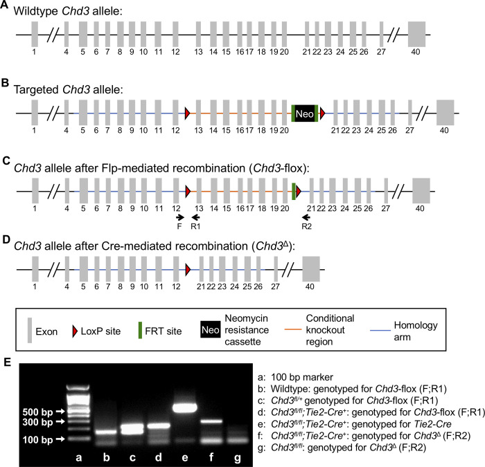
In order to generate a Chd3-flox allele to facilitate conditional deletion of the chromatin-remodeling enzyme in vivo, we contracted with Cyagen Biosciences to design a targeting vector for homologous recombination in embryonic stem (ES) cells (Fig 1A–1D). We chose to flank exons 13 through 20 of the murine Chd3 gene with loxP sites, since the CHD3 ATPase helicase—which mediates the critical function of the protein [21]—is encoded in this region. Deletion of these exons via Cre-mediated recombination was predicted to result in loss of function of the Chd3 gene by generating a frameshift mutation. Homologous recombination of the targeting vector in ES cells was confirmed by Cyagen Biosciences by Southern blotting prior to blastocyst injection. Targeting and excision events were confirmed by PCR in F1 and subsequent generations of mice (Fig 1E).
Fig 1. Generation of a conditional murine Chd3 knockout allele.
The Chd3-flox allele was generated using homologous recombination of a targeting vector in C57Bl/6 embryonic stem cells. (A): The wildtype murine Chd3 allele contains 40 exons and resides on chromosome 11. (B): The targeting vector incorporated a 5’ LoxP site (red triangle) inserted between exons 12 and 13 and a 3’ LoxP site inserted between exons 20 and 21 of the wildtype Chd3 allele. In addition, a neomycin resistance (Neo) cassette flanked by Frt sites (green bars) was inserted immediately upstream of the 3’ LoxP site for positive selection of the integrated targeting vector. (C): The Neo cassette was removed by crossing mice carrying the targeted allele to FLPe-recombinase mice, leaving a single Frt site upstream of the 3’ LoxP site in the Chd3-flox allele after recombination. Genotyping primers are indicated below the allele. (D): After crossing mice carrying the Chd3-flox allele to a line carrying a Cre recombinase, exons 13–20 were deleted (Chd3Δ). (E): The indicated mice were genotyped by PCR using the primers shown in parentheses and diagrammed in (C) to demonstrate targeting and Cre-mediated excision of the Chd3-flox allele. PCR products were run on a 2% agarose gel. See Table 1 for primer sequences and predicted amplicon sizes.
CHD3 is not essential for embryonic vascular development or for adult vascular maintenance
In order to determine whether CHD3 plays a critical role in vascular development or maintenance, we next crossed the Chd3-flox allele onto a transgenic Tie2-Cre recombinase line that is expressed in embryonic endothelial and hematopoietic cells [18]. We found that Chd3fl/fl;Tie2-Cre+ mice (hereafter, Chd3ECKO for Chd3-endothelial cell knockout) were born in expected numbers and survived past weaning and into adulthood without obvious abnormalities (Table 2).
Table 2. Live offspring at weaning from Chd3fl/+ X Chd3fl/+;Tie2-Cre+ crossesa.
| Genotype | Observed Offspringb | Expected Offspring |
|---|---|---|
| Chd3+/+ | 6 | 9.5 |
| Chd3fl/+ | 29 | 19 |
| Chd3fl/fl | 8 | 9.5 |
| Chd3+/+;Tie2-Cre+ | 9 | 9.5 |
| Chd3fl/+;Tie2-Cre+ | 15 | 19 |
| Chd3fl/fl;Tie2-Cre+ (Chd3ECKO) | 9 | 9.5 |
Chd3fl/fl;Tie2-Cre+ (Chd3ECKO) mice survive development. 76 mouse pups generated from matings between Chd3fl/+;Tie2-Cre+ males and Chd3fl/+ females were genotyped at weaning (~19 days). No lethal embryonic vascular anomalies are associated with the Chd3ECKO genotype, since the numbers of Chd3fl/fl;Tie2-Cre+ progeny observed approximate those expected.
a11 litters generated from 8 Chd3fl/+ females and 5 Chd3fl/+;Tie2-Cre+ males were genotyped.
bχ2(5dof) = 7.68, 0.5>P>0.1.
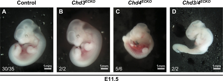
Although our genetic crosses indicated that no lethal vascular or hematopoietic abnormalities arose from Chd3 deletion with the Tie2-Cre recombinase, we visually analyzed Chd3ECKO embryos at midgestation, since that is the developmental stage at which we had previously observed significant vascular phenotypes in Chd4fl/fl;Tie2-Cre+ (hereafter Chd4ECKO) embryos [14, 15]. Specifically, we dissected Chd3ECKO and Chd4ECKO embryos at E11.5; while the Chd4ECKO embryos displayed lethal vascular rupture and hemorrhage, as we previously described, the Chd3ECKO embryos were indistinguishable from control littermates (Fig 2A–2C).
Fig 2. Chd3ECKO embryos are alive and display normal gross morphology at E11.5.
Photographs of representative E11.5 littermate embryos are shown. Each embryo was pronounced alive or dead at the time of dissection based on the presence or absence of a visible heartbeat. Numbers in the bottom left corner of each picture report the embryos resembling that picture/total embryos of each genotype dissected at E11.5. (A): Control = Tie2-Cre- (B): Chd3ECKO = Chd3fl/fl;Tie2-Cre+ (C): Chd4ECKO = Chd4fl/fl;Tie2-Cre+ (D): Chd3/4ECKO = Chd3fl/fl;Chd4fl/fl;Tie2-Cre+.
Vascular phenotypes in Chd3fl/fl;Chd4fl/fl;Tie2-Cre+ (Chd3/4ECKO) embryos resemble those seen in Chd4ECKO mutants
Because we have reported that chromatin-remodeling enzymes can co-regulate the same target genes or signaling pathways in developing blood vessels [17], we next sought to determine whether CHD3 plays a cooperative or antagonistic role with CHD4 during embryonic vascular development. Specifically, we generated embryos deficient for both endothelial Chd3 and Chd4 (Chd3fl/fl;Chd4fl/fl;Tie2-Cre+, hereafter Chd3/4ECKO) to determine whether the vascular phenotype was different from that seen in littermate Chd4ECKO embryos. We observed a comparable onset of lethality in Chd3/4ECKO and Chd4ECKO embryos between E10.5–11.5 (Table 3 and Fig 2D). Therefore, endothelial and hematopoietic cell deletion of Chd3 did not rescue lethal Chd4ECKO vascular phenotypes at midgestation. In addition, we assessed Chd3/4ECKO embryos at E10.5—one day prior to the vascular rupture seen in Chd4ECKO embryos—and saw no indication of vascular abnormalities (Fig 3). Therefore, Tie2-Cre-mediated deletion of Chd3 did not exacerbate the timing or severity of Chd4ECKO vascular phenotypes. Altogether, these genetic data indicate that CHD4 plays a more consequential role than does CHD3 in NuRD-mediated chromatin-remodeling activities during early vascular development.
Table 3. Total numbers of embryos genotyped (and % dead) at midgestation.
| Genotype | E10.5 | E11.5 |
|---|---|---|
| Control | 74 (3%) | 35 (14%) |
| Chd3ECHet and/or Chd4ECHet | 40 (1%) | 19 (0%) |
| Chd3ECKO | 26 (12%) | 2 (0%) |
| Chd4ECKO | 32 (6%) | 6 (83%) |
| Chd3/4ECKO | 8 (12%) | 2 (100%) |
The majority of Chd3/4ECKO embryos die between E10.5–11.5. Embryos were dissected at E10.5 or E11.5 from various matings that produce the following genotypes: Control (Tie2-Cre-), Chd3ECHet (Chd3fl/+;Tie2-Cre+), Chd4ECHet (Chd4fl/+;Tie2-Cre+), Chd3ECKO (Chd3fl/fl;Tie2-Cre+), Chd4ECKO (Chd4fl/fl;Tie2-Cre+), or Chd3/4ECKO (Chd3fl/fl;Chd4fl/fl;Tie2-Cre+). All embryos were deemed “alive” or “dead” at the time of dissection based on the presence or absence of a visible heartbeat. The death of most Chd4ECKO embryos between E10.5–11.5 is consistent with our previous reports [14, 15].
Fig 3. Chd3/4ECKO embryos display normal gross morphology at E10.5.
Photographs of representative live E10.5 littermate embryos are shown. Numbers in the bottom left corner of each picture report live embryos/total embryos of each genotype dissected at E10.5. (A): Control = Tie2-Cre- (B): Chd3ECKO = Chd3fl/fl;Tie2-Cre+ (C): Chd4ECKO = Chd4fl/fl;Tie2-Cre+ (D): Chd3/4ECKO = Chd3fl/fl;Chd4fl/fl;Tie2-Cre+.
Global deletion of Chd3 results in partial embryonic lethality
Since we saw no visible phenotypes associated with deletion of Chd3 in blood vessel endothelial cells and hematopoietic cells, we next sought to determine the consequence of global excision of our Chd3-flox allele. We crossed the Chd3-flox line onto a Sox2-Cre+ recombinase line that is expressed in all embryonic cells starting at the epiblast stage of development [19]. We found an underrepresentation of Chd3fl/fl;Sox2-Cre+ (hereafter called Chd3Δ/Δ;Sox2-Cre+ or Chd3-knockout) mice at weaning (Table 4). Likewise, matings between Chd3Δ/fl (Chd3-heterozygous) mice confirmed partial lethality of Chd3Δ/Δ (Chd3-knockout) mice (Table 5). Because we did not observe noticeable lethality of pups between birth and weaning when performing these crosses, we believe that Chd3-knockout embryos died before birth.
Table 4. Live offspring at weaning from Chd3fl/+;Sox2-Cre+ (Chd3Δ/+;Sox2-Cre+) X Chd3fl/+ crossesa.
| Genotype | Observed Offspringb | Expected Offspring |
|---|---|---|
| Chd3+/+ | 21 | 25.375 |
| Chd3fl/+ | 22 | 25.375 |
| Chd3Δ/+ | 26 | 25.375 |
| Chd3Δ/fl | 35 | 25.375 |
| Chd3+/+;Sox2-Cre+ | 33 | 25.375 |
| Chd3Δ/+;Sox2-Cre+ | 53 | 50.75 |
| Chd3Δ/Δ;Sox2-Cre+ | 13 | 25.375 |
Chd3Δ/Δ;Sox2-Cre+ progeny are underrepresented in offspring from Chd3Δ/+;Sox2-Cre+ males and Chd3fl/+ females. 203 mouse pups generated from matings between Chd3Δ/+;Sox2-Cre+ males and Chd3fl/+ females were genotyped at weaning (~19 days).
a19 litters generated from 5 Chd3fl/+ females and 4 Chd3Δ/+;Sox2-Cre+ males were genotyped.
bχ2(6dof) = 13.30, P<0.05.
Table 5. Live offspring at weaning from Chd3Δ/fl X Chd3Δ/fl crossesa.
| Genotype | Observed Offspringb | Expected Offspring |
|---|---|---|
| Chd3fl/fl | 19 | 15 |
| Chd3Δ/fl | 35 | 30 |
| Chd3Δ/Δ | 6 | 15 |
Chd3Δ/Δ progeny are underrepresented in offspring from Chd3Δ/fl matings. 60 mouse pups generated from matings between Chd3Δ/fl males and females were genotyped at weaning (~19 days).
a7 litters generated from 2 Chd3Δ/fl males and 2 Chd3Δ/fl females were genotyped.
bχ2(2dof) = 7.3, P<0.05.
CHD3 is predominantly expressed in adult gonads and brain tissue
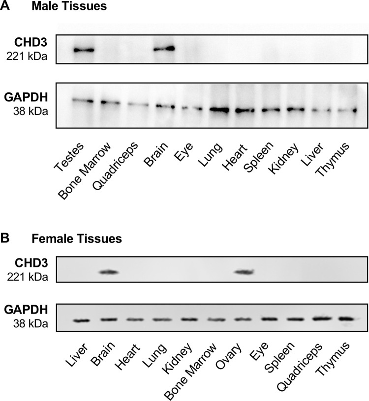
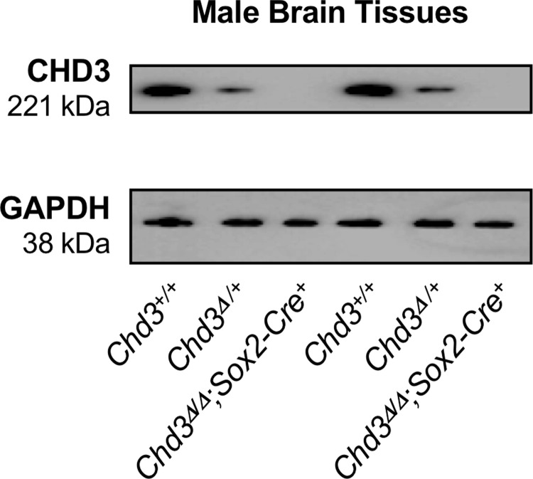
We next surveyed CHD3 expression in adult tissues, with hopes of gaining insight into possible causes of lethality for Chd3Δ/Δ animals. We harvested a variety of tissues from wildtype male and female mice and analyzed CHD3 expression by immunoblotting. We detected CHD3 protein predominantly in the brains (of both sexes), testes, and ovaries of eight-week-old mice (Fig 4). In order to validate the CHD3 antibodies we utilized for immunoblotting and to confirm the efficiency of Chd3 deletion with the Sox2-Cre recombinase, we assessed CHD3 expression in adult littermate wildtype, Chd3-heterozygous (Chd3Δ/fl), and surviving Chd3-knockout (Chd3Δ/Δ) mice (Fig 5). Immunoblotting for CHD3 protein in brain tissue revealed the expected reduction of CHD3 expression in Chd3-heterozygous samples and absence of detectable CHD3 in Chd3-knockout samples. Therefore, our Chd3-flox allele can be excised efficiently. Moreover, our expression analyses suggest that the partial pre-weaning lethality we observe on the Chd3-knockout background could be due to compromised fertility or to fatal brain defects, such as the cortical abnormalities recently described when CHD3 is knocked down in developing mouse brains [6].
Fig 4. CHD3 is predominantly expressed in the brain and gonads of adult mice.
Representative immunoblots of tissues from a single male and female wildtype mouse are shown. GAPDH was immunoblotted as a loading control. At least three mice of each gender were analyzed with comparable results.
Fig 5. Chd3 is efficiently excised with the Sox2-Cre recombinase.
Representative immunoblots of brains from two sets of wildtype (Chd3+/+), Chd3-heterozygous (Chd3Δ/+) and Chd3-knockout (Chd3Δ/Δ;Sox2-Cre+) male mice are shown. Four mice of each genotype were analyzed with comparable results.
Discussion
This study describes the first report of a conditional allele that facilitates global and cell-specific genetic analyses of CHD3 function in murine tissues. Our data indicate that CHD3 does not play a critical role in embryonic vascular development or adult blood vessel maintenance. This finding stands in contrast to our previous work with CHD4, an alternative NuRD ATPase, which transcriptionally regulates vascular development and integrity at multiple stages of embryonic life [14–17]. At face value, these results indicate that CHD4-NuRD complexes are the predominant or most consequential NuRD complexes in endothelial cells and hematopoietic cells. However, it is possible that CHD3-NuRD complexes play transcriptional regulatory roles in blood vessels under challenge conditions that were not incorporated into this study, such as ischemia, hypertension, inflammation, glucotoxicity or in solid tumors. Future work with constitutive Chd3ECKO and inducible Chd3/4ECKO mice could address this possibility. It is also interesting to consider the contribution of a third NuRD ATPase—CHD5—toward endothelial cell transcriptional regulation. As we report in this study for CHD3, CHD5 is predominantly expressed in the mouse brain and testes [22–24]. Therefore, CHD5 may also play inconsequential roles in the vasculature. Nevertheless, conditional genetic deletion studies—such as those described in the present study—would be the most rigorous way to assess possible roles for CHD5-NuRD complexes in vascular development.
Even though we found no consequential roles for CHD3 in blood vessels, our study does indicate that global deletion of Chd3 with a Sox2-Cre recombinase line results in partial embryonic lethality. We believe this lethality occurs during embryogenesis because we did not observe death of juvenile mice in our colony prior to weaning. Although we did not perform CHD3 expression analyses on embryonic tissues for this study, our immunoblotting assays indicate that CHD3 is predominantly expressed in the gonads and brains of adult mice, which could indicate a critical role for the protein in gametogenesis or fertility. However, we found Chd3-heterozygous mice (Chd3Δ/fl and Chd3Δ/+) represented at expected numbers at weaning (Tables 4 and 5), indicating that Chd3Δ gametes are viable and fertile. Moreover, a recent preprint reports that CHD3 plays no significant role in testis or sperm development when our Chd3-flox allele is combined with a primordial germ cell-specific Ddx4-Cre [25]. Alternatively, CHD3 is expressed in embryonic mouse brain cortices as early as E15.5 and has been shown through shRNA electroporation studies to influence neuronal migration and laminar identity in late stages of embryonic cortical development [6]. CHD4 likewise influences cortical development, and neuronal deletion of Chd4 with a Nestin-Cre results in lethality associated with microcephaly at birth [6]. Therefore, although the known contributions of CHD3 and CHD4 to cortical development are mechanistically different [6], it is possible that some of our Chd3-knockout embryos also die with lethal developmental brain defects.
Altogether, the Chd3-flox mice we have generated for this study provide opportunities for future genetic analysis of cell-specific roles for this NuRD-associated chromatin-remodeling enzyme. In particular, temporal neuronal-specific deletion of Chd3 could further clarify its roles in embryonic and postnatal brain development, which is clinically relevant in light of the recent report that human CHD3 mutations underlie a neurodevelopmental syndrome characterized by macrocephaly and impaired speech [21]. These and other Chd3 deletion studies would complement the numerous CHD4 cell-specific studies that have already been reported and would expand our knowledge about roles for NuRD complexes in development and disease.
Supporting information
(PDF)
Acknowledgments
We thank Tirzah Prince, Robert Pahissa, and Jocelyn Rodriguez for technical help with this project, Katia Georgopoulos (Harvard Medical School) for Chd4fl/fl mice, and Dr. Roberto Pezza and Griffin lab members for helpful discussions and advice.
Data Availability
All relevant data are within the paper.
Funding Statement
This work was supported by grants from the National Institutes of Health (https://www.nih.gov) awarded to C.T.G. (R35HL144605) and to Rodger McEver (P30GM114731). The funders had no role in study design, data collection and analysis, decision to publish, or preparation of the manuscript.
References
- 1.Xue Y, Wong J, Moreno GT, Young MK, Cote J, Wang W. NURD, a novel complex with both ATP-dependent chromatin-remodeling and histone deacetylase activities. Mol Cell. 1998;2: 851–861. 10.1016/s1097-2765(00)80299-3 [DOI] [PubMed] [Google Scholar]
- 2.Bowen NJ, Fujita N, Kajita M, Wade PA. Mi-2/NuRD: multiple complexes for many purposes. Biochim Biophys Acta. 2004;1677: 52–57. 10.1016/j.bbaexp.2003.10.010 [DOI] [PubMed] [Google Scholar]
- 3.Torchy MP, Hamiche A, Klaholz BP. Structure and function insights into the NuRD chromatin remodeling complex. Cell Mol Life Sci. 2015;72: 2491–2507. 10.1007/s00018-015-1880-8 [DOI] [PMC free article] [PubMed] [Google Scholar]
- 4.Denslow SA, Wade PA. The human Mi-2/NuRD complex and gene regulation. Oncogene. 2007;26: 5433–5438. 10.1038/sj.onc.1210611 [DOI] [PubMed] [Google Scholar]
- 5.Hoffmeister H, Fuchs A, Erdel F, Pinz S, Grobner-Ferreira R, Bruckmann A, et al. CHD3 and CHD4 form distinct NuRD complexes with different yet overlapping functionality. Nucleic Acids Res. 2017;45: 10534–10554. 10.1093/nar/gkx711 [DOI] [PMC free article] [PubMed] [Google Scholar]
- 6.Nitarska J, Smith JG, Sherlock WT, Hillege MM, Nott A, Barshop WD, et al. A Functional Switch of NuRD Chromatin Remodeling Complex Subunits Regulates Mouse Cortical Development. Cell Rep. 2016;17: 1683–1698. 10.1016/j.celrep.2016.10.022 [DOI] [PMC free article] [PubMed] [Google Scholar]
- 7.Denner DR, Rauchman M. Mi-2/NuRD is required in renal progenitor cells during embryonic kidney development. Dev Biol. 2013;375: 105–116. 10.1016/j.ydbio.2012.11.018 [DOI] [PMC free article] [PubMed] [Google Scholar]
- 8.Hung H, Kohnken R, Svaren J. The nucleosome remodeling and deacetylase chromatin remodeling (NuRD) complex is required for peripheral nerve myelination. J Neurosci. 2012;32: 1517–1527. 10.1523/JNEUROSCI.2895-11.2012 [DOI] [PMC free article] [PubMed] [Google Scholar]
- 9.Reynolds N, Latos P, Hynes-Allen A, Loos R, Leaford D, O'Shaughnessy A, et al. NuRD suppresses pluripotency gene expression to promote transcriptional heterogeneity and lineage commitment. Cell Stem Cell. 2012;10: 583–594. 10.1016/j.stem.2012.02.020 [DOI] [PMC free article] [PubMed] [Google Scholar]
- 10.Yoshida T, Hazan I, Zhang J, Ng SY, Naito T, Snippert HJ, et al. The role of the chromatin remodeler Mi-2beta in hematopoietic stem cell self-renewal and multilineage differentiation. Genes Dev. 2008;22: 1174–1189. 10.1101/gad.1642808 [DOI] [PMC free article] [PubMed] [Google Scholar]
- 11.Basta J, Rauchman M. The nucleosome remodeling and deacetylase complex in development and disease. Transl Res. 2015;165: 36–47. 10.1016/j.trsl.2014.05.003 [DOI] [PMC free article] [PubMed] [Google Scholar]
- 12.Williams CJ, Naito T, Arco PG, Seavitt JR, Cashman SM, De Souza B, et al. The chromatin remodeler Mi-2beta is required for CD4 expression and T cell development. Immunity. 2004;20: 719–733. 10.1016/j.immuni.2004.05.005 [DOI] [PubMed] [Google Scholar]
- 13.Curtis CD, Davis RB, Ingram KG, Griffin CT. Chromatin-remodeling complex specificity and embryonic vascular development. Cell Mol Life Sci. 2012;69: 3921–3931. 10.1007/s00018-012-1023-4 [DOI] [PMC free article] [PubMed] [Google Scholar]
- 14.Ingram KG, Curtis CD, Silasi-Mansat R, Lupu F, Griffin CT. The NuRD Chromatin-Remodeling Enzyme CHD4 Promotes Embryonic Vascular Integrity by Transcriptionally Regulating Extracellular Matrix Proteolysis. PLoS Genet. 2013;9: e1004031 10.1371/journal.pgen.1004031 [DOI] [PMC free article] [PubMed] [Google Scholar]
- 15.Colijn S, Gao S, Ingram KG, Menendez M, Muthukumar V, Silasi-Mansat R, et al. The NuRD chromatin-remodeling complex enzyme CHD4 prevents hypoxia-induced endothelial Ripk3 transcription and murine embryonic vascular rupture. Cell Death Differ. 2020;27: 618–631. 10.1038/s41418-019-0376-8 [DOI] [PMC free article] [PubMed] [Google Scholar]
- 16.Crosswhite PL, Podsiadlowska JJ, Curtis CD, Gao S, Xia L, Srinivasan RS, et al. CHD4-regulated plasmin activation impacts lymphovenous hemostasis and hepatic vascular integrity. J Clin Invest. 2016;126: 2254–2266. 10.1172/JCI84652 [DOI] [PMC free article] [PubMed] [Google Scholar]
- 17.Curtis CD, Griffin CT. The chromatin-remodeling enzymes BRG1 and CHD4 antagonistically regulate vascular Wnt signaling. Mol Cell Biol. 2012;32: 1312–1320. 10.1128/MCB.06222-11 [DOI] [PMC free article] [PubMed] [Google Scholar]
- 18.Kisanuki YY, Hammer RE, Miyazaki J, Williams SC, Richardson JA, Yanagisawa M. Tie2-Cre transgenic mice: a new model for endothelial cell-lineage analysis in vivo. Dev Biol. 2001;230: 230–242. 10.1006/dbio.2000.0106 [DOI] [PubMed] [Google Scholar]
- 19.Hayashi S, Lewis P, Pevny L, McMahon AP. Efficient gene modulation in mouse epiblast using a Sox2Cre transgenic mouse strain. Mech Dev. 2002;119 Suppl 1: S97–S101. [DOI] [PubMed] [Google Scholar]
- 20.Farley FW, Soriano P, Steffen LS, Dymecki SM. Widespread recombinase expression using FLPeR (flipper) mice. Genesis. 2000;28: 106–110. [PubMed] [Google Scholar]
- 21.Snijders Blok L, Rousseau J, Twist J, Ehresmann S, Takaku M, Venselaar H, et al. CHD3 helicase domain mutations cause a neurodevelopmental syndrome with macrocephaly and impaired speech and language. Nat Commun. 2018;9: 4619 10.1038/s41467-018-06014-6 [DOI] [PMC free article] [PubMed] [Google Scholar]
- 22.Thompson PM, Gotoh T, Kok M, White PS, Brodeur GM. CHD5, a new member of the chromodomain gene family, is preferentially expressed in the nervous system. Oncogene. 2003;22: 1002–1011. 10.1038/sj.onc.1206211 [DOI] [PubMed] [Google Scholar]
- 23.Zhuang T, Hess RA, Kolla V, Higashi M, Raabe TD, Brodeur GM. CHD5 is required for spermiogenesis and chromatin condensation. Mech Dev. 2014;131: 35–46. 10.1016/j.mod.2013.10.005 [DOI] [PMC free article] [PubMed] [Google Scholar]
- 24.Bergs JW, Neuendorff N, van der Heijden G, Wassenaar E, Rexin P, Elsasser HP, et al. Differential expression and sex chromosome association of CHD3/4 and CHD5 during spermatogenesis. PLoS One. 2014;9: e98203 10.1371/journal.pone.0098203 [DOI] [PMC free article] [PubMed] [Google Scholar]
- 25.de Castro RO, Goitea V, Previato L, Carbajal A, Griffin CT, Pezza RJ. CHD4-NURD controls spermatogonia survival and differentiation. bioRxiv [Preprint]. 2019. bioRxiv 687137 [posted 2019 June 29; revised 2020 Feb 27]. Available from: https://www.biorxiv.org/content/10.1101/687137v2 10.1101/687137 [DOI] [Google Scholar]


