Abstract
Purpose
Few studies have investigated the independent and joint associations of cardiorespiratory fitness (CRF) and body fat percentage (BF%) with insulin resistance in children. We investigated the independent and combined associations of CRF and BF% with fasting glycaemia and insulin resistance and their interactions with physical activity (PA) and sedentary time among 452 children aged 6–8 years.
Methods
We assessed CRF with a maximal cycle ergometer exercise test and used allometrically scaled maximal power output (Wmax) for lean body mass (LM1.13) and body mass (BM1) as measures of CRF. BF% and LM were measured by dual-energy X-ray absorptiometry, fasting glycaemia by fasting plasma glucose, and insulin resistance by fasting serum insulin and Homeostatic Model Assessment for Insulin Resistance (HOMA-IR). PA energy expenditure (PAEE), moderate-to-vigorous PA (MVPA), and sedentary time were assessed by combined movement and heart rate sensor.
Results
Wmax/LM1.13 was not associated with glucose (β=0.065, 95% CI=-0.031 to 0.161), insulin (β=-0.079, 95% CI=-0.172 to 0.015), or HOMA-IR (β=-0.065, 95% CI=-0.161 to 0.030). Wmax/BM1 was inversely associated with insulin (β=-0.289, 95% CI=-0.377 to -0.200) and HOMA-IR (β=-0.269, 95% CI=-0.359 to -0.180). BF% was directly associated with insulin (β=0.409, 95% CI=0.325 to 0.494) and HOMA-IR (β=0.390, 95% CI=0.304 to 0.475). Higher Wmax/BM1, but not Wmax/LM1.13, was associated with lower insulin and HOMA-IR in children with higher BF%. Children with higher BF% and who had lower levels of MVPA or higher levels of sedentary time had the highest insulin and HOMA-IR.
Conclusion
Children with higher BF% together with less MVPA or higher levels of sedentary time had the highest insulin and HOMA-IR. CRF appropriately controlled for body size and composition using LM was not related to insulin resistance among children.
Keywords: diabetes, youth, exercise, performance, insulin, insulin sensitivity, obesity
Introduction
The incidence of type 2 diabetes in children and adolescents has increased during this millennium (1) and represents a significant health and economic burden. Type 2 diabetes typically affects adults but has a long aetiology related to insulin resistance and impaired glucose regulation which are observed in overweight and obese youth (2). Insulin resistance during childhood may also increase the risk of atherosclerotic cardiovascular diseases in adulthood (3). Furthermore, children with a mild insulin resistance, measured by fasting plasma insulin concentration and Homeostatic Model Assessment for Insulin Resistance (HOMA-IR), have been found to be at increased risk of pre-diabetes and type 2 diabetes in adulthood (4). In addition to increased body fat content, low cardiorespiratory fitness (CRF), physical inactivity, and high levels of sedentary lifestyle have been identified as independent risk factors for insulin resistance in children (5,6). However, few studies have investigated the independent and joint associations of CRF, physical activity, and sedentary time with insulin resistance in children after accounting for body fat content (7,8).
Increased body mass index (BMI) has been found to have a graded dose-response relationship to insulin resistance and overall cardiometabolic risk in children and adolescents (9,10). Poor CRF has also been associated with increased insulin resistance in children and adolescents (11,12). Furthermore, the results of few studies suggest that higher CRF attenuates the unfavourable effects of overweight and obesity on insulin resistance in children (9,13). However, these studies have assessed CRF using measures scaled by whole body mass (BM), submaximal estimates of CRF (13), or 20 metre shuttle run test (9). Measures of CRF scaled by BM are not justified from a physiological or statistical perspective in children (14–16), because they do not remove the effect of body size and composition on CRF (16). Furthermore, body fat content explains 40% of variance in running performance during 20-metre shuttle run test (17,18). Increased body fat content, but not peak oxygen uptake (VO2peak) determined during an incremental exercise test, has been found to be strongly related to insulin resistance (19). Therefore, the assessment of CRF using measures scaled by BM using the ratio standard method or the 20-metre shuttle run test may lead to spurious associations with insulin resistance (20,21). Allometric scaling of CRF by lean body mass (LM) has been recommended to account for variation in body size and composition among children and adolescents (19,22), but few studies have utilised this approach to explore the associations of CRF with fasting plasma insulin and glucose concentrations with adjustment for adiposity.
Low CRF and adiposity are relatively stable correlates of insulin resistance, whereas low levels of moderate-to-vigorous physical activity (MVPA) and high levels of sedentary time are more modifiable risk factors for insulin resistance in children (6,23,24). A sedentary lifestyle has been found to impair insulin signalling, increase insulin resistance, reduce skeletal muscle glucose uptake, and thereby increase the risk of type 2 diabetes in adults (24). Nevertheless, few studies have investigated differences in insulin resistance among children with varying levels of CRF, adiposity, MVPA, and sedentary time (25).
Evidence on the associations of CRF appropriately adjusted for body size and composition with risk factors for type 2 diabetes is urgently required (17), because it would help develop effective strategies for the early identification of individuals at increased risk and the prevention of the disease. We therefore investigated the associations of CRF, scaled by BM or LM using ratio standard and allometric modelling, and body fat content with insulin resistance in a population sample of children. Second, we studied the joint associations of the measures of CRF and body fat content with insulin resistance. Third, we investigated whether physical activity energy expenditure (PAEE), MVPA, and sedentary time are associated with insulin resistance in children with varying CRF and body fat content. We hypothesised that CRF that is scaled using appropriate methods has weak associations with insulin resistance and that body fat content has the strongest association with insulin resistance. We also hypothesised that higher levels of MVPA or lower levels of sedentary time are related to lower insulin resistance in children with higher body fat content.
Methods
Study design and study participants
The present data are from the Physical Activity and Nutrition in Children (PANIC) Study, which is a physical activity and dietary intervention and follow-up study in a population sample of children from the city of Kuopio, Finland. Altogether 736 children 6–8 years of age from primary schools of Kuopio were invited to participate in the baseline examination in 2007–2009. A total of 512 children, who represented 70% of those invited, participated in the baseline examinations. Six children were excluded from the study at baseline because of physical disabilities that could hamper participation in the intervention or no time or motivation to attend in the study. The participants did not differ in sex distribution, age, or BMI standard deviation score (BMI-SDS) from all children who started the first grade in 2007–2009 based on data from the standard school health examinations performed for all Finnish children before the first grade (data not shown). Complete data on variables used in the analyses on the associations of CRF and body fat content with the indicators of insulin resistance were available for 452 children (236 boys, 216 girls). Complete data on variables used in the analyses on the joint associations of CRF, body fat content, PAEE, MVPA, and sedentary time with the indicators of insulin resistance were available for 388 children (196 boys, 192 girls). The study protocol was approved by the Research Ethics Committee of the Hospital District of Northern Savo. Both children and their parents gave written informed consent.
Assessment of body size, body composition, and pubertal status
Whole BM was measured twice with the children having fasted for 12 hours, emptied the bladder, and standing in light underwear by a calibrated InBody® 720 bioelectrical impedance device (Biospace, Seoul, South Korea) to an accuracy of 0.1 kg. The mean of these two values was used in the analyses. Stature was measured three times with the children standing in the Frankfurt plane without shoes using a wall-mounted stadiometer to an accuracy of 0.1 cm. The mean of the nearest two values was used in the analyses. BMI was calculated by dividing BM (kg) by body height (m) squared. BMI-SDS was calculated based on Finnish reference data (26). The prevalence of overweight and obesity was defined using the cut-off values provided by Cole et al. (27). Total fat mass, body fat percentage (BF%), and LM were measured by the Lunar® dual-energy X-ray absorptiometry device (GE Medical Systems, Madison, WI, USA) using standardised protocols.
The research physician assessed pubertal status using the 5-stage scale described by Tanner (28). The boys were defined as having entered clinical puberty if their testicular volume assessed by an orchidometer was ≥4 mL (stage ≥2). The girls were defined having entered clinical puberty if their breast development had started (stage ≥2). Maturity offset as the difference between the current age from the age at predicted peak height velocity was computed using a sex-specific formula (29).
Assessment of fasting glycaemia and insulin resistance
A research nurse took venous blood samples from the antecubital vein in the morning after a 12-hour overnight fast. Plasma glucose was measured by a hexokinase method and serum insulin was measured by an electrochemiluminescence immunoassay. Intra-assay and inter-assay coefficient of variation for the insulin analyses were 1.3–3.5% and 1.6–4.4%, respectively. Insulin resistance was also assessed using HOMA-IR and the formula fasting serum insulin x fasting plasma glucose/22.5) (30).
Assessment of cardiorespiratory fitness
We assessed CRF by a maximal exercise test using an electromagnetically braked Ergoselect 200 K® cycle ergometer coupled with a paediatric saddle module (Ergoline, Bitz, Germany) (22). The exercise test protocol included a 2.5-minute anticipatory period with the child sitting on the ergometer; a 3-minute warm-up period with a workload of 5 watts; a 1-minute steady-state period with a workload of 20 watts; an exercise period with an increase in the workload of 1 watt per 6 seconds until exhaustion, and a 4-minute recovery period with a workload of 5 watts.
The children were asked to keep the cadence stable and within 70–80 revolutions per minute. Exhaustion was defined as the inability to maintain the cadence above 65 revolutions per minute regardless of vigorous verbal exhortation. The exercise test was considered maximal by an experienced physician (TT) supervising the test, if objective and subjective criteria (heart rate >85% of predicted, sweating, flushing, inability to continue exercise test regardless of strong verbal encouragement) indicated maximal effort and maximal cardiovascular capacity (22). Heart rate was measured continuously during the last five minutes of the supine rest prior to commencing the exercise test protocol right through to the 5-minute supine post-exercise rest period using a 12-lead electrocardiogram registered by the Cardiosoft® V6.5 Diagnostic System (GE Healthcare Medical Systems, Freiburg, Germany) and the highest heart rate during the test was defined as peak heart rate (31). Maximal power output (Wmax) measured at the end of the exercise test divided by BM1 and LM1 were used as measures of CRF. We used Wmax as a measure of CRF because we did not perform respiratory gas analyses at baseline and it has been found to be a good surrogate measure of CRF in children (32).
Wmax/BM1 had a strong inverse association with BM (β = -0.498, 95% confidence interval (CI) =-0.584 to -0.412, p < 0.001) and Wmax/LM1 had a weak positive association with LM (β = 0.086, 95% CI=0.003 to 0.169, p = 0.043) indicating that ratio scaling by BM-1 or LM-1 did not completely remove the effect of body size on CRF. Therefore, allometric scaling of Wmax was performed by log-linear regression models (20). The scaling exponent for BM was 0.48 (95% CI = 0.39 to 0.57) and for it LM was 1.13 (95% CI = 1.01 to 1.26). These power function ratios removed the associations of Wmax with BM (β = -0.024, 95% CI=-0.108 to 0.059, p = 0.569) and LM (β = -0.004, 95% CI = -0.109 to 0.101, p = 0.940) suggesting the validity of scaling CRF for body size.
Assessment of physical activity and sedentary time
PA and sedentary time were assessed using a combined heart rate and movement sensor (Actiheart®, CamNtech Ltd., Papworth, UK) for a minimum of four consecutive days without interruption, including two weekdays and two weekend days, analysed in 60 second epochs (33,34). The combined heart rate and movement sensor was attached to the child´s chest with two standard eletrocardiogram electrodes (Bio Protech Inc, Wonju, South Korea). The children were asked to wear the monitor continuously, including sleep and water-based activities, and not to change their usual behaviour during the monitoring period. Data on heart rate were cleaned and individually calibrated with parameters from the maximal exercise test and combined with movement sensor data to derive PAEE. Instantaneous PAEE, i.e. PA intensity, was estimated using branched equation modelling as explained in detail earlier (35) and summarised as daily PA volume (kJ/day/kg) and time spent at certain levels of standard metabolic equivalents of task (METs) in minutes per day, weighting all hours of the day equally to reduce diurnal bias caused by imbalances in wear-time. Initially, the summarised data included 25 narrowly defined intensity categories. For the present analyses, we re-categorised these intensity categories into a broader format of sedentary time (≤1.5 METs) and M PA (>4 METs), which have been commonly applied in investigations of PA among children and youth. In order to estimate the time spent sedentary whilst awake, we subtracted average daily sleep duration from total ST. We only included children who had sufficient valid data, i.e. a recording period of at least 48 hours of wear data with the additional requirement that enough data were included from all four quadrants of a 24 hour day to avoid bias from over-representation of specific parts of days (36). This resulted in at least 12 hours of wear data from morning (3 am – 9 am), noon (9 am – 3 pm), afternoon / evening (3 pm – 9 pm), and night (9 pm – 3 am).
Assessment of diet quality
Food consumption and nutrient intake were assessed by food records administered by the parents on four pre- defined consecutive days, including two weekdays and two weekend days (99.5% of participants) or three week- days and one weekend day (0.5% of participants), as described previously (37). The food records were analysed using The Micro Nutrica dietary analysis software, Version 2.5 (The Social Insurance Institution of Finland). We used Finnish Children Healthy Eating Index (FCHEI) that summarises the consumption of vegetables, fruit, and berries; vegetable oils and vegetable oil-based margarine; foods containing high amounts of sugar; fish; and low-fat (<1%) milk based on deciles of these dietary variables in the study population. Higher scores indicate a better diet quality.
Statistical methods
Statistical analyses were performed by SPSS statistical software, version 23.0 (IBM corp. Armonk, NY, USA). Basic characteristics between the 236 boys and the 216 girls were compared using the Student’s t-test for normally distributed continuous variables, the Mann-Whitney U-test for continuous variables with skewed distributions, or the χ2-test for categorical variables. Differences in PAEE, MVPA, and sedentary time were compared between 196 boys and 192 girls. Because of skewed distribution, insulin and HOMA-IR were square-root transformed. The associations of the measures of CRF scaled by BM and LM using allometry and ratio standard and BF% with glucose, insulin, and HOMA-IR were investigated using linear regression analyses adjusted for age and sex.
Differences in glucose, insulin, and HOMA-IR between children with the combinations of lower BF% (≤ sex-specific median) and higher Wmax/LM1.13 or 1 or BM0.48 or 1 (> sex-specific median), lower BF% and lower Wmax/LM1.13 or 1 or BM0.48 or 1, higher BF% and higher Wmax/LM1.13 or 1 or BM0.48 or 1, and higher BF% and lower Wmax/LM1.13 or 1 or BM0.48 or 1 were investigated using analyses of covariance (ANCOVA) adjusted for age and sex and considering the Sidak correction for multiple comparisons. Because the results were similar for insulin and HOMA-IR, we only present the results on HOMA-IR. We found no differences in the associations of CRF with glucose, insulin, and HOMA-IR, between boys and girls (p>0.05 for interaction), and therefore performed all analyses sexes combined.
Because allometric scaling of CRF by LM has been considered the most appropriate method to express CRF (19), we used only Wmax/LM1.13 to study differences in PAEE, MVPA, and sedentary time between children with the four combinations of CRF and BF% and the joint associations of CRF, BF%, and PAEE, MVPA, or sedentary time with glucose, insulin, and HOMA-IR. Differences in PAEE, MVPA, and sedentary time between children with the combinations of lower BF% and higher Wmax/LM1.13, lower BF% and lower Wmax/LM1.13, higher BF% and higher Wmax/LM1.13, and higher BF% and lower Wmax/LM1.13 were investigated using ANCOVA adjusted for age and sex and considering the Sidak correction for multiple comparisons.
To investigate whether PAEE, MVPA, or sedentary time modified the joint associations of BF% and Wmax/LM1.13 with glucose, insulin, and HOMA-IR we compared glucose, insulin, and HOMA-IR in the four combinations of BF% and Wmax/LM1.13 in children with lower (≤ sex-specific median) or higher (> sex-specific median) levels of PAEE, MVPA, and sedentary time using ANCOVA adjusted for age and sex and considering the Sidak correction for multiple comparisons. We also investigated the independent associations of Wmax/LM1.13, BF% and PAEE, MVPA, or sedentary time with glucose, insulin, and HOMA-IR using linear regression analyses and ANCOVA adjusted for age and sex. All data were further adjusted for clinical puberty or maturity offset, or FCHEI.
Results
Basic characteristics
Boys had lower maturity offset, higher stature, less fat mass, lower BF%, and higher LM than girls (Table 1). Boys also had higher glucose, lower insulin and HOMA-IR, and higher CRF regardless of the scaling method used than girls. Furthermore, boys had higher PAEE and accumulated more MVPA than girls.
Table 1. Basic characteristics.
| All | Girls | Boys | P-value | |
|---|---|---|---|---|
| Age (years) | 7.6 (0.4) | 7.6 (0.4) | 7.7 (0.4) | 0.182 |
| Pubertal (%) | 1.6 | 2.5 | 0.9 | 0.265 |
| Maturity offset (years) | -4.0 (0.5) | -3.6 (0.3) | -4.4 (0.3) | <0.001 |
| Stature (cm) | 128.6 (5.7) | 127.5 (5.6) | 129.6 (5.5) | <0.001 |
| Body weight (kg) | 26.6 (4.7) | 26.2 (4.7) | 27.0 (4.7) | 0.071 |
| Fat mass (kg) | 4.7 (3.3–6.7) | 5.3 (3.9–7.7) | 3.9 (2.8–6.3) | <0.001 |
| Body fat percentage (%) | 19.56 (8.11) | 22.34 (7.60) | 17.02 (7.74) | <0.001 |
| Lean mass (kg) | 20.6 (2.4) | 19.4 (2.1) | 21.6 (2.2) | <0.001 |
| Body mass index standard deviation score | -0.2 (1.1) | -0.2 (1.0) | -0.3 (1.1) | 0.661 |
| Prevalence of overweight or obesity (%) | 11.1 | 12.7 | 9.7 | 0.331 |
| Fasting plasma glucose (mmol/L) | 4.81 (0.37) | 4.75 (0.37) | 4.87 (0.37) | 0.001 |
| Fasting serum insulin (mU/L) | 4.49 (2.36) | 4.81 (2.22) | 4.19 (2.46) | 0.006 |
| Homeostatic model assessment for insulin resistance | 0.98 (0.56) | 1.04 (0.52) | 0.93 (0.59) | 0.040 |
| Maximal power output (Watts) | 76.3 (15.4) | 69.4 (13.0) | 82.5 (14.7) | <0.001 |
| Maximal power output (W/kg of lean body mass1.13) | 2.5 (0.3) | 2.4 (0.3) | 2.6 (0.3) | <0.001 |
| Maximal power output (W/kg of lean body mass1) | 3.69 (0.51) | 3.56 (0.50) | 3.81 (0.50) | <0.001 |
| Maximal power output (W/kg of body weight0.48) | 15.8 (2.8) | 14.5 (2.3) | 17.0 (2.7) | <0.001 |
| Maximal power output (W/kg of body weight1) | 2.87 (0.54) | 2.67 (0.47) | 3.09 (0.53) | <0.001 |
| Peak heart rate during maximal exercise test (beats/min) | 195 (8.8) | 195 (9.2) | 196 (8.4) | 0.413 |
| PAEE (kJ/body mass/d) | 99.1 (32.9) | 90.6 (27.7) | 107 (35.4) | <0.001 |
| Moderate-to-vigorous physical activity (min/d) | 116 (63.9) | 96.9 (53.9) | 135 (67.3) | <0.001 |
| Sedentary time (min/d) | 233 (127) | 240 (127) | 225 (126) | 0.255 |
Data are from the Student t-test or Mann-Whitney U test for continuous variables and from the Chi-square test for categorical variables and are displayed as means (SD), medians (IQR), or percentages (%). PAEE = Physical activity energy expenditure
Independent associations of measures of CRF, BF%, PA, and sedentary time with fasting glycaemia and insulin resistance
Wmax/LM-1 and Wmax/LM1.13 were not associated with glucose, insulin, or HOMA-IR after adjustment for age and sex (Table 2). Wmax/BM1 was inversely and BF% was directly associated with insulin and HOMA-IR. Wmax/BM0.48 was inversely associated with insulin but the inverse association with HOMA-IR was not statistically significant. Further adjustments had no effect on these associations.
Table 2.
Associations of the measures of cardiorespiratory fitness, body fat percentage, physical activity, and sedentary behaviour with fasting glycaemia and insulin resistance in children
| Fasting plasma glucose (mmol/L) | Fasting serum insulin (mU/L) | HOMA-IR | |
|---|---|---|---|
| Cardiorespiratory fitness and body fat content (N=452) | |||
| Maximal power output (W/kg of lean body mass1.13) | 0.065 (-0.031 to 0.161) | -0.079 (-0.172 to 0.015) | -0.065 (-0.161 to 0.030) |
| Maximal power output (W/kg of lean body mass1) | 0.074 (-0.02 to 0.168) | -0.063 (-0.158 to 0.031) | -0.050 (-0.144 to 0.045) |
| Maximal power output (W/kg of body weight0.48) | 0.059 (-0.047 to 0.166) | -0.119 (-0.221 to -0.014)* | -0.105 (-0.210 to 0.001) |
| Maximal power output (W/kg of body weight1) | -0.015 (-0.108 to 0.078) | -0.289 (-0.377 to -0.200)*** | -0.269 (-0.359 to -0.180)*** |
| Body fat percentage (%) | 0.083 (-0.010-0.176) | 0.409 (0.325 to 0.494)*** | 0.390 (0.304 to 0.475)*** |
| Physical activity and sedentary time (N=388) | |||
| Moderate to vigorous physical activity (min/d) | -0.023 (-0.126 to 0.081) | -0.261 (-0.356 to -0.165)*** | -0.249 (-0.345 to -0.153)*** |
| Sedentary time (min/d) | 0.099 (0.000 to 0.197) | 0.272 (0.181 to 0.363)*** | 0.271 (0.176 to 0.369)*** |
| Physical activity energy expenditure (kJ/body mass/d) | -0.060 (-0.159 to 0.040) | -0.269 (-0.360 to -0.178)*** | -0.260 (-0.351 to -0.169)*** |
Data are standardised regression coefficient and their 95% confidence intervals from multivariate linear regression analyses adjusted for age and sex. *p<0.05; **p<0.01; ***p<0.001. HOMA-IR = Homeostatic model assessment for insulin resistance
MVPA and PAEE were inversely and sedentary time was directly associated with insulin and HOMA-IR (Table 2). These associations remained statistically significant after further adjustment for BF% or Wmax/LM1.13 and other measures of CRF. Further adjustments had no effect on these associations.
Joint associations of CRF and BF% with HOMA-IR
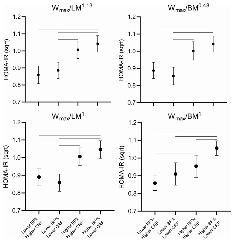
Children with higher BF% and higher Wmax/LM1.13 and those with higher BF% and lower Wmax/LM1.13 had higher HOMA-IR than children with lower BF% and higher Wmax/LM1.13 and those with lower BF% and lower Wmax/LM1.13 (Figure 1). The results were similar when medians of Wmax/LM1 was used. Further adjustments had no effect on these differences.
Figure 1.
Differences in HOMA-IR among children with different levels of body fat percentage (BF%) and cardiorespiratory fitness scaled by lean body mass (LM) or body mass (BM). N in Wmax/LM-1.13 or Wmax/LM1 = Lower BF%/higher CRF=121; Lower BF%/lower CRF=105; Higher CRF/higher BF%=121; Higher BF%/lower CRF=120. N in Wmax/BM0.48 or Wmax/LM1 = Lower BF%/higher CRF=158; Lower BF%/lower CRF=68; Higher CRF/higher BF%=68; Higher BF%/lower CRF=158. Lines between groups denotes a statistically significant difference between groups at p<0.05.
Children with lower BF% and lower Wmax/BM0.48 and those with lower BF% and higher Wmax/BM0.48 had lower HOMA-IR than children with higher BF% and lower Wmax/BM0.48 and those with higher BF% and higher Wmax/BM0.48 (Figure 1). Furthermore, children with higher BF% and lower Wmax/BM1 had higher HOMA-IR than those with other three combinations of BF% and Wmax/BM1 (Figure 1). Children with higher BF% and higher Wmax/BM-1 also had higher HOMA-IR than those with lower BF% and higher Wmax/BM-1. Further adjustments had no effect on these differences.
Joint associations of CRF and BF% with PA and sedentary time
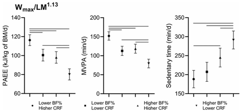
Children with lower BF% and higher Wmax/LM1.13 had higher PAEE and they accumulated more MVPA than those with other three combinations of BF% and Wmax/LM1.13 (Figure 2). Children with lower BF% and lower Wmax/LM1.13 and those with higher BF% and higher Wmax/LM1.13 had higher PAEE and more MVPA than their peers with higher BF% and lower Wmax/LM1.13. Furthermore, children with lower BF% and higher Wmax/LM1.13 and those with lower BF% and lower Wmax/LM1.13 had less sedentary time than children with higher BF% and lower Wmax x LM1.13. All these differences remained statistically significant after further adjustment for clinical puberty or maturity offset, or FCHEI.
Figure 2.
Differences in physical activity energy expenditure (PAEE), moderate to vigorous physical activity (MVPA), and sedentary time (ST) among children with different levels of body fat percentage and cardiorespiratory fitness normalised for lean mass (LM1.13). N = Lower BF%/higher CRF=121; Lower BF%/lower CRF=105; Higher CRF/higher BF%=121; Higher BF%/lower CRF=120. Lines between groups denotes a statistically significant difference between groups at p<0.05.
Joint associations of CRF, BF%, PA, and sedentary time with HOMA-IR
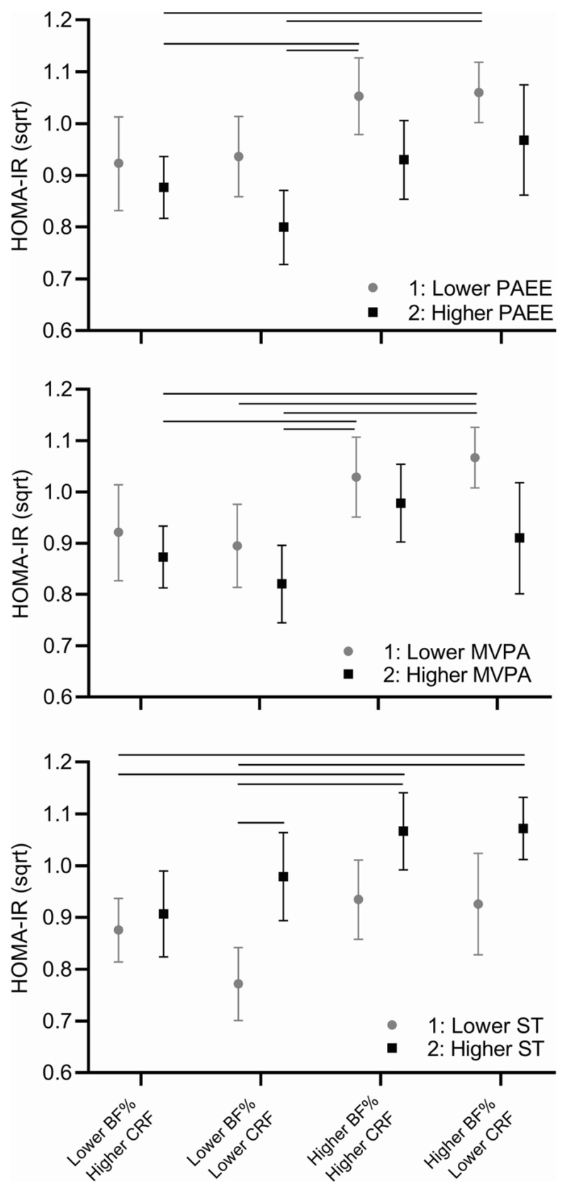
Children with higher BF%, higher Wmax/LM1.13, and lower PAEE and those with higher BF%, lower Wmax/LM1.13, and lower PAEE had higher HOMA-IR than children with lower BF%, higher Wmax/LM1.13, and higher PAEE and those with lower BF%, lower Wmax/LM1.13, and higher PAEE (Figure 3). These differences remained similar when PAEE was replaced by MVPA (Figure 3). Moreover, children with higher BF%, lower Wmax/LM1.13, and lower MVPA also had higher HOMA-IR than children with lower BF%, lower Wmax/LM-1.13, and lower MVPA. Further adjustments had no effect on these differences.
Figure 3.
Differences in HOMA-IR among children with different levels of body fat percentage (BF%), allometrically scaled cardiorespiratory fitness for lean mass (LM1.13), and physical activity energy expenditure (PAEE), moderate to vigorous physical activity (MVPA), or sedentary time (ST). N=lower BF%/higher CRF/lower PA or higher ST = 32; lower BF%/higher CRF/higher PA or lower ST = 75; lower BF%/lower CRF/lower PA or higher ST = 37; lower BF%/lower CRF/higher PA or lower ST = 52; higher BF%/higher CRF/lower PA or higher ST = 47; higher BF%/higher CRF/higher PA or lower ST = 45; higher BF%/lower CRF/lower PA or higher ST = 79; higher BF%/higher CRF/higher PA or lower ST = 45. Lines between groups denotes a statistically significant difference between groups at p<0.05.
Children with lower BF%, higher Wmax/LM1.13, and less sedentary time and those with lower BF%, lower Wmax/LM1.13, and less sedentary time had lower HOMA-IR than children with higher BF%, higher Wmax/LM1.13, and more sedentary time and those with higher BF%, lower Wmax/LM1.13, and more sedentary time (Figure 3). Moreover, children with lower BF%, lower Wmax/LM1.13, and less sedentary time had lower HOMA-IR than those with lower BF%, lower Wmax/LM1.13, and more sedentary time. Further adjustments had no effect on these differences.
Discussion
Our main finding was that Wmax scaled by LM using either allometry or ratio standard was not related to fasting glucose, fasting insulin, or HOMA-IR. However, Wmax scaled by BM1 had an inverse association with insulin and HOMA-IR, but these associations were attenuated when body size was controlled for using allometry. We also observed that higher Wmax/BM1, but not Wmax/LM1.13, Wmax/LM1, or Wmax/BM0.48, attenuated the association between higher BF% and insulin resistance. Moreover, children with higher BF% and lower Wmax/LM1.13 were physically less active and more sedentary than other children, and lower levels of PA and higher levels of sedentary time magnified the direct associations of BF% with insulin and HOMA-IR.
In conjunction with previous studies (20,38,39), we observed that children with lower CRF scaled by BM were more insulin resistant than their more fit peers. However, these findings are likely to be mediated by body size and composition because CRF scaled by BM also has a strong inverse association with BM and fat mass (22) and therefore dividing CRF by BM does not fully remove the effect of body size and adipose tissue on CRF (15). In addition, we found a strong direct association of BF% with insulin and HOMA-IR. Furthermore, our observations that Wmax scaled by LM was not associated with insulin resistance is supported by the findings of few previous studies showing that scaling CRF by LM reduced the magnitude of the association between CRF and insulin resistance (21,40,41). However, Ekelund and coworkers found that Wmax divided by fat-free mass, which was estimated using skinfold thickness, had a weak inverse association with insulin and clustered cardiometabolic risk independent of waist circumference (12). One reason for the discrepancy between the results of our study and the study by Ekelund et al. (12) may be that the participants of their study were older than those in the present study. It is possible that the role of CRF in insulin resistance increases with increasing age and maturation (7). Their larger study population also resulted in better statistical power and thereby increased the likelihood of observing statistically significant associations between variables of interest. However, there is some evidence that CRF scaled by fat-free mass derived from skinfold thickness may not completely remove the influence of body size and composition on CRF (42). We also found that allometrically scaled CRF was not associated with insulin resistance further suggesting that an inverse association between CRF and insulin resistance in previous studies is largely confounded by body size and composition.
We found that higher CRF divided by BM1 was related to lower insulin resistance in children with higher BF%. This observation agrees with the results of some previous studies suggesting that higher CRF provides health benefits particularly in overweight and obese youth (9,13). We are not aware of previous studies on the joint associations of BF% and CRF scaled by LM or BM using allometric methods with insulin resistance in children. Children with higher BF% had higher insulin resistance than those with lower BF% regardless of CRF scaled by LM1.13 or 1 or BM0.48. These results suggested that the inverse association of CRF scaled by BM-1 with insulin resistance is largely explained by body composition. However, CRF may have even stronger association with insulin resistance among individuals with obesity (7,9). Most children in our study were normal weight, and the mean BF% was 17% in boys and 22% in girls. However, weight status has been found to be a more important correlate of fasting insulin and HOMA-IR than 20 metre endurance shuttle run test performance (43). Consistent with this observation, our results together with others (21,23,40,41) suggest that body fat content is a stronger determinant of insulin resistance than CRF in children.
Higher levels of PA and lower levels of sedentary time have been related to lower cardiometabolic risk in children (6,23,44). The results of randomised controlled trials also suggest that exercise training has beneficial effects on insulin resistance especially in overweight and obese youth (45). In our study, there were no marked differences in fasting insulin or HOMA-IR between children with higher PA or lower sedentary time levels with varying levels of BF% and CRF. Nevertheless, physically less active and sedentary children with higher BF% were more insulin resistant than their more active and less sedentary peers with lower BF%.
There are few studies on the joint associations of CRF, adiposity, PA, and sedentary time with insulin resistance in children (25). We found that children with higher BF% and lower CRF had the lowest PA and highest sedentary time levels. They also had the highest fasting insulin and HOMA-IR, especially when CRF was scaled by BM. Our findings suggest that lower PA and higher sedentary time at least partly explain the increased insulin resistance in children with lower CRF and higher BF%. The accumulation of free fatty acids in skeletal muscle and liver impairing insulin signalling has been suggested as the underlying mechanisms in obesity-induced insulin resistance (46), although this explanation has been questioned recently (47). In contrast, regular PA upregulates insulin-independent GLUT 4 pathway for glucose disposal (48), whereas a sedentary lifestyle may impair insulin-regulated glucose disposal by down-regulating the insulin signalling pathway to translocate GLUT 4 and by reducing GLUT 4 protein content (24). Furthermore, previous studies have suggested that changes in insulin signalling (24) or alterations in serum metabolome (49) induced by physical exercise may explain the associations between higher CRF and lower insulin resistance. However, our results together with others (50) suggest that the relationship between CRF and insulin resistance is weak and is likely to be due to adiposity due to the inappropriate scaling of CRF. Nevertheless, we cannot rule out the possibility, that CRF has tissue specific associations that has not been covered in our or in previous studies. The present findings together with the previous studies suggest that higher levels of PA and lower levels of sedentary time, but not necessarily CRF, attenuate the harmful effects of increased BF% on insulin resistance.
There are strengths and weakness in the present study. The strengths of the present study include the valid and reproducible measurements of CRF using an exercise test until exhaustion, body composition using whole-body DXA, and insulin resistance using fasting insulin and HOMA-IR in a population sample of children. We also assessed free-living PA and sedentary time using individually calibrated movement and heart rate sensing. Nevertheless, the measurement of free-living behaviours is not as precise as that of CRF and body fat content, and therefore the magnitude of the associations of PA and sedentary time with insulin resistance may be underestimated. In addition, our sample was relatively small for the analysis of differences in insulin resistance between children with higher and lower PA and sedentary time. Furthermore, we did not directly measure maximal oxygen uptake which is considered the gold standard for measuring CRF. However, Wmax determined from an exercise test until exhaustion has been found to be a valid measure of CRF in children (32). It is possible that the associations of CRF, BF%, PA, and sedentary time with insulin resistance in different tissues (51,52) or using more dynamic measures of insulin resistance and insulin sensitivity, such as Cederholm index (53) may not be similar to those that we observed by assessing whole-body insulin resistance using HOMA-IR calculated by fasting serum and plasma glucose concentrations. Our study showed interesting results on the associations of CRF, BF%, PA, and sedentary time in a sample of children who had comparable levels of insulin resistance than other European children (54). Nevertheless, it would be interesting to see whether these relations exist in populations with higher insulin resistance or a higher prevalence of overweight and obesity. Because of a relatively small sample size for combined analyses, we dichotomised CRF, BF%, PA, and sedentary time using sex-specific medians instead of the lowest percentiles from international or national reference values which may have affected on our results. However, our findings on the associations of Wmax/BM1 with insulin resistance are comparable to those of studies in North American cohorts of children 7-13 years of age that utilised CRF and BMI dichotomized at median (13,55). Finally, our study was cross-sectional which limits our ability to make causal inferences.
In conclusion, we found that CRF had negligible role in insulin resistance among children aged 6–8 years when body size and composition were appropriately controlled for. Our results also suggest that higher levels of PA and lower levels of sedentary time may be more important than higher levels of CRF in improving insulin sensitivity in childhood. Additional research is warranted to investigate whether these results are similar in other age and maturation groups and using different cut-offs for CRF and body fat content. Finally, more longitudinal studies on the associations of changes in different measures of CRF, such as maximal oxygen uptake and Wmax, scaled by LM with insulin resistance during growth and maturation are needed to understand the long-term consequences of changes in CRF with regard to the development of metabolic and cardiovascular diseases.
Acknowledgements
The PANIC Study has financially been supported by Ministry of Education and Culture of Finland, Ministry of Social Affairs and Health of Finland, Research Committee of the Kuopio University Hospital Catchment Area (State Research Funding), Finnish Innovation Fund Sitra, Social Insurance Institution of Finland, Finnish Cultural Foundation, Foundation for Paediatric Research, Diabetes Research Foundation in Finland, Finnish Foundation for Cardiovascular Research, Juho Vainio Foundation, Paavo Nurmi Foundation, Yrjö Jahnsson Foundation, and the city of Kuopio. Moreover, the PhD students and postdoctoral researchers of The PANIC Study have been supported by Program for Clinical Research and Program for Health Sciences of Doctoral School of University of Eastern Finland, Finnish Doctoral Programs in Public Health, Päivikki and Sakari Sohlberg Foundation, Paulo Foundation, Jalmari and Rauha Ahokas Foundation, Aarne and Aili Turunen Foundation, Finnish Medical Foundation, Jenny and Antti Wihuri Foundation, Kuopio Naturalists' Society, Olvi Foundation, and the city of Kuopio. SB was supported by UK Medical Research Council (MC_UU_12015/3) and the NIHR Biomedical Research Centre Cambridge [IS-BRC-1215-20014]. The sponsors had no role in designing the study, the collection, analysis, or interpretation of the data, the writing of the report, or the decision to submit the manuscript for publication.
Footnotes
Conflict of Interest
Authors declare no conflicts of interest,
The results of the present study do not constitute endorsement by ACSM.
Authors declare that the results of the study are presented clearly, honestly, and without fabrication, falsification, or inappropriate data manipulation.
Author Contributions
EAH and TK analysed the data. EAH and PW drafted the manuscripts. NL, TT, JV, UE, SB, and TAL collected and processed the data for analyses. AB, TF, IMT contributed to planning the manuscript and interpreting the results and reviewed the manuscript. All authors provided significant intellectual contribution to the manuscript.
References
- 1.Mayer-Davis EJ, Lawrence JM, Dabelea D, et al. Incidence Trends of Type 1 and Type 2 Diabetes among Youths, 2002–2012. N Engl J Med. 2017;376(15):1419–29. doi: 10.1056/NEJMoa1610187. [DOI] [PMC free article] [PubMed] [Google Scholar]
- 2.Reaven GM. Insulin Resistance: The Link Between Obesity and Cardiovascular Disease. Med Clin North Am. 2011;95(5):875–92. doi: 10.1016/j.mcna.2011.06.002. [DOI] [PubMed] [Google Scholar]
- 3.Steinberger J, Daniels SR, Eckel RH, et al. Progress and challenges in metabolic syndrome in children and adolescents: a scientific statement from the American Heart Association Atherosclerosis, Hypertension, and Obesity in the Young Committee of the Council on Cardiovascular Disease in the Young. Circulation. 2009;119(4):628–47. doi: 10.1161/CIRCULATIONAHA.108.191394. [DOI] [PubMed] [Google Scholar]
- 4.Nguyen QM, Srinivasan SR, Xu JH, Chen W, Kieltyka L, Berenson GS. Utility of childhood glucose homeostasis variables in predicting adult diabetes and related cardiometabolic risk factors: The Bogalusa Heart Study. Diabetes Care. 2010;33(3):670–5. doi: 10.2337/dc09-1635. [DOI] [PMC free article] [PubMed] [Google Scholar]
- 5.Ortega FB, Cadenas-sanchez C, Lee D, Ruiz JR, Blair SN, Sui X. State of the Science Fitness and Fatness as Health Markers through the Lifespan : An Overview of Current Knowledge. PROGREVMED. 2018;3:e0013. doi: 10.1097/pp9.0000000000000013. [DOI] [PMC free article] [PubMed] [Google Scholar]
- 6.Poitras VJ, Gray CE, Borghese MM, et al. Systematic review of the relationships between objectively measured physical activity and health indicators in school-aged children and youth. Appl Physiol Nutr Metab. 2016;41:197–239. doi: 10.1139/apnm-2015-0663. [DOI] [PubMed] [Google Scholar]
- 7.Kennedy AB, Lavie CJ, Blair SN. Fitness or Fatness: Which Is More Important? JAMA. 2018;319(3):231–2. doi: 10.1001/jama.2017.21649. [DOI] [PubMed] [Google Scholar]
- 8.Ortega FB, Ruiz JR, Labayen I, Lavie CJ, Blair SN. The Fat but Fit paradox: What we know and don’t know about it. Br J Sports Med. 2018;52(3):151–3. doi: 10.1136/bjsports-2016-097400. [DOI] [PubMed] [Google Scholar]
- 9.Nyström CD, Henriksson P, Martínez-Vizcaíno V, et al. Does cardiorespiratory fitness attenuate the adverse effects of severe/morbid obesity on cardiometabolic risk and insulin resistance in children? A pooled analysis. Diabetes Care. 2017;40(11):1580–7. doi: 10.2337/dc17-1334. [DOI] [PubMed] [Google Scholar]
- 10.Skinner AC, Perrin EM, Moss LA, Skelton JA. Cardiometabolic Risks and Severity of Obesity in Children and Young Adults. N Engl J Med. 2015;373(14):1307–17. doi: 10.1056/NEJMoa1502821. [DOI] [PubMed] [Google Scholar]
- 11.Lang JJ, Tremblay MS, Ortega FB, Ruiz JR, Tomkinson GR. Review of criterion-referenced standards for cardiorespiratory fitness: what percentage of 1 142 026 international children and youth are apparently healthy? Br J Sports Med. 2019;53(15):953–8. doi: 10.1136/bjsports-2016-096955. [DOI] [PubMed] [Google Scholar]
- 12.Ekelund U, Anderssen SA, Froberg K, Sardinha LB, Andersen LB, Brage S. Independent associations of physical activity and cardiorespiratory fitness with metabolic risk factors in children: The European youth heart study. Diabetologia. 2007;50(9):1832–40. doi: 10.1007/s00125-007-0762-5. [DOI] [PubMed] [Google Scholar]
- 13.DuBose KD, Eisenmann JC, Donnelly JE. Aerobic fitness attenuates the metabolic syndrome score in normal-weight, at-risk-for-overweight, and overweight children. Pediatrics. 2007;120(5):e1262–8. doi: 10.1542/peds.2007-0443. [DOI] [PubMed] [Google Scholar]
- 14.Graves LEF, Batterham AM, Foweather L, Mcwhannell N, Hopkins ND, Boddy LM, et al. Scaling of peak oxygen uptake in children: A comparison of three body size index models. Med Sci Sports Exerc. 2013;45(12):2341–5. doi: 10.1249/MSS.0b013e31829bfa79. [DOI] [PubMed] [Google Scholar]
- 15.Tanner J. Fallacy of per-weigh and per-surface area standards, and their relation to spurious correlation. J Appl Physiol. 1949;2(1):2–15. doi: 10.1152/jappl.1949.2.1.1. [DOI] [PubMed] [Google Scholar]
- 16.Welsman J, Armstrong N. Interpreting Aerobic Fitness in Youth : The Fallacy of Ratio Scaling. Pediatr Exerc Sci. 2019;31(2):184–90. doi: 10.1123/pes.2018-0141. [DOI] [PubMed] [Google Scholar]
- 17.Armstrong N. Top 10 Research Questions Related to Youth Aerobic Fitness. Res Q Exerc Sport. 2017;88(2):130–48. doi: 10.1080/02701367.2017.1303298. [DOI] [PubMed] [Google Scholar]
- 18.Mayorga-Vega D, Aguilar-Soto P, Viciana J. Criterion-Related Validity of the 20-M Shuttle Run Test for Estimating Cardi-orespiratory Fitness: A Meta-Analysis. J Sport Sci Med. 2015;14:536–47. [PMC free article] [PubMed] [Google Scholar]
- 19.Loftin M, Sothern M, Abe T, Bonis M. Expression of VO2peak in Children and Youth, with Special Reference to Allometric Scaling. Sports Med [Internet] 2016;46:1451–60. doi: 10.1007/s40279-016-0536-7. [DOI] [PubMed] [Google Scholar]
- 20.Agbaje AO, Haapala EA, Lintu N, et al. Peak oxygen uptake cut-points to identify children at increased cardiometabolic risk - The PANIC Study. Scand J Med Sci Sports. 2019;29(1):16–24. doi: 10.1111/sms.13307. [DOI] [PubMed] [Google Scholar]
- 21.Shaibi G, Cruz M, Ball G, Weigensberg M, Kobaissi H, Salem G, et al. Cardiovascular fitness and the metabolic syndrome in overweight latino youths. Med Sci Sport Exerc. 2005;37(6):922–8. [PubMed] [Google Scholar]
- 22.Tompuri T, Lintu N, Savonen K, Laitinen T, Laaksonen D, Jääskeläinen J, et al. Measures of cardiorespiratory fitness in relation to measures of body size and composition among children. Clin Physiol Funct Imaging. 2015;35(6):469–77. doi: 10.1111/cpf.12185. [DOI] [PubMed] [Google Scholar]
- 23.Väistö J, Haapala EA, Viitasalo A, et al. Longitudinal associations of physical activity and sedentary time with cardiometabolic risk factors in children. Scand J Med Sci Sports. 2019;29:113–23. doi: 10.1111/sms.13315. [DOI] [PMC free article] [PubMed] [Google Scholar]
- 24.Booth FW, Roberts CK, Thyfault JP, Ruegsegger GN, Toedebusch RG. Role of Inactivity in Chronic Diseases: Evolutionary Insight and Pathophysiological Mechanisms. Physiol Rev. 2017;97(4):1351–402. doi: 10.1152/physrev.00019.2016. [DOI] [PMC free article] [PubMed] [Google Scholar]
- 25.Barker AR, Gracia-Marco L, Ruiz JR, et al. Physical activity, sedentary time, TV viewing, physical fitness and cardiovascular disease risk in adolescents: The HELENA study. Int J Cardiol. 2018;254:303–9. doi: 10.1016/j.ijcard.2017.11.080. [DOI] [PubMed] [Google Scholar]
- 26.Saari A, Sankilampi U, Hannila M-L, Kiviniemi V, Kesseli K, Dunkel L. New Finnish growth references for children and adolescents aged 0 to 20 years: Length/height-for-age, weight-for-length/height, and body mass index-for-age. Ann Med. 2011;43(3):235–48. doi: 10.3109/07853890.2010.515603. [DOI] [PubMed] [Google Scholar]
- 27.Cole T, Bellizzi M, Flegal K, Dietz W. Establishing a standard definition for child overweight and obesity worldwide: international survey. BMJ. 2000;320(7244):1240–3. doi: 10.1136/bmj.320.7244.1240. [DOI] [PMC free article] [PubMed] [Google Scholar]
- 28.Malina RM, Bouchard C, Bar-Or O. Growth, Maturation, and Physical Activity. 2nd ed. Champaign: Human Kinetics; 2004. pp. 283–90. 350–357. [Google Scholar]
- 29.Moore SA, McKay HA, Macdonald H, et al. Enhancing a somatic maturity prediction model. Med Sci Sports Exerc. 2015;47(8):1755–64. doi: 10.1249/MSS.0000000000000588. [DOI] [PubMed] [Google Scholar]
- 30.Matthews DR, Hosker JP, Rudenski AS, Naylor BA, Treacher DF, Turner RC. Homeostasis model assessment: insulin resistance and beta-cell function from fasting plasma glucose and insulin concentrations in man. Diabetologia. 1985;28(7):412–9. doi: 10.1007/BF00280883. [DOI] [PubMed] [Google Scholar]
- 31.Lintu N, Viitasalo A, Tompuri T, et al. Cardiorespiratory fitness, respiratory function and hemodynamic responses to maximal cycle ergometer exercise test in girls and boys aged 9– 11 years: the PANIC Study. Eur J Appl Physiol. 2015;115(2):235–43. doi: 10.1007/s00421-014-3013-8. [DOI] [PubMed] [Google Scholar]
- 32.Dencker M, Thorsson O, Karlsson MK, Lindén C, Wollmer P, Andersen LB. Maximal oxygen uptake versus maximal power output in children. J Sports Sci. 2008;26(13):1397–402. doi: 10.1080/02640410802199789. [DOI] [PubMed] [Google Scholar]
- 33.Collings PJ, Westgate K, Väistö J, et al. Cross-Sectional Associations of Objectively-Measured Physical Activity and Sedentary Time with Body Composition and Cardiorespiratory Fitness in Mid-Childhood: The PANIC Study. Sport Med. 2017;47:769–80. doi: 10.1007/s40279-016-0606-x. [DOI] [PMC free article] [PubMed] [Google Scholar]
- 34.Brage S, Brage N, Franks PW, Ekelund U, Wareham NJ. Reliability and validity of the combined heart rate and movement sensor Actiheart. Eur J Clin Nutr. 2005;59:561–70. doi: 10.1038/sj.ejcn.1602118. [DOI] [PubMed] [Google Scholar]
- 35.Brage S, Brage N, Franks PW, et al. Branched equation modeling of simultaneous accelerometry and heart rate monitoring improves estimate of directly measured physical activity energy expenditure. J Appl Physiol. 2004;96(1):343–51. doi: 10.1152/japplphysiol.00703.2003. [DOI] [PubMed] [Google Scholar]
- 36.Brage S, Westgate K, Wijndaele K, Godinho J, Griffin S, Wareham N. Evaluation of a method for minimising diurnal information bias in objective sensor data. Int Conf Amb Mon Phys Act Mov; 2013. (Conference Proceeding) [Google Scholar]
- 37.Eloranta a M, Lindi V, Schwab U, Kiiskinen S, Kalinkin M, Lakka HM, et al. Dietary factors and their associations with socioeconomic background in Finnish girls and boys 6-8 years of age: the PANIC Study. Eur J Clin Nutr. 2011;65(11):1211–8. doi: 10.1038/ejcn.2011.113. [DOI] [PubMed] [Google Scholar]
- 38.Ferreira I, Twisk JWR, van Mechelen W, Kemper HCG, Stehouwer CDA. Development of Fatness, Fitness, and Lifestyle From Adolescence to the Age of 36 Years. Arch Intern Med. 2005;165(1):42. doi: 10.1001/archinte.165.1.42. [DOI] [PubMed] [Google Scholar]
- 39.Steele RM, Brage S, Corder K, Wareham NJ, Ekelund U. Physical activity, cardiorespiratory fitness, and the metabolic syndrome in youth. J Appl Physiol. 2008;105(1):342–51. doi: 10.1152/japplphysiol.00072.2008. [DOI] [PMC free article] [PubMed] [Google Scholar]
- 40.McMurray RG, Hosick PA, Bugge A. Importance of proper scaling of aerobic power when relating to cardiometabolic risk factors in children. Ann Hum Biol. 2011;38(5):647–54. doi: 10.3109/03014460.2011.598561. [DOI] [PubMed] [Google Scholar]
- 41.Ahn B, McMurray R, Harrell J. Scaling of VO2max and Its Relationship With Insulin Resistance in Children. Pediatr Exerc Sci. 2013;25:43–51. doi: 10.1123/pes.25.1.43. [DOI] [PubMed] [Google Scholar]
- 42.Tolfrey K. Scaling of maximal oxygen uptake by lower leg muscle volume in boys and men. J Appl Physiol. 2006;100(6):1851–6. doi: 10.1152/japplphysiol.01213.2005. [DOI] [PubMed] [Google Scholar]
- 43.Moschonis G, Mougios V, Papandreou C, et al. “Leaner and less fit” children have a better cardiometabolic profile than their “heavier and more fit” peers: The Healthy Growth Study. Nutr Metab Cardiovasc Dis. 2013;23(11):1058–65. doi: 10.1016/j.numecd.2012.11.010. [DOI] [PubMed] [Google Scholar]
- 44.Ekelund U, Luan J, Sherar LB, Esliger DW, Griew P, Cooper A. Moderate to vigorous physical activity and sedentary time and cardiometabolic risk factors in children and adolescents. JAMA. 2012 Feb 15;307(7):704–12. doi: 10.1001/jama.2012.156. [DOI] [PMC free article] [PubMed] [Google Scholar]
- 45.Fedewa MV, Gist NH, Evans EM, Dishman RK. Exercise and Insulin Resistance in Youth: A Meta-Analysis. Pediatrics. 2014;133(1):e163–74. doi: 10.1542/peds.2013-2718. [DOI] [PubMed] [Google Scholar]
- 46.Galgani JE, Moro C, Ravussin E. Metabolic flexibility and insulin resistance. Am J Physiol - Endocrinol Metab. 2008;295(5):1009–17. doi: 10.1152/ajpendo.90558.2008. [DOI] [PMC free article] [PubMed] [Google Scholar]
- 47.Karpe F, Dickmann JR, Frayn KN. Fatty acids, obesity, and insulin resistance: Time for a reevaluation. Diabetes. 2011;60(10):2441–9. doi: 10.2337/db11-0425. [DOI] [PMC free article] [PubMed] [Google Scholar]
- 48.Richter EA, Hargreaves M. Exercise, GLUT4, and Skeletal Muscle Glucose Uptake. Physiol Rev. 2013;93(3):993–1017. doi: 10.1152/physrev.00038.2012. [DOI] [PubMed] [Google Scholar]
- 49.Kujala UM, Vaara JP, Kainulainen H, Vasankari T, Vaara E, Kyröläinen H. Associations of Aerobic Fitness and Maximal Muscular Strength With Metabolites in Young Men. JAMA Netw Open. 2019;2(8):e198265. doi: 10.1001/jamanetworkopen.2019.8265. [DOI] [PMC free article] [PubMed] [Google Scholar]
- 50.Ball GDC, Shaibi GQ, Cruz ML, et al. Insulin Sensitivity, Cardiorespiratory Fitness, and Physical Activity in Overweight Hispanic Youth. Obes Reserarch. 2004;12(1):77–85. doi: 10.1038/oby.2004.11. [DOI] [PubMed] [Google Scholar]
- 51.Honkala SM, Johansson J, Motiani KK, et al. Short-term interval training alters brain glucose metabolism in subjects with insulin resistance. J Cereb Blood Flow Metab. 2018;38(10):1828–38. doi: 10.1177/0271678X17734998. [DOI] [PMC free article] [PubMed] [Google Scholar]
- 52.Eskelinen JJ, Heinonen I, Löyttyniemi E, Saunavaara V, Kirjavainen A, Virtanen KA, et al. Muscle-specific glucose and free fatty acid uptake after sprint interval and moderate-intensity training in healthy middle-aged men. J Appl Physiol. 2015;118(9):1172–80. doi: 10.1152/japplphysiol.01122.2014. [DOI] [PubMed] [Google Scholar]
- 53.Cockcroft EJ, Williams CA, Weaver H, O’Connor A, Jackman SR, Armstrong N, et al. Acute Exercise and Insulin Sensitivity in Boys: A Time-Course Study. Int J Sports Med. 2017;38(13):967–74. doi: 10.1055/s-0043-118007. [DOI] [PubMed] [Google Scholar]
- 54.Peplies J, Jiménez-Pavón D, Savva SC, Buck C, Günther K, Fraterman A, et al. Percentiles of fasting serum insulin, glucose, HbA1c and HOMA-IR in pre-pubertal normal weight European children from the IDEFICS cohort. Int J Obes. 2014;38:S39–47. doi: 10.1038/ijo.2014.134. [DOI] [PubMed] [Google Scholar]
- 55.Eisenmann JC, Katzmarzyk PT, Perusse L, Tremblay A, Després J-P, Bouchard C. Aerobic fitness, body mass index, and CVD risk factors among adolescents: the Québec family study. Int J Obes. 2005;29(9):1077–83. doi: 10.1038/sj.ijo.0802995. [DOI] [PubMed] [Google Scholar]


