Dear Editor,
A 40-year-old man presented with complaints of dry cough for two days. The patient had a previous history of contact with a coronavirus disease-19 (COVID-19) positive patient. Physical examination revealed no abnormality. The following vital signs were obtained on triage: a temperature of 37.7°C, heart rate of 82 beats per minute, respiratory rate of 22 breaths per minute, and blood pressure of 125/80 mmHg. Laboratory tests revealed a total leukocyte count of 4.44 ×10⁹/L, hemoglobin level of 13.9 g/dL, C-reactive protein level (CRP) of 79.88 mg/L, platelet count of 120 ×109/L and normal levels of serum amylase, bilirubin, and D-dimer.
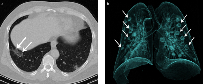
The patient underwent unenhanced chest computed tomography (CT) with a preliminary diagnosis of COVID-19 pneumonia. Chest CT demonstrated peripheral, multilobar areas of ground-glass opacity. Chest CT and 3D reconstruction of chest CT demonstrated multiple, multilobar peripherally placed lesions with a “reversed halo sign” (Fig.). Nasopharyngeal swab, obtained 1 hour after CT, was positive for COVID-19 with real-time polymerase chain reaction (RT-PCR), and the diagnosis of COVID-19 pneumonia was confirmed. The patient was hospitalized and treated with hydroxychloroquine and oseltamivir. On day 8 of hospitalization, the patient’s laboratory results and clinical symptoms improved and patient was discharged.
Figure. a, b.
Axial (a) and volume-rendered 3D reconstruction (b) chest CT images (RadiAnt DICOM Viewer 5.5.1; Medixant) demonstrate multiple, multilobar peripherally located lesions with “reversed halo sign” (arrows). See also the 3D Movie (available online).
“Reversed halo sign”, also known as the “atoll sign”, is defined as central ground-glass opacity surrounded by a more or less complete ring-like consolidation (1). It is strongly suggestive but not specific for cryptogenic organizing pneumonia, and it can be found in various infectious and noninfectious pulmonary diseases (2). This sign was recently reported in several COVID-19 studies and was found to be related with more severe and critical cases (2). We aimed to show this sign on 3D CT with a demonstrative illustration. Radiologists should be familiar with this sign to help with timely and accurate management of COVID-19 pneumonia.
Supplementary Information
Volume-rendered 3D reconstruction chest CT image (RadiAnt DICOM Viewer 5.5.1; Medixant) demonstrates multiple, multilobar peripherally located lesions with “reversed halo sign”.
Footnotes
Conflict of interest disclosure
The authors declared no conflicts of interest.
References
- 1.Ye Z, Zhang Y, Wang Y, et al. Chest CT manifestations of new coronavirus disease 2019 (COVID-19): a pictorial review. Eur Radiol. 2020:1–9. doi: 10.1007/s00330-020-06801-0. [DOI] [PMC free article] [PubMed] [Google Scholar]
- 2.Zhao H, Liang T, Wu C, et al. The reversed halo sign in COVID-19 pneumonia. Research Square. 2020. Mar 31, Available at: https://www.researchsquare.com/article/rs-20394/v1.
Associated Data
This section collects any data citations, data availability statements, or supplementary materials included in this article.
Supplementary Materials
Volume-rendered 3D reconstruction chest CT image (RadiAnt DICOM Viewer 5.5.1; Medixant) demonstrates multiple, multilobar peripherally located lesions with “reversed halo sign”.


