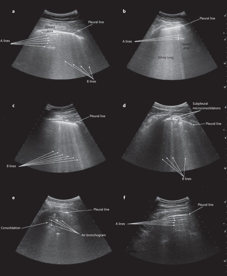
Fig. 1.
Appearance of COVID-19-related alveolar-interstitial pneumonia at bedside lung ultrasound. a Nonconfluent B lines (comet-tail artifacts) with spared areas of normal lung parenchyma showing A lines (horizontal artifacts). b Confluent B lines with “white lung” pattern and spared areas of normal lung parenchyma showing A lines. c Diffuse, nonconfluent B lines reflecting homogeneous interstitial involvement of lung parenchyma. d Subpleural microconsolidations with indentation of pleural line, associated with a nonconfluent focal B-line pattern. e Overt subpleural consolidation with air bronchograms. f Spared area showing A lines corresponding to a region of normally ventilated lung parenchyma without alveolar-interstitial involvement.

