Dear Editor,
1.
In December 2019, an unexplained outbreak occurred in China. 1 In Turkey, the first case was announced on March 2020. 2
This was a retrospective, observational study. We reviewed electronic medical records and collected data on their age, sex, comorbidities, and typical symptoms. Patients demographic and clinical characteristics are shown in Table 1.
TABLE 1.
Demographic and clinical characteristics of patients
| Id | Sex | Age | Systemic symptoms | Skin lesions | Localization | Skin symptoms | Nasopharyngeal swab | Additional pathology | Radiologic findings | Course |
|---|---|---|---|---|---|---|---|---|---|---|
| 1 | M | 32 | Cough, coryza, hyposmia, hypogeusia, weakness | Annular erythema | Trunk | No itching | Positive | No | Negative | Resolution |
| 2 | M | 42 | No | Erythema plaques | Trunk | Itching | Positive | No | Negative | Resolution |
| 3 | M | 29 | Sore throat, weakness, anosmia, myalgia | Erythematous maculer and papular lesions | Trunk | No itching | Positive | No | Negative | Resolution |
| 4 | M | 18 | Fever, headache, sinus pressure, anosmia, | Erythematousviolet macules | Foot | Mild itching | Positive | No | Negative | Resolution |
| 5 | M | 10 months | No | Erythematous rash | Trunk | Mild itching | Positive | No | Negative |
2. CASE 1
A 32‐year‐old man presented with mild dry cough, nasal congestion, flu‐like symptoms without fever. He reported loss of taste and smell. At the same time, a small red annular rash appeared on the left side of his trunk. This lesion on his trunk continued to expand. A giant erythematous macular lesion was observed on the left trunk extending in the anterior‐posterior direction (Figure 1).
FIGURE 1.
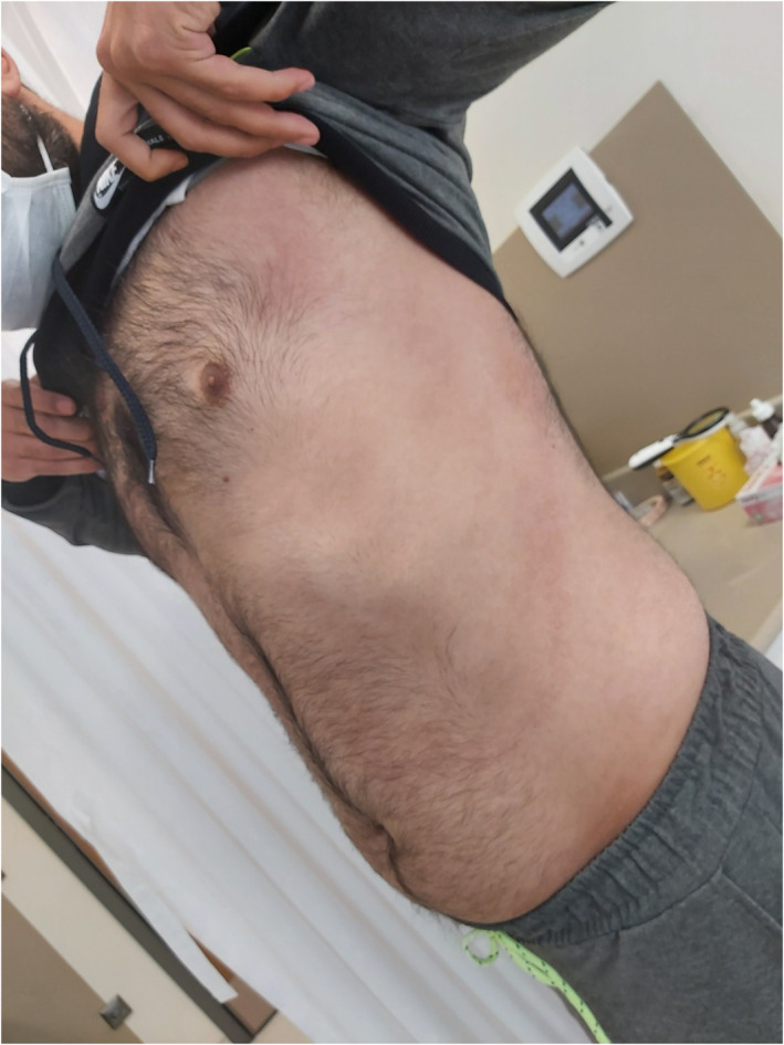
Erythematous macular lesion on the trunk
3. CASE 2
A 42‐year‐old man with COVID‐19 presented with extensive urticariform rash on his trunk (Figure 2). He was detected by the test performed because of having a Covid‐19 positive patient in his family. He has an appearance of urticarial eruption when diagnosed. He had no additional symptoms. Routine tests were normal.
FIGURE 2.
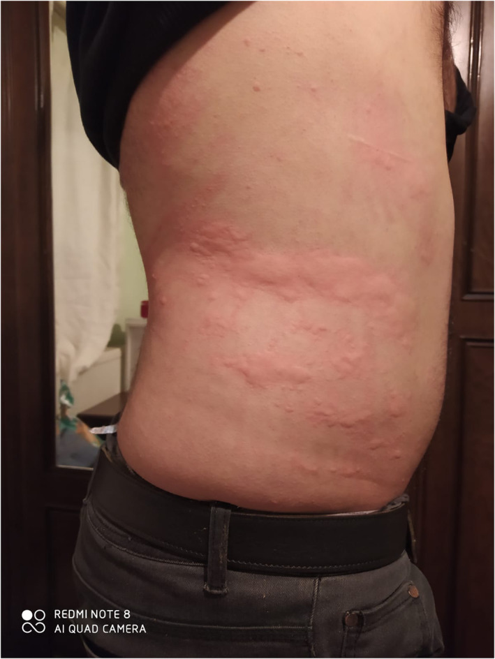
Urticariform rash on the trunk
4. CASE 3
A 29‐year‐old man presented with an acute eruption on his trunk. The number of lesions increased during the quarantine period. Tens of erythematous macular and papular lesions 1 to 15 mm in diameter with sporadic fine scales were observed on physical examination (Figures 3).
FIGURE 3.
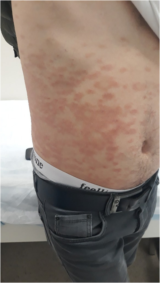
Erythematous macular and papular lesions 1 to 15 mm in diameter with sporadic fine scales
5. CASE 4
We present a healthy teenager with no history of any other skin problems. His symptoms began with a mild headache, anosmia, and low‐grade fever. A few days after the onset of these symptoms, his cutaneous lesions appeared. Purple, erythematous macules were seen on the left four toes, involving the metatarsophalangeal joints (Figure 4). He showed complete clinical recovery within a few days.
FIGURE 4.
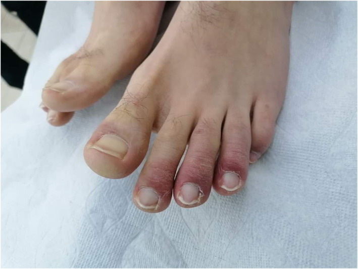
Purple and erythematous macules on 4 toes
6. CASE 5
The last patient is a 10‐month‐old baby. He exhibited a widespread erythematous rash on his body and arm (Figure 5). The rash disappeared on day 10 without treatment.
FIGURE 5.
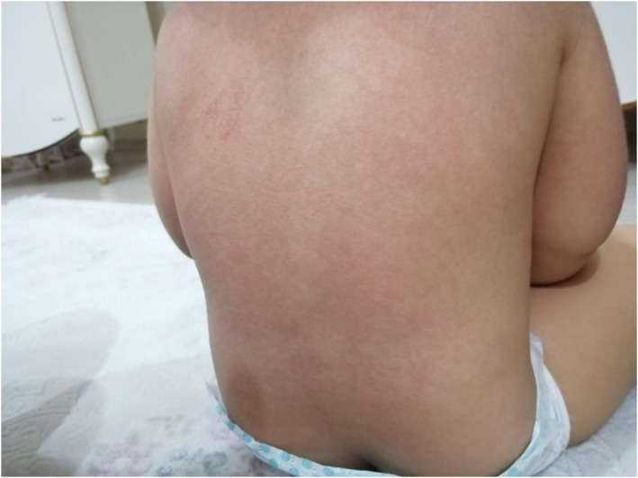
Erythematous rash on body
The first report of cutaneous manifestations in COVID‐19 was from Italy. They concluded that the manifestations are similar to skin findings seen in other common viral infections. 1
In another review on the clinical features of COVID‐19 from China, rash was observed in 0.2% of cases. However, no details about skin findings were described in that study. 3 There were also publications reporting that two infants had rashes at birth. 4
It is possible for COVID‐19 patients to initially present with a skin rash that may be misdiagnosed as another disease. In a case report, erythematous rashes similar to drug reactions were observed, suggesting that dermatologists must also consider COVID‐19 in the differential diagnosis of cutaneous drug reactions. 5 , 6
COVID‐19 can feature signs of small blood vessel occlusion. In the literature, there are some reports describing ischemic and ecchymotic lesions of the fingers, dusky acrocyanosis and dry gangrene and more frequently of the toes in patients suffering from very severe, often lethal forms of COVID‐19. 1 , 7 , 8 , 9
In brief, consistent with Recalcati's report, the lesions we describe often look like erythematous, urticaria, or morbilliform rash. The trunk was the most frequently involved region, itching was mild or absent, and the lesions usually healed within a few days. The COVID‐19 patients who presented with skin findings were relatively young and most had a mild clinical course, which we believe is a result of their younger age and lack of additional pathologies. The cutaneous manifestations could be related to the patients' immune response. Indeed, the observed skin manifestations were generally non‐specific and similar to those viral infections. Reporting cutaneous manifestations of COVID‐19 may help us to detect and treat mild or asymptomatic cases and prevent further transmission.
7. CONFLICT OF INTEREST
The authors declare no conflict of interest.
8. ETHICS STATEMENT
The study was approved by the Turkish Ministry of Health Ethics Committee and written informed consent was obtained from the patients or their families.
REFERENCES
- 1. Recalcati S. Cutaneous manifestations in COVID‐19: a first perspective. J Eur Acad Dermatol Venereol. 2020;26:e212‐e213. 10.1111/jdv.16387. [DOI] [PubMed] [Google Scholar]
- 2. https://covid19.saglik.gov.tr/#
- 3. Wei‐Jie G, Zheng‐Yi N, Yu H, Liang W, Ou C, et al. Clinical characteristics of coronavirus disease 2019 in China. N Engl J Med. 2020;382(18):1708‐1720. 10.1056/NEJMoa2002032. [DOI] [PMC free article] [PubMed] [Google Scholar]
- 4. Chen Y, Peng H, Wang L, et al. Infants born to mothers with a new coronavirus (COVID‐19). Front Pediatr. 2020;8:104. 10.3389/fped.2020.00104. [DOI] [PMC free article] [PubMed] [Google Scholar]
- 5. Fernandez‐Nieto D, Ortega‐Quijano D, Segurado‐Miravalles G, Pindado‐Ortega C, Prieto‐Barrios M, Jimenez‐Cauhe J. Comment on: cutaneous manifestations in COVID‐19: a first perspective. Safety concerns of clinical images and skin biopsies. J Eur Acad Dermatol Venereol. 2020;34:e252‐e254. 10.1111/jdv.16470. [DOI] [PMC free article] [PubMed] [Google Scholar]
- 6. Mahé A, Birckel E, Krieger S, Merklen C, Bottlaender L. A distinctive skin rash associated with coronavirus disease 2019 ? J Eur Acad Dermatol Venereol. 2020;34:e246‐e247. 10.1111/jdv.16471. [DOI] [PMC free article] [PubMed] [Google Scholar]
- 7. Zhang Y, Cao W, Xiao M, et al. Clinical and coagulation characteristics of 7 patients with critical COVID‐2019 pneumonia and acro‐ischemia. Zhonghua Xue Ye Xue Za Zhi. 2020;41:E006. 10.3760/cma.j.issn.0253-2727.2020.0006. [DOI] [PubMed] [Google Scholar]
- 8. Zhang Y, Xiao M, Zhang S, et al. Coagulopathy and antiphospholipid antibodies in patients with Covid‐19. N Engl J Med. 2020;382(17):e38. 10.1056/NEJMc2007575. [DOI] [PMC free article] [PubMed] [Google Scholar]
- 9. Mazzotta F, Troccoli T. Acute acro‐ischemia in the child at the time of COVID‐19. Eur J Pediat Dermatol. 2020;30:71‐74. https://www.ejpd.com/images/nuova-vasculite-covid-ENG.pdf. [Google Scholar]


